User login
CVH in pregnant women: Ample room for improvement
Cardiovascular disease is both common and chronic, and it remains the leading cause of death in women. Because it is a life-long condition, cardiovascular disease must be managed over the entire lifespan. In recognition of the important role of obstetricians and gynecologists in monitoring women’s health, the American Heart Association/American College of Obstetricians and Gynecologists 2018 guidelines1 promoted the use of “Life’s Simple 7”2 for assessing cardiovascular health (CVH) in women.

These seven metrics include diet, physical activity, smoking status, body mass index (BMI), blood pressure, total cholesterol, and fasting blood glucose levels. They have been shown to predict positive health outcomes in nonpregnant adults. However, until now, CVH had not been assessed in pregnant women.
Perak et al. recently performed the first cross-sectional study of the prevalence of CVH metrics in pregnant women using the AHA definition.3 Using data from the National Health and Nutrition Examination Surveys (NHANES), they used the Life’s Simple 7 metrics to assess CVH in 1,117 pregnant and 8,200 nonpregnant women in the United States aged 20-44 years. Each of the Life’s Simple 7 metrics was scored 0, 1, or 2 points, corresponding to a rating of poor, intermediate, or ideal, respectively. Thus, the total CVH score ranged from 0-14 points, with total scores of 0-7 indicating low CVH, 8-11 indicating moderate CVH, and 12-14 indicating high CVH.
which was even worse than in nonpregnant women, of whom only 13% were scored as having ideal CVH. Ideal scores were observed for 0.1% of pregnant women for diet, 27% for physical activity, 39% for cholesterol levels, 51% for BMI, 78% for smoking, 90% for blood pressure, and 92% for fasting blood glucose. Physical activity and cholesterol levels appeared to be the major drivers of the lower CVH scores in pregnant women.
Although further studies are warranted to determine the relevance of CVH during pregnancy to outcomes for both mother and offspring, the study by Perak et al. is an important step toward the development of pregnancy-specific guidelines and definitions for CVH metrics. These are stated goals of the AHA/ACOG that will help promote CVH in women across their lifespans, but which have not been possible due to scant data.
Emerging data suggest that cumulative lifetime exposure is a significant factor in cardiovascular disease outcomes; therefore, earlier intervention would have a more significant impact. Just as gestational diabetes is a predictor of future type 2 diabetes, CVH earlier in a woman’s life predicts cardiovascular disease later in life.4-7 The best data in this regard come from genetic and other studies of hyperlipidemia, which suggest that lowering lipid levels before symptoms develop may prevent cardiovascular disease. In contrast, treatment of patients with clinically manifest disease neither offers a cure nor prevents the occurrence of most cardiovascular events.
It is a particularly salient point in this regard that there currently are no guidelines on treatment of hypercholesterolemia during pregnancy. Notably, the study by Perak et al. suggested that cholesterol levels may have a significant impact on CVH in pregnant women. There also is emerging data supporting the importance of controlling blood pressure across the lifespan,7,8 including during pregnancy.9
For many women, their ob.gyn. is their primary care physician, and pregnancy is often the first time that a woman will have a substantial interaction with the health care system. The AHA/ACOG advisory panel described pregnancy as a “physiological stress test” for women that offers the opportunity to identify those at increased risk of cardiovascular disease.1
As pregnancy is a time when women particularly are motivated to improve their health,10 it also presents a valuable opportunity for physicians, including ob.gyns., to make a lifelong impact on the CVH of their patients through early identification, education, and intervention.
Dr. Charles Hong is the Melvin Sharoky, MD, Professor of Medicine and director of cardiovascular research in the department of medicine at the University of Maryland School of Medicine. Dr. E. Albert Reece, who specializes in maternal-fetal medicine, is executive vice president for medical affairs at the University of Maryland School of Medicine as well as the John Z. and Akiko K. Bowers Distinguished Professor and dean of the school of medicine. Neither physician had any relevant financial disclosures. Contact him at obnews@mdedge.com.
References
1. Circulation. 2018;137:e843–e852.
2. Circulation. 2010 Jan 20;121(4):586–613.
3. J Am Heart Assoc. 2020 Feb 17;9:e015123.
4. J Am Coll Cardiol. 2018 Sep 4;72(10):1141-56.
5. N Engl J Med. 2016 Dec 1;375:2144-53.
6. Nat Rev Cardiol. 2011 Nov 1;8(12):721-5.
7. J Am Coll Cardiol. 2019 Jul 23;74(3):330-41.
8. Circulation. 2020 Mar 2:141:725-7.
9. Circulation. 2013 Feb 12;127(6):681-90.
10. Nutrients. 2018 Aug 8. doi: 10.3390/nu10081032.
Cardiovascular disease is both common and chronic, and it remains the leading cause of death in women. Because it is a life-long condition, cardiovascular disease must be managed over the entire lifespan. In recognition of the important role of obstetricians and gynecologists in monitoring women’s health, the American Heart Association/American College of Obstetricians and Gynecologists 2018 guidelines1 promoted the use of “Life’s Simple 7”2 for assessing cardiovascular health (CVH) in women.
These seven metrics include diet, physical activity, smoking status, body mass index (BMI), blood pressure, total cholesterol, and fasting blood glucose levels. They have been shown to predict positive health outcomes in nonpregnant adults. However, until now, CVH had not been assessed in pregnant women.
Perak et al. recently performed the first cross-sectional study of the prevalence of CVH metrics in pregnant women using the AHA definition.3 Using data from the National Health and Nutrition Examination Surveys (NHANES), they used the Life’s Simple 7 metrics to assess CVH in 1,117 pregnant and 8,200 nonpregnant women in the United States aged 20-44 years. Each of the Life’s Simple 7 metrics was scored 0, 1, or 2 points, corresponding to a rating of poor, intermediate, or ideal, respectively. Thus, the total CVH score ranged from 0-14 points, with total scores of 0-7 indicating low CVH, 8-11 indicating moderate CVH, and 12-14 indicating high CVH.
which was even worse than in nonpregnant women, of whom only 13% were scored as having ideal CVH. Ideal scores were observed for 0.1% of pregnant women for diet, 27% for physical activity, 39% for cholesterol levels, 51% for BMI, 78% for smoking, 90% for blood pressure, and 92% for fasting blood glucose. Physical activity and cholesterol levels appeared to be the major drivers of the lower CVH scores in pregnant women.
Although further studies are warranted to determine the relevance of CVH during pregnancy to outcomes for both mother and offspring, the study by Perak et al. is an important step toward the development of pregnancy-specific guidelines and definitions for CVH metrics. These are stated goals of the AHA/ACOG that will help promote CVH in women across their lifespans, but which have not been possible due to scant data.
Emerging data suggest that cumulative lifetime exposure is a significant factor in cardiovascular disease outcomes; therefore, earlier intervention would have a more significant impact. Just as gestational diabetes is a predictor of future type 2 diabetes, CVH earlier in a woman’s life predicts cardiovascular disease later in life.4-7 The best data in this regard come from genetic and other studies of hyperlipidemia, which suggest that lowering lipid levels before symptoms develop may prevent cardiovascular disease. In contrast, treatment of patients with clinically manifest disease neither offers a cure nor prevents the occurrence of most cardiovascular events.
It is a particularly salient point in this regard that there currently are no guidelines on treatment of hypercholesterolemia during pregnancy. Notably, the study by Perak et al. suggested that cholesterol levels may have a significant impact on CVH in pregnant women. There also is emerging data supporting the importance of controlling blood pressure across the lifespan,7,8 including during pregnancy.9
For many women, their ob.gyn. is their primary care physician, and pregnancy is often the first time that a woman will have a substantial interaction with the health care system. The AHA/ACOG advisory panel described pregnancy as a “physiological stress test” for women that offers the opportunity to identify those at increased risk of cardiovascular disease.1
As pregnancy is a time when women particularly are motivated to improve their health,10 it also presents a valuable opportunity for physicians, including ob.gyns., to make a lifelong impact on the CVH of their patients through early identification, education, and intervention.
Dr. Charles Hong is the Melvin Sharoky, MD, Professor of Medicine and director of cardiovascular research in the department of medicine at the University of Maryland School of Medicine. Dr. E. Albert Reece, who specializes in maternal-fetal medicine, is executive vice president for medical affairs at the University of Maryland School of Medicine as well as the John Z. and Akiko K. Bowers Distinguished Professor and dean of the school of medicine. Neither physician had any relevant financial disclosures. Contact him at obnews@mdedge.com.
References
1. Circulation. 2018;137:e843–e852.
2. Circulation. 2010 Jan 20;121(4):586–613.
3. J Am Heart Assoc. 2020 Feb 17;9:e015123.
4. J Am Coll Cardiol. 2018 Sep 4;72(10):1141-56.
5. N Engl J Med. 2016 Dec 1;375:2144-53.
6. Nat Rev Cardiol. 2011 Nov 1;8(12):721-5.
7. J Am Coll Cardiol. 2019 Jul 23;74(3):330-41.
8. Circulation. 2020 Mar 2:141:725-7.
9. Circulation. 2013 Feb 12;127(6):681-90.
10. Nutrients. 2018 Aug 8. doi: 10.3390/nu10081032.

Low fitness level linked to higher risk of heart failure in diabetes
PHOENIX – Lower baseline fitness and greater decline in fitness over time are independently associated with a higher risk of heart failure in patients with diabetes, results from a large analysis showed.
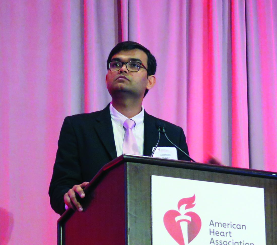
“Diabetes is an important risk factor for the development of heart failure, and the diagnosis of diabetes in newly diagnosed cases of heart failure has been increasing,” Ambarish Pandey, MD, said at the Epidemiology and Prevention/Lifestyle and Cardiometabolic Health meeting. “Type 2 diabetes is associated with increased burden of traditional risk factors such as hypertension, kidney dysfunction, and dyslipidemia – each of which in turn increase the risk of both atherothrombotic disease as well as heart failure.”
Recent data from the Swedish National Diabetes Register have shown that optimal management of these risk factors in patients with type 2 diabetes can actually mitigate the risk of atherosclerotic events such as acute MI, but the risk of heart failure does not significantly lower with optimal management of these traditional cardiovascular risk factors (N Engl J Med. 2018;379:633-44). “These findings highlight that novel approaches that go beyond just managing traditional cardiovascular risk factors are needed for prevention of heart failure in patients with type 2 diabetes,” said Dr. Pandey, of the division of cardiology at the University of Texas Southwestern Medical Center, Dallas. “Our group has demonstrated that physical inactivity and low levels of fitness are associated with a higher risk of heart failure. We have also shown that the protective effect of physical activity against heart failure risk is stronger against heart failure with preserved ejection fraction, which is a subtype of heart failure that is increasing in prevalence and has no effective therapies.”
Dr. Pandey and his colleagues set out to test the research hypothesis that fitness decline and increases in body mass index over time are significantly associated with a higher risk of heart failure. To do this, they drew from the LookAHEAD Trial, a multicenter analysis of 5,145 overweight or obese patients with type 2 diabetes who were randomized to an intensive lifestyle intervention or to usual care. The intervention consisted of a caloric intake goal of 1,200 to 1,800 kcal per day and engaging in at least 175 minutes per week of physical activity. Participants were stratified into one of three fitness group levels: low, moderate, and high, from 5 metabolic equivalents (METs) in the lowest fitness tertile to 9 METs in the highest fitness tertile. The primary outcome of the trial was adverse cardiovascular events. The intervention was implemented for almost 10 years, and patients were followed for up to 12 years from baseline.
The heart failure outcomes were not systematically adjudicated in the primary LookAHEAD trial, so Dr. Pandey and colleagues conducted an ancillary study of all incident hospitalizations in the study and followed them for 2 additional years. Overall, the researchers identified 257 incident heart failure events. The cumulative incidence of heart failure for the usual care versus the intensive lifestyle intervention arm was not statistically different (an event rate of 4.53 vs. 4.32 per 1,000 person-years, respectively; hazard ratio, 0.96). “This demonstrated that the intensive lifestyle intervention in the LookAHEAD trial did not significantly modify the risk of heart failure,” Dr. Pandey said.
However, an adjusted analysis revealed that the risk of heart failure was 39% lower in the moderately fit group and 62% lower in the high fit group, compared with the low-fitness group. Among heart failure subtypes, the risk of heart failure with preserved ejection fraction (HFpEF) was 40% lower in the moderately fit group and 77% lower in the high-fitness group. On the other hand, baseline level of fitness level was not associated with risk of heart failure reduced ejection fraction (HFrEF) after the researchers adjusted for cardiovascular risk factors.
Next, Dr. Pandey and his colleagues used Cox modeling to examine the association of baseline and longitudinal changes in fitness and BMI with risk of heart failure. For change in fitness and BMI analysis, they used the 4-year follow-up data in 3,092 participants who underwent repeat fitness testing and had available data on BMI. They excluded patients who developed heart failure within the first 4 years of the study.
The mean age of the ancillary study population was about 60 years, and there was a lower proportion of women in the high fitness tertile (41%). The researchers observed a graded, inverse association between higher fitness levels and lower risk of heart failure such that increasing fitness from baseline was associated with a substantial decrease in the risk of heart failure. Specifically, a 10% decline in fitness over the 4 years of follow-up was associated with a 11% increase in the overall risk of heart failure (HR, 1.11). “This was largely consistent with the two heart failure subtypes,” he said. Similarly, a 10% increase in BMI over the 4 years of follow-up was associated with a 25% increase in the overall risk of heart failure (HR 1.25). On the other hand, a 10% decrease BMI was associated with a 20% decrease in the risk of heart failure (HR .80). This was also largely consistent for both heart failure subtypes. According to co-lead investigator Kershaw Patel, MD, “these findings suggest that therapies targeting large and sustained improvements in fitness and weight loss may modify the risk of heart failure among patients with diabetes.”
“Lower fitness at baseline was more strongly associated with the risk of HFpEF vs. HFrEF, and greater weight loss over follow-up is associated with a lower risk of heart failure independent of changes in other risk factors,” Dr. Pandey concluded at the meeting, which was sponsored by the American Heart Association.
In an interview, session moderator Joshua J. Joseph, MD, said that it remains unclear what type of setting is ideal for carrying out cardiorespiratory fitness in this patient population. “What is the supervision needed for that to occur?” asked Dr. Joseph, of The Ohio State University, Columbus. “Can patients do this on their own, or do they need guidance? What is the best approach? That’s the question we all have to answer individually in our own communities.”
Dr. Pandey reported having no disclosures.
SOURCE: Pandey A. Epi/Lifestyle 2020, Abstract 16.
PHOENIX – Lower baseline fitness and greater decline in fitness over time are independently associated with a higher risk of heart failure in patients with diabetes, results from a large analysis showed.
“Diabetes is an important risk factor for the development of heart failure, and the diagnosis of diabetes in newly diagnosed cases of heart failure has been increasing,” Ambarish Pandey, MD, said at the Epidemiology and Prevention/Lifestyle and Cardiometabolic Health meeting. “Type 2 diabetes is associated with increased burden of traditional risk factors such as hypertension, kidney dysfunction, and dyslipidemia – each of which in turn increase the risk of both atherothrombotic disease as well as heart failure.”
Recent data from the Swedish National Diabetes Register have shown that optimal management of these risk factors in patients with type 2 diabetes can actually mitigate the risk of atherosclerotic events such as acute MI, but the risk of heart failure does not significantly lower with optimal management of these traditional cardiovascular risk factors (N Engl J Med. 2018;379:633-44). “These findings highlight that novel approaches that go beyond just managing traditional cardiovascular risk factors are needed for prevention of heart failure in patients with type 2 diabetes,” said Dr. Pandey, of the division of cardiology at the University of Texas Southwestern Medical Center, Dallas. “Our group has demonstrated that physical inactivity and low levels of fitness are associated with a higher risk of heart failure. We have also shown that the protective effect of physical activity against heart failure risk is stronger against heart failure with preserved ejection fraction, which is a subtype of heart failure that is increasing in prevalence and has no effective therapies.”
Dr. Pandey and his colleagues set out to test the research hypothesis that fitness decline and increases in body mass index over time are significantly associated with a higher risk of heart failure. To do this, they drew from the LookAHEAD Trial, a multicenter analysis of 5,145 overweight or obese patients with type 2 diabetes who were randomized to an intensive lifestyle intervention or to usual care. The intervention consisted of a caloric intake goal of 1,200 to 1,800 kcal per day and engaging in at least 175 minutes per week of physical activity. Participants were stratified into one of three fitness group levels: low, moderate, and high, from 5 metabolic equivalents (METs) in the lowest fitness tertile to 9 METs in the highest fitness tertile. The primary outcome of the trial was adverse cardiovascular events. The intervention was implemented for almost 10 years, and patients were followed for up to 12 years from baseline.
The heart failure outcomes were not systematically adjudicated in the primary LookAHEAD trial, so Dr. Pandey and colleagues conducted an ancillary study of all incident hospitalizations in the study and followed them for 2 additional years. Overall, the researchers identified 257 incident heart failure events. The cumulative incidence of heart failure for the usual care versus the intensive lifestyle intervention arm was not statistically different (an event rate of 4.53 vs. 4.32 per 1,000 person-years, respectively; hazard ratio, 0.96). “This demonstrated that the intensive lifestyle intervention in the LookAHEAD trial did not significantly modify the risk of heart failure,” Dr. Pandey said.
However, an adjusted analysis revealed that the risk of heart failure was 39% lower in the moderately fit group and 62% lower in the high fit group, compared with the low-fitness group. Among heart failure subtypes, the risk of heart failure with preserved ejection fraction (HFpEF) was 40% lower in the moderately fit group and 77% lower in the high-fitness group. On the other hand, baseline level of fitness level was not associated with risk of heart failure reduced ejection fraction (HFrEF) after the researchers adjusted for cardiovascular risk factors.
Next, Dr. Pandey and his colleagues used Cox modeling to examine the association of baseline and longitudinal changes in fitness and BMI with risk of heart failure. For change in fitness and BMI analysis, they used the 4-year follow-up data in 3,092 participants who underwent repeat fitness testing and had available data on BMI. They excluded patients who developed heart failure within the first 4 years of the study.
The mean age of the ancillary study population was about 60 years, and there was a lower proportion of women in the high fitness tertile (41%). The researchers observed a graded, inverse association between higher fitness levels and lower risk of heart failure such that increasing fitness from baseline was associated with a substantial decrease in the risk of heart failure. Specifically, a 10% decline in fitness over the 4 years of follow-up was associated with a 11% increase in the overall risk of heart failure (HR, 1.11). “This was largely consistent with the two heart failure subtypes,” he said. Similarly, a 10% increase in BMI over the 4 years of follow-up was associated with a 25% increase in the overall risk of heart failure (HR 1.25). On the other hand, a 10% decrease BMI was associated with a 20% decrease in the risk of heart failure (HR .80). This was also largely consistent for both heart failure subtypes. According to co-lead investigator Kershaw Patel, MD, “these findings suggest that therapies targeting large and sustained improvements in fitness and weight loss may modify the risk of heart failure among patients with diabetes.”
“Lower fitness at baseline was more strongly associated with the risk of HFpEF vs. HFrEF, and greater weight loss over follow-up is associated with a lower risk of heart failure independent of changes in other risk factors,” Dr. Pandey concluded at the meeting, which was sponsored by the American Heart Association.
In an interview, session moderator Joshua J. Joseph, MD, said that it remains unclear what type of setting is ideal for carrying out cardiorespiratory fitness in this patient population. “What is the supervision needed for that to occur?” asked Dr. Joseph, of The Ohio State University, Columbus. “Can patients do this on their own, or do they need guidance? What is the best approach? That’s the question we all have to answer individually in our own communities.”
Dr. Pandey reported having no disclosures.
SOURCE: Pandey A. Epi/Lifestyle 2020, Abstract 16.
REPORTING FROM EPI/LIFESTYLE 2020
Dramatic rise in hypertension-related deaths in the United States
There has been a dramatic rise in hypertension-related deaths in the United States between 2007 and 2017, a new study shows. The authors, led by Lakshmi Nambiar, MD, Larner College of Medicine, University of Vermont, Burlington, analyzed data from the Centers for Disease Control and Prevention, which collates information from every death certificate in the country, amounting to more than 10 million deaths.
They found that age-adjusted hypertension-related deaths had increased from 18.3 per 100,000 in 2007 to 23.0 per 100,000 in 2017 (P < .001 for decade-long temporal trend).
Nambiar reported results of the study at an American College of Cardiology 2020/World Congress of Cardiology press conference on March 19. It was also published online on the same day in the Journal of the American College of Cardiology.
She noted that death rates due to cardiovascular disease have been falling over the past 20 years largely attributable to statins to treat high cholesterol and stents to treat coronary artery disease. But since 2011, the rate of decline in cardiovascular deaths has slowed. One contributing factor is an increase in heart failure-related deaths but there hasn’t been any data in recent years on hypertension-related deaths.
“Our data show an increase in hypertension-related deaths in all age groups, in all regions of the United States, and in both sexes. These findings are alarming and warrant further investigation, as well as preventative efforts,” Nambiar said. “This is a public health emergency that has not been fully recognized,” she added.
“We were surprised to see how dramatically these deaths were increasing, and we think this is related to the rise in diabetes, obesity, and the aging of the population. We need targeted public health measures to address some of those factors,” Nambiar told Medscape Medical News.
“We are winning the battle against coronary artery disease with statins and stents but we are not winning the battle against hypertension,” she added.
Worst Figures in Rural South
Results showed that hypertension-related deaths increased in both rural and urban regions, but the increase was much steeper in rural areas — a 72% increase over the decade compared with a 20% increase in urban areas.
The highest death risk was identified in the rural South, which demonstrated an age-adjusted 2.5-fold higher death rate compared with other regions (P < .001).
The urban South also demonstrated increasing hypertension-related cardiovascular death rates over time: age-adjusted death rates in the urban South increased by 27% compared with all other urban regions (P < .001).
But the absolute mortality rates and slope of the curves demonstrate the highest risk in patients in the rural South, the researchers report. Age-adjusted hypertension-related death rates increased in the rural South from 23.9 deaths per 100,000 in 2007 to 39.5 deaths per 100,000 in 2017.
Nambiar said the trends in the rural South could be related to social factors and lack of access to healthcare in the area, which has been exacerbated by failure to adopt Medicaid expansion in many of the states in this region.
“When it comes to the management of hypertension you need to be seen regularly by a primary care doctor to get the best treatment and regular assessments,” she stressed.
Chair of the ACC press conference at which the data were presented, Martha Gulati, MD, University of Arizona School of Medicine, Phoenix, said: “In this day and time, there is less smoking, which should translate into lower rates of hypertension, but these trends reported here are very different from what we would expect and are probably associated with the rise in other risk factors such as diabetes and obesity, especially in the rural South.”
Nambiar praised the new ACC/AHA hypertension guidelines that recommend a lower diagnostic threshold, “so more people now fit the criteria for raised blood pressure and need treatment.”
“It is important for all primary care physicians and cardiologists to recognize the new threshold and treat people accordingly,” she said. “High blood pressure is the leading cause of cardiovascular disease. If we can control it better, we may be able to control some of this increased mortality we are seeing.”
This article first appeared on Medscape.com.

AFib-related cardiovascular deaths on the rise
PHOENIX, ARIZ. – Cardiovascular deaths and death rates related to atrial fibrillation have risen since 1999, with significant acceleration following 2009, results from a cross-sectional analysis of national data show.

“AFib is the most common arrhythmia disorder in the United States and it is estimated that it will effect more than 12 million Americans by 2030,” Yoshihiro Tanaka, MD, PhD, said at the Epidemiology and Prevention/Lifestyle and Cardiometabolic Health meeting. “The predicted lifetime risk ranges from 25% to 35%, and AFib is associated with an increased risk for heart failure, stroke, and death.”
A recent review reported that declines in total heart disease mortality rates in the United States have plateaued since 2011 (JAMA 2019;322[8]:780-2). However, it is not well understood what factors such as AFib contribute to this rate of plateau. In an effort to quantify U.S. trends in AFib-related CVD death rates, Dr. Tanaka and colleagues conducted a serial cross-sectional analysis of death certificate data from the Centers for Disease Control and Prevention’s Wide-Ranging Online Data for Epidemiologic Research (WONDER) database during 1999-2017.
Outcomes included age-adjusted mortality per 100,000 based on the 2000 U.S. standard population. The researchers also used joinpoint regression to calculate the average annual percentage change over time and conducted subgroup analyses by race and sex and across two age groups: 35-64 years and 65-84 years.
In all, 522,104 AFib-related CVD deaths were identified during 1999-2017. Dr. Tanaka reported that age-adjusted mortality increased from 16.0 per 100,000 persons in 1999 to 22.2 per 100,000 person in 2017, with an acceleration following an inflection point in 2009. Specifically, the average annual percentage change in AFib-related CVD deaths rose from 0.4% in 2009 to 3.5% in 2017 (P < .001). “These increases were consistent across all race-sex subgroups,” said Dr. Tanaka, of the department of preventive medicine at Northwestern University, Chicago. “Relative increases were also greater in younger compared with older adults, although the absolute number of deaths in younger adults was less.”
The researchers observed that age-adjusted mortality increased across blacks and whites in both age groups, with a more pronounced increase among black and white men. Black men had the highest age-adjusted mortality among persons aged 35-64 (6.5 per 100,000 persons, compared with 4.2 among white men, 2.8 in black women, and 1.6 in white women 1.6 per 100,000). At the same time, white men had the highest age-adjusted mortality rate among those aged 65-84 years (112.5 per 100,000 persons, compared with 87.7 in black men, 77.4 in white women, and 61.3 in black women).
In an interview, one of the session’s moderators, Alvaro Alonso, MD, PhD, said that the study’s reliance on mortality data is a limitation. “You have to be careful with that, because it’s not the whole picture,” said Dr. Alonso, professor of epidemiology at the Rollins School of Public Health at Emory University, Atlanta. “It could be an underestimation of what is going on. The increase in recent years is probably due to a higher awareness of AFib as a risk factor for stroke; it’s more on the radar. Also, around 2009-2010, we started having new anticoagulants for AFib. It’s getting diagnosed more. When you look at coronary heart disease and stroke, there has been a decrease over time. In mortality and incidence of AFib, we don’t have that. That’s probably because we don’t know very much about what the risk factors for AFib are and how to prevent it.”
Dr. Tanaka said that the cause of increase in AFib-related CVD mortality can be classified into two major categories: a balance between case fatality of AFib and the prevalence of AFib. “The case fatality rate should have decreased over the last years,” he said at the meeting, which was sponsored by the American Heart Association. “In contrast, in the context of the aging of the population, the prevalence of AFib increased over the past years. Contributing factors include increasing awareness of AFib, a change in coding between ICD-9 and ICD-10, and a change in coding practices by physicians.”
Strengths of the study, he said, include its large sample size and the fact that the researchers were able to capture data from all death certificates filed in the United States. Limitations include the fact that the data “do not identify if changes in age-adjusted mortality rates are due to changing incidence or to case fatality rates,” he said. “CDC WONDER does not allow us to explore causes of these descriptive findings, but this would be an important next step.”
Dr. Tanaka reported having no financial disclosures.
SOURCE: Tanaka Y. EPI/Lifestyle 2020, Session 5, Abstract 15.
PHOENIX, ARIZ. – Cardiovascular deaths and death rates related to atrial fibrillation have risen since 1999, with significant acceleration following 2009, results from a cross-sectional analysis of national data show.
“AFib is the most common arrhythmia disorder in the United States and it is estimated that it will effect more than 12 million Americans by 2030,” Yoshihiro Tanaka, MD, PhD, said at the Epidemiology and Prevention/Lifestyle and Cardiometabolic Health meeting. “The predicted lifetime risk ranges from 25% to 35%, and AFib is associated with an increased risk for heart failure, stroke, and death.”
A recent review reported that declines in total heart disease mortality rates in the United States have plateaued since 2011 (JAMA 2019;322[8]:780-2). However, it is not well understood what factors such as AFib contribute to this rate of plateau. In an effort to quantify U.S. trends in AFib-related CVD death rates, Dr. Tanaka and colleagues conducted a serial cross-sectional analysis of death certificate data from the Centers for Disease Control and Prevention’s Wide-Ranging Online Data for Epidemiologic Research (WONDER) database during 1999-2017.
Outcomes included age-adjusted mortality per 100,000 based on the 2000 U.S. standard population. The researchers also used joinpoint regression to calculate the average annual percentage change over time and conducted subgroup analyses by race and sex and across two age groups: 35-64 years and 65-84 years.
In all, 522,104 AFib-related CVD deaths were identified during 1999-2017. Dr. Tanaka reported that age-adjusted mortality increased from 16.0 per 100,000 persons in 1999 to 22.2 per 100,000 person in 2017, with an acceleration following an inflection point in 2009. Specifically, the average annual percentage change in AFib-related CVD deaths rose from 0.4% in 2009 to 3.5% in 2017 (P < .001). “These increases were consistent across all race-sex subgroups,” said Dr. Tanaka, of the department of preventive medicine at Northwestern University, Chicago. “Relative increases were also greater in younger compared with older adults, although the absolute number of deaths in younger adults was less.”
The researchers observed that age-adjusted mortality increased across blacks and whites in both age groups, with a more pronounced increase among black and white men. Black men had the highest age-adjusted mortality among persons aged 35-64 (6.5 per 100,000 persons, compared with 4.2 among white men, 2.8 in black women, and 1.6 in white women 1.6 per 100,000). At the same time, white men had the highest age-adjusted mortality rate among those aged 65-84 years (112.5 per 100,000 persons, compared with 87.7 in black men, 77.4 in white women, and 61.3 in black women).
In an interview, one of the session’s moderators, Alvaro Alonso, MD, PhD, said that the study’s reliance on mortality data is a limitation. “You have to be careful with that, because it’s not the whole picture,” said Dr. Alonso, professor of epidemiology at the Rollins School of Public Health at Emory University, Atlanta. “It could be an underestimation of what is going on. The increase in recent years is probably due to a higher awareness of AFib as a risk factor for stroke; it’s more on the radar. Also, around 2009-2010, we started having new anticoagulants for AFib. It’s getting diagnosed more. When you look at coronary heart disease and stroke, there has been a decrease over time. In mortality and incidence of AFib, we don’t have that. That’s probably because we don’t know very much about what the risk factors for AFib are and how to prevent it.”
Dr. Tanaka said that the cause of increase in AFib-related CVD mortality can be classified into two major categories: a balance between case fatality of AFib and the prevalence of AFib. “The case fatality rate should have decreased over the last years,” he said at the meeting, which was sponsored by the American Heart Association. “In contrast, in the context of the aging of the population, the prevalence of AFib increased over the past years. Contributing factors include increasing awareness of AFib, a change in coding between ICD-9 and ICD-10, and a change in coding practices by physicians.”
Strengths of the study, he said, include its large sample size and the fact that the researchers were able to capture data from all death certificates filed in the United States. Limitations include the fact that the data “do not identify if changes in age-adjusted mortality rates are due to changing incidence or to case fatality rates,” he said. “CDC WONDER does not allow us to explore causes of these descriptive findings, but this would be an important next step.”
Dr. Tanaka reported having no financial disclosures.
SOURCE: Tanaka Y. EPI/Lifestyle 2020, Session 5, Abstract 15.
REPORTING FROM EPI/LIFESTYLE 2020
Cardiovascular risk varies between black ethnic subgroups
PHOENIX, ARIZ. – Cardiovascular disease risk factors differ significantly between three black ethnic subgroups in the United States, compared with whites, results from a large, long-term cross-sectional study show.

“Race alone does not account for health disparities in CVD risk factors,” lead author Diana Baptiste, DNP, RN, CNE, said at the Epidemiology and Prevention/Lifestyle and Cardiometabolic Health meeting. “We must consider the environmental, psychosocial, and social factors that may play a larger role in CVD risk among these populations.”
Dr. Baptiste, of the Johns Hopkins University School of Nursing Center for Cardiovascular and Chronic Care in Baltimore, noted that blacks bear a disproportionately greater burden of CVD than that of any other racial group. “Blacks living in the U.S. are not monolithic and include different ethnic subgroups: African Americans, Afro-Caribbeans, defined as black persons who are born in the Caribbean islands, and African immigrants, defined as black persons who are born in Africa,” she said. “It is unclear how Afro-Caribbeans and African immigrants compare to African Americans and whites with regard to CVD risk factors.”
To examine trends in CVD risk factors among the three black ethnic subgroups compared with whites, she and her colleagues performed a cross-sectional analysis of 452,997 adults who participated in the 2010-2018 National Health Interview Survey (NHIS). Of these, 82% were white and 18% were black. Among blacks, 89% were African Americans, 6% were Afro-Caribbeans, and 5% were African immigrants. Outcomes of interest were four self-reported CVD risk factors: hypertension, diabetes, overweight/obesity, and smoking. The researchers used generalized linear models with Poisson distribution to calculate predictive probabilities of CVD risk factors, adjusted for age and sex.
Dr. Baptiste reported that African immigrants represented the youngest subgroup, with an average age of 41 years, compared with an average age of 50 among whites. They were also less likely to have health insurance (76%), compared with Afro-Caribbeans (81%), African Americans (83%), and whites (91%; P < .001). Disparities were observed in the proportion of individuals living below the poverty level. This was led by African Americans (24%), followed by African immigrants (22%), Afro-Caribbeans (18%), and whites (9%).
African immigrants were most likely to be college educated (36%), compared with whites (32%), Afro-Caribbeans (23%), and African Americans (17%; P =.001). In addition, only 33% of African Americans were married, compared with more than 50% of participants in the other ethnic groups.
African Americans had the highest prevalence of hypertension over the time period (from 44% in 2010 to 42% in 2018), while African immigrants had the lowest (from 19% to 17%). African Americans also had the highest prevalence of diabetes over the time period (from 14% to 15%), while African immigrants had the lowest (from 9% to 7%). The prevalence of overweight and obesity was highest among African Americans (from 74% to 76%), while African immigrants had the lowest (63% to 60%). Finally, smoking prevalence was highest in whites and African Americans compared with African immigrants and Afro-Caribbeans, but the prevalence decreased significantly between 2010 and 2018 (P for trend < .001).
In an interview, one of the meeting session’s moderators, Sherry-Ann Brown, MD, PhD, said that the study’s findings underscore the importance of heterogeneity when counseling patients about CVD risk factors. “Everybody comes from a different cultural background,” said Dr. Brown, a cardiologist and physician scientist at Mayo Clinic, Rochester, Minn. “Cultural backgrounds have an impact on when people eat, how they eat, who they eat with, when they exercise, and whether obesity is valued or not. It’s important to recognize that those cultural underpinnings can contribute to heterogeneity. Other factors – whether they are psychosocial or socioeconomic or environmental – also contribute.”
Strengths of the study, Dr. Baptiste said, included the use of a large, nationally representative dataset. Limitations included its cross-sectional design and the National Health Interview Survey’s reliance on self-reported data. “There were also small sample sizes for African immigrants and Afro-Caribbeans,” she said.
The study was supported by Johns Hopkins University School of Nursing Center for Cardiovascular and Chronic Care. Dr. Baptiste reported having no financial disclosures.
The meeting was sponsored by the American Heart Association.
SOURCE: Baptiste D et al. EPI/Lifestyle 2020, Session 4, Abstract 8.
PHOENIX, ARIZ. – Cardiovascular disease risk factors differ significantly between three black ethnic subgroups in the United States, compared with whites, results from a large, long-term cross-sectional study show.
“Race alone does not account for health disparities in CVD risk factors,” lead author Diana Baptiste, DNP, RN, CNE, said at the Epidemiology and Prevention/Lifestyle and Cardiometabolic Health meeting. “We must consider the environmental, psychosocial, and social factors that may play a larger role in CVD risk among these populations.”
Dr. Baptiste, of the Johns Hopkins University School of Nursing Center for Cardiovascular and Chronic Care in Baltimore, noted that blacks bear a disproportionately greater burden of CVD than that of any other racial group. “Blacks living in the U.S. are not monolithic and include different ethnic subgroups: African Americans, Afro-Caribbeans, defined as black persons who are born in the Caribbean islands, and African immigrants, defined as black persons who are born in Africa,” she said. “It is unclear how Afro-Caribbeans and African immigrants compare to African Americans and whites with regard to CVD risk factors.”
To examine trends in CVD risk factors among the three black ethnic subgroups compared with whites, she and her colleagues performed a cross-sectional analysis of 452,997 adults who participated in the 2010-2018 National Health Interview Survey (NHIS). Of these, 82% were white and 18% were black. Among blacks, 89% were African Americans, 6% were Afro-Caribbeans, and 5% were African immigrants. Outcomes of interest were four self-reported CVD risk factors: hypertension, diabetes, overweight/obesity, and smoking. The researchers used generalized linear models with Poisson distribution to calculate predictive probabilities of CVD risk factors, adjusted for age and sex.
Dr. Baptiste reported that African immigrants represented the youngest subgroup, with an average age of 41 years, compared with an average age of 50 among whites. They were also less likely to have health insurance (76%), compared with Afro-Caribbeans (81%), African Americans (83%), and whites (91%; P < .001). Disparities were observed in the proportion of individuals living below the poverty level. This was led by African Americans (24%), followed by African immigrants (22%), Afro-Caribbeans (18%), and whites (9%).
African immigrants were most likely to be college educated (36%), compared with whites (32%), Afro-Caribbeans (23%), and African Americans (17%; P =.001). In addition, only 33% of African Americans were married, compared with more than 50% of participants in the other ethnic groups.
African Americans had the highest prevalence of hypertension over the time period (from 44% in 2010 to 42% in 2018), while African immigrants had the lowest (from 19% to 17%). African Americans also had the highest prevalence of diabetes over the time period (from 14% to 15%), while African immigrants had the lowest (from 9% to 7%). The prevalence of overweight and obesity was highest among African Americans (from 74% to 76%), while African immigrants had the lowest (63% to 60%). Finally, smoking prevalence was highest in whites and African Americans compared with African immigrants and Afro-Caribbeans, but the prevalence decreased significantly between 2010 and 2018 (P for trend < .001).
In an interview, one of the meeting session’s moderators, Sherry-Ann Brown, MD, PhD, said that the study’s findings underscore the importance of heterogeneity when counseling patients about CVD risk factors. “Everybody comes from a different cultural background,” said Dr. Brown, a cardiologist and physician scientist at Mayo Clinic, Rochester, Minn. “Cultural backgrounds have an impact on when people eat, how they eat, who they eat with, when they exercise, and whether obesity is valued or not. It’s important to recognize that those cultural underpinnings can contribute to heterogeneity. Other factors – whether they are psychosocial or socioeconomic or environmental – also contribute.”
Strengths of the study, Dr. Baptiste said, included the use of a large, nationally representative dataset. Limitations included its cross-sectional design and the National Health Interview Survey’s reliance on self-reported data. “There were also small sample sizes for African immigrants and Afro-Caribbeans,” she said.
The study was supported by Johns Hopkins University School of Nursing Center for Cardiovascular and Chronic Care. Dr. Baptiste reported having no financial disclosures.
The meeting was sponsored by the American Heart Association.
SOURCE: Baptiste D et al. EPI/Lifestyle 2020, Session 4, Abstract 8.
REPORTING FROM EPI/LIFESTYLE 2020
COVID-19 will test medical supply stocks
In a JAMA Live Stream interview, in the United States.

Dr. Fauci got into the details of what is known, what is unknown, what is being done in laboratories, and what clinical elements are still not understood about this disease.
The next several weeks, he said, are likely to tell the tale of whether our health care system is up to the challenge of care for the most ill among those who will be affected by COVID-19.
“It shouldn’t panic or frighten us, but we have to know we’re dealing with a very serious problem that we have to address, and we have to deal with it in a very bold way,” said Dr. Fauci, director of the National Institute of Allergy and Infectious Diseases at the National Institutes of Health.
Speaking in an interview with JAMA Editor in Chief Howard Bauchner, MD, Dr. Fauci said the situation favors action over fear. “Let’s apply that energy to doing the things that we know can mitigate this.”
He added that he heard the message loud and clear from health care leaders in Italy and France during a World Health Organization coronavirus call earlier in the day. Officials in those countries, he said, were “almost pleading with the rest of the world to please take this very seriously, because it happens all of a sudden – very abruptly. ... The best time to mitigate is before that happens, because if you wait until after it happens you’re playing catch-up.”
Dr. Bauchner, noting that strict social distancing has been underway in many parts of the United States for several days, posited that, by early April, “We’ll really have a sense if we can manage in terms of serious illness.” Seattle, New York, Boston, and the San Francisco Bay Area may experience demand that outstrips ICU capacity at that point, but the rest of the country, he said, “is doing relatively well.”
Stress test on the health care system
Dr. Fauci agreed with this statement and added: “We’re going to know – for better or worse – whether we have enough of what it takes to be able to practice the kind of medicine that we optimally would want to practice.
In the matter of a week or 2 ... I think we’ll get a feel for whether or not we really have enough of the supplies that it takes.”
The well-publicized regional shortages in personal protective equipment (PPE) are forcing tough choices in some areas. As expedited – and even drive-through – testing begins, some of the demand for testing-related PPE may abate, especially if protocols include self-administration of nasal swabs, he noted.
Dr. Fauci added that the strategic national stockpile of medical supplies and equipment has not yet been tapped, “but you need to backfill that as quickly as you can once you start drawing from the strategic national stockpile.”
Returning to work after COVID-19 infection
Regarding the thorny question of when health care workers should be permitted to return to work after coronavirus infection, “it’s an evolving story,” said Dr. Fauci. Current guidance advises that health care providers stay away from work until two negative tests after resolution of fever and improvement of respiratory symptoms, or 3 fever-free days.
“We are approaching a point where you’re going to get enough people who are getting infected that we aren’t going to be able to do that,” he said. Depending on the stress to the health care system in a given locality, he said that facilities are going to have to “decide with good judgment” when health care workers go back on the job after coronavirus infection.
Asked how soon an individual would reliably test positive for COVID-19 after exposure, Dr. Fauci said, “We don’t know the answer to that. ... We can surmise it ...” He noted that it’s a median of about 5 days with a range of 2 to 14 days, before an infected individual becomes symptomatic. “I can say it’s not going to happen immediately,” he added, noting that he wouldn’t expect to see a positive test until about 2 days after exposure at the earliest. “When you get to the point where you are symptomatic, you’re almost certainly going to be positive then. ... This is just an extrapolation,” rather than conclusions drawn from solid data, he emphasized.
Higher risk reported in cardiac patients
Dr. Bauchner, who was relaying questions sent in from physicians during the live-streamed interview, asked about a newly issued joint statement from the American Heart Association, American College of Cardiology, and the Heart Failure Society of America, which on March 17 affirmed that individuals on ACE inhibitors and angiotensin receptor blockers (ARBs) continue that therapy if they should become ill with COVID-19. The European Society of Cardiology issued a similar recommendation a few days prior.
Despite these societies’ statements, Dr. Fauci pointed to population-level data in Italy as suggesting that the case isn’t yet closed. “We really need to get data, and we need to get data fast. There’s a mechanistic rationale for the concern. It’s there, and it’s firm,” he said. The theoretical concern is that ACE inhibitors can upregulate expression of the ACE-2 protein on cell membranes, which is the entry point for SARS-Cov-2 to enter cells.
He added that he remains concerned about the number of coronavirus fatalities of patients in Italy who had hypertension as their only, or primary, underlying health problem.“That to me was a bit of a red flag,” he said. “Patients with hypertension almost certainly had a physician, and the physician almost certainly treated that person with medication. Why should someone who has hypertension that was well controlled have a much greater chance of dying?” he asked, noting that “I look at a person with well-controlled hypertension as a relatively healthy person. I don’t know what the answer is, but somebody has to look very carefully,” ideally by means of a natural history study that identifies medications used by those who died from coronavirus.
Potential therapies
Regarding potential therapies for COVID-19, Dr. Fauci acknowledged the social media buzz and flurry of medical letters and case reports about the use of hydroxychloroquine (Plaquenil) to treat active infection. He said that he and other researchers are “in active discussion” about how best to study the efficacy and safety of hydroxychloroquine, but he also acknowledged that many treating clinicians will use hydroxychloroquine empirically in the absence of other treatments with proven efficacy.
Clinical trials underway in China for antiviral medication are facing some enrollment challenges currently “because people want to get the drug,” said Dr. Fauci. “They don’t want to be in the trial; they just want to get the drug.” Though each of two trials has targeted approximately 500 participants as the number needed for sufficient statistical power, Dr. Fauci urged Chinese data safety monitoring boards to “take a close look” at the data already accrued for the several hundred patients who have already enrolled for the studies “to see if there’s any hint of efficacy.”
In a JAMA Live Stream interview, in the United States.
Dr. Fauci got into the details of what is known, what is unknown, what is being done in laboratories, and what clinical elements are still not understood about this disease.
The next several weeks, he said, are likely to tell the tale of whether our health care system is up to the challenge of care for the most ill among those who will be affected by COVID-19.
“It shouldn’t panic or frighten us, but we have to know we’re dealing with a very serious problem that we have to address, and we have to deal with it in a very bold way,” said Dr. Fauci, director of the National Institute of Allergy and Infectious Diseases at the National Institutes of Health.
Speaking in an interview with JAMA Editor in Chief Howard Bauchner, MD, Dr. Fauci said the situation favors action over fear. “Let’s apply that energy to doing the things that we know can mitigate this.”
He added that he heard the message loud and clear from health care leaders in Italy and France during a World Health Organization coronavirus call earlier in the day. Officials in those countries, he said, were “almost pleading with the rest of the world to please take this very seriously, because it happens all of a sudden – very abruptly. ... The best time to mitigate is before that happens, because if you wait until after it happens you’re playing catch-up.”
Dr. Bauchner, noting that strict social distancing has been underway in many parts of the United States for several days, posited that, by early April, “We’ll really have a sense if we can manage in terms of serious illness.” Seattle, New York, Boston, and the San Francisco Bay Area may experience demand that outstrips ICU capacity at that point, but the rest of the country, he said, “is doing relatively well.”
Stress test on the health care system
Dr. Fauci agreed with this statement and added: “We’re going to know – for better or worse – whether we have enough of what it takes to be able to practice the kind of medicine that we optimally would want to practice.
In the matter of a week or 2 ... I think we’ll get a feel for whether or not we really have enough of the supplies that it takes.”
The well-publicized regional shortages in personal protective equipment (PPE) are forcing tough choices in some areas. As expedited – and even drive-through – testing begins, some of the demand for testing-related PPE may abate, especially if protocols include self-administration of nasal swabs, he noted.
Dr. Fauci added that the strategic national stockpile of medical supplies and equipment has not yet been tapped, “but you need to backfill that as quickly as you can once you start drawing from the strategic national stockpile.”
Returning to work after COVID-19 infection
Regarding the thorny question of when health care workers should be permitted to return to work after coronavirus infection, “it’s an evolving story,” said Dr. Fauci. Current guidance advises that health care providers stay away from work until two negative tests after resolution of fever and improvement of respiratory symptoms, or 3 fever-free days.
“We are approaching a point where you’re going to get enough people who are getting infected that we aren’t going to be able to do that,” he said. Depending on the stress to the health care system in a given locality, he said that facilities are going to have to “decide with good judgment” when health care workers go back on the job after coronavirus infection.
Asked how soon an individual would reliably test positive for COVID-19 after exposure, Dr. Fauci said, “We don’t know the answer to that. ... We can surmise it ...” He noted that it’s a median of about 5 days with a range of 2 to 14 days, before an infected individual becomes symptomatic. “I can say it’s not going to happen immediately,” he added, noting that he wouldn’t expect to see a positive test until about 2 days after exposure at the earliest. “When you get to the point where you are symptomatic, you’re almost certainly going to be positive then. ... This is just an extrapolation,” rather than conclusions drawn from solid data, he emphasized.
Higher risk reported in cardiac patients
Dr. Bauchner, who was relaying questions sent in from physicians during the live-streamed interview, asked about a newly issued joint statement from the American Heart Association, American College of Cardiology, and the Heart Failure Society of America, which on March 17 affirmed that individuals on ACE inhibitors and angiotensin receptor blockers (ARBs) continue that therapy if they should become ill with COVID-19. The European Society of Cardiology issued a similar recommendation a few days prior.
Despite these societies’ statements, Dr. Fauci pointed to population-level data in Italy as suggesting that the case isn’t yet closed. “We really need to get data, and we need to get data fast. There’s a mechanistic rationale for the concern. It’s there, and it’s firm,” he said. The theoretical concern is that ACE inhibitors can upregulate expression of the ACE-2 protein on cell membranes, which is the entry point for SARS-Cov-2 to enter cells.
He added that he remains concerned about the number of coronavirus fatalities of patients in Italy who had hypertension as their only, or primary, underlying health problem.“That to me was a bit of a red flag,” he said. “Patients with hypertension almost certainly had a physician, and the physician almost certainly treated that person with medication. Why should someone who has hypertension that was well controlled have a much greater chance of dying?” he asked, noting that “I look at a person with well-controlled hypertension as a relatively healthy person. I don’t know what the answer is, but somebody has to look very carefully,” ideally by means of a natural history study that identifies medications used by those who died from coronavirus.
Potential therapies
Regarding potential therapies for COVID-19, Dr. Fauci acknowledged the social media buzz and flurry of medical letters and case reports about the use of hydroxychloroquine (Plaquenil) to treat active infection. He said that he and other researchers are “in active discussion” about how best to study the efficacy and safety of hydroxychloroquine, but he also acknowledged that many treating clinicians will use hydroxychloroquine empirically in the absence of other treatments with proven efficacy.
Clinical trials underway in China for antiviral medication are facing some enrollment challenges currently “because people want to get the drug,” said Dr. Fauci. “They don’t want to be in the trial; they just want to get the drug.” Though each of two trials has targeted approximately 500 participants as the number needed for sufficient statistical power, Dr. Fauci urged Chinese data safety monitoring boards to “take a close look” at the data already accrued for the several hundred patients who have already enrolled for the studies “to see if there’s any hint of efficacy.”
REPORTING FROM JAMA LIVE STREAM

COVID-19: U.S. cardiology groups reaffirm continued use of RAAS-active drugs
Controversy continued over the potential effect of drugs that interfere with the renin-angiotensin-aldosterone system via the angiotensin-converting enzymes (ACE) may have on exacerbating infection with the SARS-CoV-2 virus that causes COVID-19.
A joint statement from the American Heart Association, American College of Cardiology, and the Heart Failure Society of America on March 17 gave full, unqualified support to maintaining patients on drugs that work this way, specifically the ACE inhibitors and angiotensin-receptor blockers (ARBs), which together form a long-standing cornerstone of treatment for hypertension, heart failure, and ischemic heart disease.
The three societies “recommend continuation” of ACE inhibitors or ARBs “for all patients already prescribed.” The statement went on to say that patients already diagnosed with a COVID-19 infection “should be fully evaluated before adding or removing any treatments, and any changes to their treatment should be based on the latest scientific evidence and shared decision making with their physician and health care team.”
“We understand the concern – as it has become clear that people with cardiovascular disease are at much higher risk of serious complications including death from COVID-19. However, we have reviewed the latest research – the evidence does not confirm the need to discontinue ACE inhibitors or ARBs, and we strongly recommend all physicians to consider the individual needs of each patient before making any changes to ACE-inhibitor or ARB treatment regimens,” said Robert A. Harrington, MD, president of the American Heart Association and professor and chair of medicine at Stanford (Calif.) University, in the statement.
“There are no experimental or clinical data demonstrating beneficial or adverse outcomes among COVID-19 patients using ACE-inhibitor or ARB medications,” added Richard J. Kovacs, MD, president of the American College of Cardiology and professor of cardiology at Indiana University in Indianapolis.
The “latest research” referred to in the statement likely focuses on a report that had appeared less than a week earlier in a British journal that hypothesized a possible increase in the susceptibility of human epithelial cells of the lungs, intestine, kidneys, and blood vessels exposed to these or certain other drugs, like the thiazolidinedione oral diabetes drugs or ibuprofen, because they cause up-regulation of the ACE2 protein in cell membranes, and ACE2 is the primary cell-surface receptor that allows the SARS-CoV-2 virus to enter.
“We therefore hypothesize that diabetes and hypertension treatment with ACE2-stimulating drugs increases the risk of developing severe and fatal COVID-19,” wrote Michael Roth, MD, and his associates in their recent article (Lancet Resp Med. 2020 Mar 11. doi: 10.1016/S2213-2600[20]30116-8). While the potential clinical impact of an increase in the number of ACE2 molecules in a cell’s surface membrane remains uninvestigated, the risk this phenomenon poses should mean that patients taking these drugs should receive heightened monitoring for COVID-19 disease, suggested Dr. Roth, a professor of biomedicine who specializes in studying inflammatory lung diseases including asthma, and associates.
However, others who have considered the impact that ACE inhibitors and ARBs might have on ACE2 and COVID-19 infections have noted that the picture is not simple. “Higher ACE2 expression following chronically medicating SARS‐CoV‐2 infected patients with AT1R [angiotensin receptor 1] blockers, while seemingly paradoxical, may protect them against acute lung injury rather than putting them at higher risk to develop SARS. This may be accounted for by two complementary mechanisms: blocking the excessive angiotensin‐mediated AT1R activation caused by the viral infection, as well as up-regulating ACE2, thereby reducing angiotensin production by ACE and increasing the production” of a vasodilating form of angiotensin, wrote David Gurwitz, PhD, in a recently published editorial (Drug Dev Res. 2020 Mar 4. doi: 10.1002/ddr.21656). A data-mining approach may allow researchers to determine whether patients who received drugs that interfere with angiotensin 1 function prior to being diagnosed with a COVID-19 infection had a better disease outcome, suggested Dr. Gurwitz, a molecular geneticist at Tel Aviv University in Jerusalem.
The statement from the three U.S. cardiology societies came a few days following a similar statement of support for ongoing use of ACE inhibitors and ARBs from the European Society of Cardiology’s Council on Hypertension.
Dr. Harrington, Dr. Kovacs, Dr. Roth, and Dr. Gurwitz had no relevant disclosures.
FDA advises stopping SGLT2 inhibitor treatment prior to surgery
The new changes affect canagliflozin, dapagliflozin, empagliflozin, and ertugliflozin, and were made because surgery may put patients being treated with SGLT2 inhibitors at a higher risk of ketoacidosis. Canagliflozin, dapagliflozin, and empagliflozin should be discontinued 3 days before scheduled surgery, and ertugliflozin should be stopped at least 4 days before, the agency noted in a press release. Blood glucose should be monitored after drug discontinuation and appropriately managed before surgery.
“The SGLT2 inhibitor may be restarted once the patient’s oral intake is back to baseline and any other risk factors for ketoacidosis are resolved,” the agency added.
SGLT2 inhibitors lower blood sugar by causing the kidney to remove sugar from the body through urine. Side effects for the drugs vary, but include urinary tract infections and genital mycotic infection. Patients with severe renal impairment or end-stage renal disease, who are on dialysis treatment, or who have a known hypersensitivity to the medication should not take SGLT2 inhibitors, the FDA said.
African Americans with SLE face increased risk of CVD hospitalizations
PHOENIX – African Americans with systemic lupus erythematosus are more likely to experience recurrent hospitalizations for cardiovascular disease, compared with other racial/ethnic groups, results from a single-state registry study found.

“SLE is an autoimmune disease that causes inflammation affecting multiple organ systems including the cardiovascular system,” Meghan Angley, MPH, said at the Epidemiology and Prevention/Lifestyle and Cardiometabolic Health meeting. “Therefore, individuals with SLE are at risk for early CVD. African Americans represent the racial group at greatest risk for SLE.”
According to Ms. Angley, with the department of epidemiology at Emory University, Atlanta, white women with SLE have CVD associated mortality 12 years earlier than their non-SLE counterparts, while African American women with SLE have CVD-associated mortality 19 years earlier than their non-SLE counterparts. “We know that recurrent hospitalizations for CVD are associated with mortality,” she said. “These represent potential points of identification of high-risk individuals and also points of interventions.”
In order to study racial disparities across recurrent hospitalizations for cardiovascular disease in an SLE population, Ms. Angley and her colleagues drew from the Georgia Lupus Registry, which is a population-based registry of patients with validated SLE in two Georgia counties. They included all cases diagnosed between 2000 and 2004. The registry was linked to records of all inpatient hospitalizations in Georgia between 2000 and 2013. The researchers used ICD-9 codes to identify hospitalizations for coronary heart disease, peripheral artery disease, cerebrovascular disease, and heart failure and used the Prentice-Williams-Peterson model for recurrent time-to-event analysis. Specifically, they looked at the total time scale from the point of diagnosis to each of the subsequent CVD hospitalizations and truncated the number of hospitalizations at three to maintain stable modeling estimates. The analysis was censored at the time of patient death or at the end of 2013 and adjusted for sex and age at diagnosis.
The sample included 417 African Americans with SLE and 149 non–African Americans with the disease. Most (86%) were female, and the non–African American group was slightly more likely to have been diagnosed with SLE after the age of 45 years, compared with the African American group (36% vs. 30%, respectively).
Ms. Angley and her colleagues found that 24% of African Americans had at least one CVD hospitalization, and 14% had at least two, while 13% of non–African Americans had at least one CVD hospitalization, and 5% had at least two. Among those in the African American group, reasons for hospitalizations were congestive heart failure, (58%), cerebrovascular disease (27%), coronary heart disease (18%), and peripheral artery disease (2%). Among those in the non–African American group, reasons for hospitalizations were congestive heart failure (38%), coronary heart disease (38%), cerebrovascular disease (25%), and peripheral artery disease (6%).
Overall, African American race was associated with recurrent hospitalizations (adjusted hazard ratio, 1.9). In an event-specific stratified analysis, the association between African American race and the hazard of recurrence became even more pronounced with each event (hospitalization 1 aHR, 1.2; hospitalization 2 aHR, 1.5; hospitalization 3 aHR, 1.9). The researchers also observed that African Americans were hospitalized sooner, compared with non–African Americans: a median of 3.68 versus 4.61 years for hospitalization 1, 3.73 years versus 5.98 years for hospitalization 2, and 4.84 years versus 8.14 years for hospitalization 3.
“African Americans with SLE are more likely to experience recurrent hospitalizations for CVD,” Ms. Angley concluded at the meeting, which was sponsored by the American Heart Association. “The events occur sooner after diagnosis than in non–African Americans, suggesting that African Americans may be more vulnerable to the cardiovascular complications of SLE. Our next steps include examining potential reasons for these disparities, such as looking at primary care patterns over time, SLE severity over time, and treatment at CVD hospitalizations.”
In an interview, one of the meeting session’s moderators, Sherry-Ann Brown, MD, called for additional research to determine the reasons for disparities that were observed between African Americans with SLE and their non–African American counterparts. “We need to figure out why and address it,” said Dr. Brown, who is a cardiologist and physician-scientist at Mayo Clinic, Rochester, Minn. “We recognize that social determinants of health, such as insurance, socioeconomic factors, and psychosocial factors, can contribute. We need to figure out the additional steps we need to take in order to close that gap.”
Ms. Angley reported having no disclosures. The study was funded by grants from the Centers for Disease Control and Prevention and by the National Institutes of Health.
SOURCE: Angley M et al. Epi/Lifestyle 2020, Abstract 5.
PHOENIX – African Americans with systemic lupus erythematosus are more likely to experience recurrent hospitalizations for cardiovascular disease, compared with other racial/ethnic groups, results from a single-state registry study found.
“SLE is an autoimmune disease that causes inflammation affecting multiple organ systems including the cardiovascular system,” Meghan Angley, MPH, said at the Epidemiology and Prevention/Lifestyle and Cardiometabolic Health meeting. “Therefore, individuals with SLE are at risk for early CVD. African Americans represent the racial group at greatest risk for SLE.”
According to Ms. Angley, with the department of epidemiology at Emory University, Atlanta, white women with SLE have CVD associated mortality 12 years earlier than their non-SLE counterparts, while African American women with SLE have CVD-associated mortality 19 years earlier than their non-SLE counterparts. “We know that recurrent hospitalizations for CVD are associated with mortality,” she said. “These represent potential points of identification of high-risk individuals and also points of interventions.”
In order to study racial disparities across recurrent hospitalizations for cardiovascular disease in an SLE population, Ms. Angley and her colleagues drew from the Georgia Lupus Registry, which is a population-based registry of patients with validated SLE in two Georgia counties. They included all cases diagnosed between 2000 and 2004. The registry was linked to records of all inpatient hospitalizations in Georgia between 2000 and 2013. The researchers used ICD-9 codes to identify hospitalizations for coronary heart disease, peripheral artery disease, cerebrovascular disease, and heart failure and used the Prentice-Williams-Peterson model for recurrent time-to-event analysis. Specifically, they looked at the total time scale from the point of diagnosis to each of the subsequent CVD hospitalizations and truncated the number of hospitalizations at three to maintain stable modeling estimates. The analysis was censored at the time of patient death or at the end of 2013 and adjusted for sex and age at diagnosis.
The sample included 417 African Americans with SLE and 149 non–African Americans with the disease. Most (86%) were female, and the non–African American group was slightly more likely to have been diagnosed with SLE after the age of 45 years, compared with the African American group (36% vs. 30%, respectively).
Ms. Angley and her colleagues found that 24% of African Americans had at least one CVD hospitalization, and 14% had at least two, while 13% of non–African Americans had at least one CVD hospitalization, and 5% had at least two. Among those in the African American group, reasons for hospitalizations were congestive heart failure, (58%), cerebrovascular disease (27%), coronary heart disease (18%), and peripheral artery disease (2%). Among those in the non–African American group, reasons for hospitalizations were congestive heart failure (38%), coronary heart disease (38%), cerebrovascular disease (25%), and peripheral artery disease (6%).
Overall, African American race was associated with recurrent hospitalizations (adjusted hazard ratio, 1.9). In an event-specific stratified analysis, the association between African American race and the hazard of recurrence became even more pronounced with each event (hospitalization 1 aHR, 1.2; hospitalization 2 aHR, 1.5; hospitalization 3 aHR, 1.9). The researchers also observed that African Americans were hospitalized sooner, compared with non–African Americans: a median of 3.68 versus 4.61 years for hospitalization 1, 3.73 years versus 5.98 years for hospitalization 2, and 4.84 years versus 8.14 years for hospitalization 3.
“African Americans with SLE are more likely to experience recurrent hospitalizations for CVD,” Ms. Angley concluded at the meeting, which was sponsored by the American Heart Association. “The events occur sooner after diagnosis than in non–African Americans, suggesting that African Americans may be more vulnerable to the cardiovascular complications of SLE. Our next steps include examining potential reasons for these disparities, such as looking at primary care patterns over time, SLE severity over time, and treatment at CVD hospitalizations.”
In an interview, one of the meeting session’s moderators, Sherry-Ann Brown, MD, called for additional research to determine the reasons for disparities that were observed between African Americans with SLE and their non–African American counterparts. “We need to figure out why and address it,” said Dr. Brown, who is a cardiologist and physician-scientist at Mayo Clinic, Rochester, Minn. “We recognize that social determinants of health, such as insurance, socioeconomic factors, and psychosocial factors, can contribute. We need to figure out the additional steps we need to take in order to close that gap.”
Ms. Angley reported having no disclosures. The study was funded by grants from the Centers for Disease Control and Prevention and by the National Institutes of Health.
SOURCE: Angley M et al. Epi/Lifestyle 2020, Abstract 5.
REPORTING FROM EPI/LIFESTYLE 2020
PARAGON-HF: Optimal systolic pressure in HFpEF is 120-129 mm Hg
A target systolic blood pressure (SBP) of 120-129 mm Hg in patients with heart failure with preserved ejection fraction proved to be the sweet spot with the lowest rates of major adverse cardiovascular and renal events in a new analysis from the landmark PARAGON-HF trial.
This finding from the largest-ever randomized, controlled study in heart failure with preserved ejection fraction (HFpEF) strengthens support for current U.S. joint hypertension guidelines, which call for a target SBP less than 130 mm Hg in patients with HFpEF (J Am Coll Cardiol. 2017 Aug 8;70[6]:776-803), a recommendation based upon weak evidence until now. That’s because the SPRINT trial, the major impetus for adoption of intensive blood pressure control in the current guidelines, excluded patients with symptomatic HF, Scott D. Solomon, MD, and coinvestigators noted in their new analysis. The study was published in the Journal of the American College of Cardiology and had been planned for presentation during the joint scientific sessions of the American College of Cardiology and the World Heart Federation. ACC organizers chose to present parts of the meeting virtually after COVID-19 concerns caused them to cancel the meeting.
The new analysis from PARAGON-HF (Prospective Comparison of ARNI with ARB Global Outcomes in HFpEF) also ruled out the SBP-lowering effect of sacubitril/valsartan (Entresto) as the explanation for the combination drug’s demonstrated beneficial impact on outcomes in the subgroup with an SBP of 120-129 mm Hg. That wasn’t actually a surprise. Indeed, the new study had two hypotheses: one, that the relationship between SBP and cardiovascular and renal outcomes in HFpEF would follow a J-shaped curve, and two, that sacubitril/valsartan’s blood pressure–lowering effect would not account for the drug’s outcome benefits in the subset of HFpEF patients with an SBP in the sweet spot of 120-129 mm Hg. Both hypotheses were borne out, noted Dr. Solomon, professor of medicine at Harvard Medical School and director of noninvasive cardiology at Brigham and Women’s Hospital, both in Boston.
“These data strongly support that additional mechanisms other than blood pressure–lowering account for the benefit. But this is not surprising. The same can be said for most of the therapies that work in heart failure,” he said in an interview.
Take, for example, spironolactone. In TOPCAT (Treatment of Preserved Cardiac Function Heart Failure with an Aldosterone Antagonist), another major trial in which Dr. Solomon played a leadership role, the beneficial effect of spironolactone on clinical outcomes also proved unrelated to the drug’s blood pressure–lowering effect.
Other known effects of sacubitril/valsartan, a novel angiotensin receptor–neprilysin inhibitor, or ARNI, might in theory account for the observed clinical benefits in ARNI-treated patients with an on-treatment SBP of 120-129 mm Hg in PARAGON-HF. These include improved left atrial remodeling, an increase in natriuretic peptides, and improved myocardial relaxation. However, the current lack of understanding of the basic mechanistic processes underlying the varied clinical expressions of HFpEF is a major factor contributing to the lack of any proven-effective therapy for this extremely common and costly disorder, according to Dr. Solomon and coinvestigators.
In contrast to HFpEF, for which to date there is no proven treatment, heart failure with reduced ejection fraction sacubitril/valsartan has a class I recommendation on the strength of its performance in significantly reducing cardiovascular deaths and heart failure hospitalizations in the PARADIGM-HF trial (N Engl J Med. 2014 Sep 11;371:993-1004).
PARAGON-HF included 4,822 patients with symptomatic HFpEF who were randomized to sacubitril/valsartan at 97/103 mg b.i.d. or valsartan at 160 mg b.i.d. As previously reported (N Engl J Med. 2019 Oct 24;381[17]:1609-20), at an average follow-up of 35 months, the primary outcome – a composite of total hospitalizations for heart failure and cardiovascular death – occurred at a rate of 12.8 events per 100 patient-years in the sacubitril/valsartan group and 14.6 per 100 patient-years in the valsartan arm, for a 13% relative risk reduction that narrowly missed statistical significance (P = .059).
However, sacubitril/valsartan showed significant benefit on some prespecified secondary endpoints, including worsening renal function, change in New York Heart Association class, and quality of life. Women, who notably accounted for 52% of study participants, appeared to benefit from sacubitril/valsartan more than men as evidenced by their 27% relative risk reduction in the primary endpoint. Also, in the roughly half of PARAGON-HF participants with a baseline left ventricular ejection fraction of 45%-57%, treatment with sacubitril/valsartan resulted in a statistically significant 22% relative risk reduction in the primary endpoint, compared with valsartan alone.
SBP and cardiovascular outcomes in HFpEF
In the new analysis, Dr. Solomon and coworkers examined outcomes based on baseline and mean achieved SBP quartiles regardless of treatment arm. In an unadjusted analysis, the primary composite endpoint occurred at a rate of 15.2 events/100 patient-years in HFpEF patients with an achieved SBP below 120 mm Hg, 11.4/100 patient-years at 120-129 mm Hg, 12.2/100 patient-years at 130-139 mm Hg, and 15.6/100 patient-years at 140 mm Hg or more. Further, in a multivariate regression analysis extensively adjusted for atrial fibrillation, sex, race, and numerous other potential confounders, the group with an achieved SBP of 120-129 mm Hg continued to fare best. The adjusted risks for the primary endpoint were 11% and 21% higher in patients in the first and third quartiles of achieved SBP, compared with those at 120-129 mm Hg, although neither trend reached statistical significance. But patients in the top quartile, with an achieved SBP of 140 mm Hg or more, had a highly significant 56% increase in risk, compared with patients in the second-lowest SBP quartile.
Change in blood pressure from baseline to week 48 had no impact on quality of life or high-sensitivity troponin T. However, each 10–mm Hg lowering of SBP was associated with a modest 2.1% reduction in log-transformed N-terminal of the prohormone brain natriuretic peptide.
Sacubitril/valsartan reduced SBP by an average of 5.2 mm Hg more than valsartan alone at 4 weeks regardless of baseline SBP. And the combo drug had a significantly greater SBP-lowering effect in women than men, by a margin of 6.3 mm Hg versus 4.0 mm Hg. But a Cox regression analysis showed that in women, as in the study population as a whole, sacubitril/valsartan’s SBP-lowering effects didn’t account for the drug’s impact on outcomes.
In an editorial accompanying publication of the new PARAGON-HF blood pressure analysis (J Am Coll Cardiol. 2020 Mar 16. doi: 10.1016/j.jacc.2020.02.024), Hector O. Ventura, MD, and colleagues at the Ochsner Clinic in New Orleans observed that the study results “lend some credence to the prognostic relationship of blood pressure in HFpEF, but whether they should serve as a therapeutic target or are merely a prognostic surrogate determined by other pathogenic factors, such as vascular ventricular uncoupling or aortic stiffness on one hand when blood pressure is greater than 140 mm Hg, or a reduced cardiac performance indicated by reduced blood pressure to less than 120 mm Hg, remains uncertain.”
“What is certain, however, is that the relationship and contributions of hypertension in manifest HFpEF are complex, multifactorial and likely go well beyond a simplistic framework of hemodynamic influences,” they added.
Dr. Solomon has received research grants from and serves as a consultant to Novartis, which funded PARAGON-HF, and has similar financial relationships with more than a dozen other pharmaceutical companies. Dr. Ventura reported having no relevant financial interests.
SOURCE: Solomon SD et al. J Am Coll Cardiol. 2020 Mar 16. doi: 10.1016/j.jacc.2020.02.009.
FROM ACC 2020