User login
Phil Robinson: Rheumatologist, colleague, huntsman spider rescuer
Helen Tanner remembers stealing glimpses of her husband, Philip (“Phil”) Robinson, MBChB, PhD, associate professor at the University of Queensland (Australia), catching and rehoming huge Huntsman spiders. Robinson made the extra effort because he didn’t want to hurt them; he wasn’t a big fan of the large spider with a potential leg span of 6 inches that’s commonly found in Australia, per the Australian Museum.
Robinson also relished taking his children, Eddie, 4, and Tommy, 7, on roller coaster rides, which they enjoyed, despite the experience typically giving him motion sickness, Tanner said.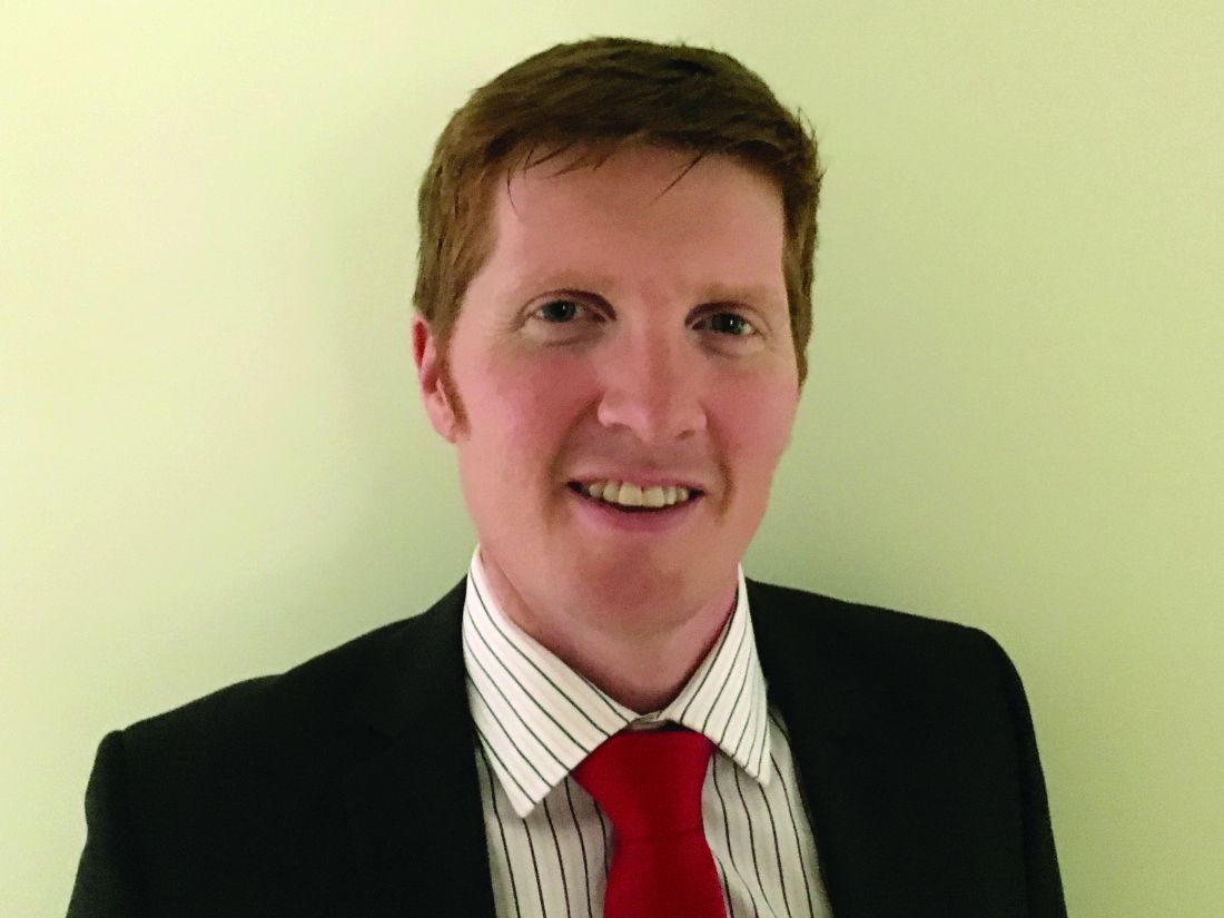
“He would do anything to make the children happy,” she said. “His children meant the world to him.”
Robinson died Jan. 3 as a result of diffuse gastric adenocarcinoma, according to his wife, who added that it was a short, 2-week-long illness.
A leader of global effort to understand COVID-19 and rheumatic disease
Jinoos Yazdany, MD, MPH, chief of the division of rheumatology at Zuckerberg San Francisco General Hospital and professor of medicine at the University of California, San Francisco, described Robinson as “one of the hardest-working people I have ever known. ... [still] his deep love and dedication for his family and kids was always present.”
Robinson would wake up early, even on weekends, to lead international calls across multiple time zones, Yazdany said. “He was driven by a deep curiosity and an intense desire to generate scholarship that would help people with rheumatic diseases.”
Yazdany added that Robinson had a full research portfolio in gout and spondyloarthritis and a busy clinical practice.
She often caught glimpses of Robinson’s young children during Zoom calls. “He was also a talented baker and loved to bake with his kids, often posting pictures of his creations on social media,” Yazdany said.
A mutual colleague compiled some of Robinson’s baking successes. That includes “ ‘probably about to be locked down’ cookies” on July 17, 2021, and “Queensland lockdown cookies!!!” on July 2, 2021. Reuters reported on July 21, 2021, that Australia was witnessing an alarming increase in COVID-19 cases.
Robinson also worked his social media skills to rally support for the COVID-19 Global Rheumatology Alliance, according to an article published by his colleagues in The Rheumatologist. Yazdany collaborated with him in this effort.
Launched on March 12, 2020, the Global Rheumatology Alliance’s mission is to “collect, analyze, and disseminate information about COVID-19 and rheumatology to patients, physicians, and other relevant groups to improve the care of patients with rheumatic disease.” Robinson served as chair of governance and policy for the collaborative effort.
Inspired by a conversation on Twitter by Leonard Calabrese, DO, a rheumatologist at the Cleveland Clinic in Ohio, about an outcomes registry created by gastroenterologists specific to patients with inflammatory bowel disease, Robinson launched a discussion about a similar effort for rheumatology on Twitter, they write.
Along with colleagues and within a single Zoom conference call, Yazdany and Robinson had a plan to organize the registry, Robinson’s colleagues write.
Two projects of the Global Rheumatology Alliance are a health care provider–entered registry for providers to enter data about rheumatology patients with COVID-19 infections, and their COVID-19 Vax Survey, which is available in 12 languages, including English.
Yazdany had never met Robinson before she started working with him on the Global Rheumatology Alliance. They started chatting on Twitter, then moved to Zoom conference calls, and subsequently had weeks when they talked by phone and “emailed constantly,” she said.
“As I reflect on our initial interactions, I am struck by how brilliantly we got along and trusted each other,” Yazdany told this news organization. “We both liked to think big, believed in inclusive collaborations, and were committed to helping people with rheumatic diseases during a scary and uncertain time.”
Still, Yazdany noted that she and Phil brought different strengths to their collaborations. She brought her skills related to the technical aspects of research databases, while “Phil worked his magic in mobilizing friends and colleagues from all over the world,” she said. “He served as a wonderful leader, one whom people believed in and would follow.”
The two colleagues, who spent much of their collaborations over Zoom calls, email, and Slack, while living more than 7,000 miles apart, finally met in person at ACR Convergence 2022, which took place in Philadelphia that year. “It felt like the best kind of reunion with a dear friend,” Yazdany remembered.
A mentor who created a platform for ‘good people to do great things’
David Liew, MBBS, PhD, consultant rheumatologist and clinical pharmacologist at Austin Health in Melbourne, marveled at Robinson’s ability to “distill things simply and cleanly, with clarity but without losing detail.” Liew, who collaborated with Robinson throughout the COVID-19 pandemic, likens the experience to “jamming with [jazz musician John] Coltrane.”
“What I think was particularly remarkable was the capacity to not only have those thoughts himself, but to facilitate others to have that springboard,” said Liew, who added that Robinson “took enormous pleasure in facilitating others’ success.”
“I think the greatest joy he drew out of the COVID-19 Global Rheumatology Alliance, apart from facing up to the challenge that needed to be faced, was creating a platform for good people to do great things,” said Liew, who recalled one situation where Robinson gently challenged him. Looking back now, Liew can “now see he very clearly was laying me up, giving me the best chance to shine.”
He describes Robinson as “a deep soul who loved his wife and two sons enormously” and “a whiskey aficionado.” “[Whiskey] suited his contemplative style,” Liew recalled. “Some of my fondest conversations with him were over a whiskey, either in person or virtually, pondering the ‘big issues.’”
A ‘friend and a colleague and so much more’
Claire Barrett, MBBS, president of the Australian Rheumatology Association, described Robinson as a “friend and a colleague and so much more. ... [He was] someone who I worked, laughed, ate, drank, danced, and had fun with,” she told this news organization. They served together as volunteers for the Australian Rheumatology Association and its Queensland branch, as well as Arthritis Queensland; they were also colleagues at Metro North Hospital and Health Service in Brisbane, Queensland.
Robinson was her “go to” for insightful comment on a variety of topics, she said. That could be advice on managing a patient with difficult gout, challenging spondyloarthritis, or the best treatment for a patient with COVID-19.
“[Phil] was a friend I could ask about anything, knowing I would not be judged,” Barrett said. “His kids and our grandkids are similar ages, so we would swap stories and photos and laugh about how cute/funny/cuddly/busy/etc. they were. My heart is broken he won’t get the chance to enjoy their future and the excitement of having Phil continuing to be such an active dad.”
Tanner, Robinson’s wife, said, “he loved everything.” That included the academic side of medicine, and working out what was wrong with his patients and helping them get better. “He was dedicated to this,” she added.
“He also loved the camaraderie of the job – all the people he met and interacted with. [Phil] loved sharing his ideas for research and also discussing complex patients with colleagues. He was driven by finding the answers to problems and doing this as part of a team of researchers/clinicians. He wasn’t interested in personal success.”
Robinson received his medical degree from Otago Medical School in Dunedin, New Zealand, according to the University of Queensland. His specialty training in general and acute care medicine and rheumatology was completed in Wellington, New Zealand, and Dunedin. Robinson also achieved a PhD in human genetics at the University of Queensland Diamantina Institute and had a postdoctoral fellowship at the Queensland Brain Institute at the University of Queensland.
Before his death, he worked at the Royal Brisbane and Women’s Hospital in Herston, Queensland, and at St. Andrew’s War Memorial Hospital in Spring Hill in Brisbane.
A version of this article first appeared on Medscape.com.
Best estimates made for hydroxychloroquine retinopathy risk
A new study likely makes the best estimate yet of the degree of retinopathy risk that patients who take the antimalarial drug hydroxychloroquine (HCQ) can expect, deriving mainly from the cumulative dose taken during the first 5 years of use, according to a study published in Annals of Internal Medicine.
HCQ works to decrease activity in a patient’s immune system, which is effective in many cases of systemic lupus erythematosus, one of the most common indications for the drug. However, an adverse outcome of treatment can be HCQ retinopathy, a progressive form of vision loss in patients taking HCQ over an extended period (mostly for longer than 5 years). The disease is often asymptomatic, although some patients do present a paracentral scotoma and a decrease in color vision. Patients may also notice flashing shapes in their vision and find that they have difficulty reading. Eventually, HCQ retinopathy can lead to loss of visual acuity, loss of peripheral vision, and loss of night vision.
Researchers from Kaiser Permanente Northern California and Harvard Medical School analyzed 3,325 persons who received HCQ for 5 or more years between 2004 and 2020. Their goal was to both characterize the long-term risk for incident HCQ retinopathy and examine the degree to which average HCQ dose within the first 5 years of treatment serves as a prediction of the risk.
The researchers then estimated the risk for developing retinopathy after 15 years, according to patients’ average dosing levels during the first 5 years of therapy. Overall, 81 participants developed HCQ retinopathy with overall cumulative incidences of 2.5% after 10 years and 8.6% after 15 years; the risk was greater for those given a higher dose during the first 5 years of treatment.
The mechanism of how HCQ toxicity may occur is still not completely known. There is evidence that toxicity happens because HCQ binds to melanin in both the retinal pigment epithelium and uvea in high concentrations. HCQ can interfere with lysosomal function, leading to oxidation and accumulation of lysosomes, which can cause dysfunction of the retinal pigment epithelium.
Progressive retinopathy can continue even after the drug is stopped. “It’s thought to be a very mild but important risk,” said Nilanjana Bose, MD, MBA, a rheumatologist with Memorial Hermann Health System in Houston. “Patients taking HCQ must be screened for retinal issues, most certainly elderly patients and patients with any kind of comorbidities.”

A 2021 joint position statement from the American College of Rheumatology, American Academy of Dermatology, the Rheumatologic Dermatology Society, and the American Academy of Ophthalmology recommends a baseline eye exam within a few months after starting therapy, then additional screening at 5 years on HCQ and annually thereafter.
“Early detection of retinopathy is important in overall visual prognosis, because toxicity can continue even after discontinuation of the medication,” said Rukhsana G. Mirza, MD, professor of ophthalmology and medical education at Northwestern University in Chicago.

“Examination alone is not sufficient to evaluate early changes, and specialized testing must be done. These include color photos, visual field tests, optical coherence tomography, fundus autofluorescence and in some cases, multifocal electroretinogram. Also, the AAO [American Academy of Ophthalmology] has specific recommendations related to Asian patients as they may have a different pattern of retinopathy that must also be considered.”
More accurate risk measurements
This news organization asked study coauthor April Jorge, MD, assistant professor of medicine in the division of rheumatology, allergy, and immunology at Massachusetts General Hospital and Harvard Medical School, Boston, to discuss the study, how it correlates to past research, and what it adds that’s new and useful to rheumatologists and ophthalmologists:
Question: Your research found that a higher dose of HCQ in the first 5 years of treatment led to a greater risk of retinopathy. Is there any indication that a lower dose given more frequently, either within that 5-year period or longer, would pose a similar risk?
Answer: In our study, we assessed the HCQ dose in the first 5 years of use but followed patients who continued the medication longer than 5 years, through up to 15 years of use. Therefore, we compared the risk of HCQ retinopathy associated with different HCQ dosages but for the same duration of use. We found that for any dose of HCQ, the risk of retinopathy increases the longer the medication is used. However, patients who used a higher dose of HCQ had a higher risk of developing retinopathy over time.

Although current guidelines recommend avoiding any HCQ dose over 5 mg/kg per day to reduce the risk of retinopathy, we found a higher risk of retinopathy associated with dosing over 6 mg/kg per day than between 5 and 6 mg/kg per day and the lowest risk with dosing under 5 mg/kg per day.
Q: How does your study align with and/or expand upon previous research regarding HCQ risk?
A: An important prior study of hydroxychloroquine retinopathy was the 2014 study by Ronald B. Melles, MD, and Michael F. Marmor, MD, published in JAMA Ophthalmology. Prior to our present study, that was the largest study to use the modern screening method (optical coherence tomography) to detect HCQ retinopathy. That screening tool is more sensitive than older methods, so it can detect early/mild cases of retinopathy that are typically asymptomatic. Compared to older studies, that 2014 study found a much higher risk of HCQ retinopathy than was previously appreciated.
However, that 2014 study did have some key limitations that could affect the risk estimates, such as using prevalent cases. A key feature of our present study is that we took several important steps to generate more accurate risk estimates. This included using an incident user cohort and detecting incident retinopathy cases through serial review of optical coherence tomography (screening) studies.
To achieve a high degree of methodologic rigor in correctly identifying retinopathy outcomes, we had expert ophthalmologists perform masked adjudication of all screening studies, and we assessed the intra-rater reliability of these study interpretations. Therefore, our study adds to the literature more accurate estimates of retinopathy risk. We found a lower cumulative incidence of retinopathy than was identified in the 2014 study, but the risk is still noteworthy.
Also unique to our study, we graded the severity of HCQ retinopathy outcomes. This was important, as we found that the majority of retinopathy cases detected through routine screening are mild and presumed to be asymptomatic. This will likely be reassuring news for patients that we can screen for this adverse event to detect it early and prevent vision loss.
Another important difference was that we assessed the risk of retinopathy associated with using over 6 mg/kg per day, between 5 and 6 mg/kg per day, and less than 5 mg/kg per day, whereas the highest dosing group assessed in the 2014 study included all patients using over 5 mg/kg per day. The risk was considerably higher in the > 6 mg/kg per day group than in the 5-6 mg/kg per day group.
Q: How can rheumatologists and ophthalmologists use this new information specifically to better treat their patients?
A: Our study provides more accurate estimates of the risk of HCQ retinopathy than in prior studies. These risk estimates can be used when rheumatologists (and other clinicians who prescribe HCQ) consider the risks and benefits of this otherwise important and well-tolerated medication. The risk associated with different dose ranges could also inform dosing decisions, since dosing over 6 mg/kg per day may be more of a concern than using doses in the 5-6 mg/kg range. Ophthalmologists can also use these new risk estimates to counsel patients of the importance of HCQ retinopathy screening and can also hopefully provide some reassurance to patients that the risk of severe retinopathy is low as long as they are being monitored.
The study authors were supported by grants from the National Institute of Arthritis and Musculoskeletal and Skin Diseases and the Rheumatology Research Foundation. The authors report no relevant financial relationships. Dr. Bose and Dr. Mirza had no relevant disclosures.
A version of this article first appeared on Medscape.com.
A new study likely makes the best estimate yet of the degree of retinopathy risk that patients who take the antimalarial drug hydroxychloroquine (HCQ) can expect, deriving mainly from the cumulative dose taken during the first 5 years of use, according to a study published in Annals of Internal Medicine.
HCQ works to decrease activity in a patient’s immune system, which is effective in many cases of systemic lupus erythematosus, one of the most common indications for the drug. However, an adverse outcome of treatment can be HCQ retinopathy, a progressive form of vision loss in patients taking HCQ over an extended period (mostly for longer than 5 years). The disease is often asymptomatic, although some patients do present a paracentral scotoma and a decrease in color vision. Patients may also notice flashing shapes in their vision and find that they have difficulty reading. Eventually, HCQ retinopathy can lead to loss of visual acuity, loss of peripheral vision, and loss of night vision.
Researchers from Kaiser Permanente Northern California and Harvard Medical School analyzed 3,325 persons who received HCQ for 5 or more years between 2004 and 2020. Their goal was to both characterize the long-term risk for incident HCQ retinopathy and examine the degree to which average HCQ dose within the first 5 years of treatment serves as a prediction of the risk.
The researchers then estimated the risk for developing retinopathy after 15 years, according to patients’ average dosing levels during the first 5 years of therapy. Overall, 81 participants developed HCQ retinopathy with overall cumulative incidences of 2.5% after 10 years and 8.6% after 15 years; the risk was greater for those given a higher dose during the first 5 years of treatment.
The mechanism of how HCQ toxicity may occur is still not completely known. There is evidence that toxicity happens because HCQ binds to melanin in both the retinal pigment epithelium and uvea in high concentrations. HCQ can interfere with lysosomal function, leading to oxidation and accumulation of lysosomes, which can cause dysfunction of the retinal pigment epithelium.
Progressive retinopathy can continue even after the drug is stopped. “It’s thought to be a very mild but important risk,” said Nilanjana Bose, MD, MBA, a rheumatologist with Memorial Hermann Health System in Houston. “Patients taking HCQ must be screened for retinal issues, most certainly elderly patients and patients with any kind of comorbidities.”
A 2021 joint position statement from the American College of Rheumatology, American Academy of Dermatology, the Rheumatologic Dermatology Society, and the American Academy of Ophthalmology recommends a baseline eye exam within a few months after starting therapy, then additional screening at 5 years on HCQ and annually thereafter.
“Early detection of retinopathy is important in overall visual prognosis, because toxicity can continue even after discontinuation of the medication,” said Rukhsana G. Mirza, MD, professor of ophthalmology and medical education at Northwestern University in Chicago.
“Examination alone is not sufficient to evaluate early changes, and specialized testing must be done. These include color photos, visual field tests, optical coherence tomography, fundus autofluorescence and in some cases, multifocal electroretinogram. Also, the AAO [American Academy of Ophthalmology] has specific recommendations related to Asian patients as they may have a different pattern of retinopathy that must also be considered.”
More accurate risk measurements
This news organization asked study coauthor April Jorge, MD, assistant professor of medicine in the division of rheumatology, allergy, and immunology at Massachusetts General Hospital and Harvard Medical School, Boston, to discuss the study, how it correlates to past research, and what it adds that’s new and useful to rheumatologists and ophthalmologists:
Question: Your research found that a higher dose of HCQ in the first 5 years of treatment led to a greater risk of retinopathy. Is there any indication that a lower dose given more frequently, either within that 5-year period or longer, would pose a similar risk?
Answer: In our study, we assessed the HCQ dose in the first 5 years of use but followed patients who continued the medication longer than 5 years, through up to 15 years of use. Therefore, we compared the risk of HCQ retinopathy associated with different HCQ dosages but for the same duration of use. We found that for any dose of HCQ, the risk of retinopathy increases the longer the medication is used. However, patients who used a higher dose of HCQ had a higher risk of developing retinopathy over time.
Although current guidelines recommend avoiding any HCQ dose over 5 mg/kg per day to reduce the risk of retinopathy, we found a higher risk of retinopathy associated with dosing over 6 mg/kg per day than between 5 and 6 mg/kg per day and the lowest risk with dosing under 5 mg/kg per day.
Q: How does your study align with and/or expand upon previous research regarding HCQ risk?
A: An important prior study of hydroxychloroquine retinopathy was the 2014 study by Ronald B. Melles, MD, and Michael F. Marmor, MD, published in JAMA Ophthalmology. Prior to our present study, that was the largest study to use the modern screening method (optical coherence tomography) to detect HCQ retinopathy. That screening tool is more sensitive than older methods, so it can detect early/mild cases of retinopathy that are typically asymptomatic. Compared to older studies, that 2014 study found a much higher risk of HCQ retinopathy than was previously appreciated.
However, that 2014 study did have some key limitations that could affect the risk estimates, such as using prevalent cases. A key feature of our present study is that we took several important steps to generate more accurate risk estimates. This included using an incident user cohort and detecting incident retinopathy cases through serial review of optical coherence tomography (screening) studies.
To achieve a high degree of methodologic rigor in correctly identifying retinopathy outcomes, we had expert ophthalmologists perform masked adjudication of all screening studies, and we assessed the intra-rater reliability of these study interpretations. Therefore, our study adds to the literature more accurate estimates of retinopathy risk. We found a lower cumulative incidence of retinopathy than was identified in the 2014 study, but the risk is still noteworthy.
Also unique to our study, we graded the severity of HCQ retinopathy outcomes. This was important, as we found that the majority of retinopathy cases detected through routine screening are mild and presumed to be asymptomatic. This will likely be reassuring news for patients that we can screen for this adverse event to detect it early and prevent vision loss.
Another important difference was that we assessed the risk of retinopathy associated with using over 6 mg/kg per day, between 5 and 6 mg/kg per day, and less than 5 mg/kg per day, whereas the highest dosing group assessed in the 2014 study included all patients using over 5 mg/kg per day. The risk was considerably higher in the > 6 mg/kg per day group than in the 5-6 mg/kg per day group.
Q: How can rheumatologists and ophthalmologists use this new information specifically to better treat their patients?
A: Our study provides more accurate estimates of the risk of HCQ retinopathy than in prior studies. These risk estimates can be used when rheumatologists (and other clinicians who prescribe HCQ) consider the risks and benefits of this otherwise important and well-tolerated medication. The risk associated with different dose ranges could also inform dosing decisions, since dosing over 6 mg/kg per day may be more of a concern than using doses in the 5-6 mg/kg range. Ophthalmologists can also use these new risk estimates to counsel patients of the importance of HCQ retinopathy screening and can also hopefully provide some reassurance to patients that the risk of severe retinopathy is low as long as they are being monitored.
The study authors were supported by grants from the National Institute of Arthritis and Musculoskeletal and Skin Diseases and the Rheumatology Research Foundation. The authors report no relevant financial relationships. Dr. Bose and Dr. Mirza had no relevant disclosures.
A version of this article first appeared on Medscape.com.
FROM ANNALS OF INTERNAL MEDICINE
Belimumab for pregnant women with lupus: B-cell concerns remain
The largest combined analysis of birth outcome data for women with systemic lupus erythematosus (SLE) who took belimumab (Benlysta) during pregnancy appears to indicate that the biologic is “unlikely to cause very frequent birth defects,” but the full extent of possible risk remains unknown. The drug’s effect on B cells, immune function, and infections in exposed offspring were not captured in the data, but a separate case report published after the belimumab pregnancy data report indicates that the drug does cross the placenta and builds up in the blood of the newborn, reducing B cells at birth.
Children of women with SLE have increased birth defect risks, and standard SLE therapeutic agents (for example, cyclophosphamide, methotrexate, mycophenolate mofetil) have been implicated in birth defects and pregnancy loss, but birth defect data for biologic drugs such as belimumab are limited. While belimumab animal data revealed no evidence of fetal harm or pregnancy loss rates, there was evidence of immature and mature B-cell count reductions.
Belimumab is approved by the Food and Drug Administration for use in patients aged 5 years and older with active, autoantibody-positive SLE who are taking standard therapy, and also for those with lupus nephritis.
Michelle Petri, MD, of Johns Hopkins University, Baltimore, and coauthors reported in Annals of the Rheumatic Diseases on data they compiled through March 8, 2020, from belimumab clinical trials, the Belimumab Pregnancy Registry (BPR), and postmarketing/spontaneous reports that encompassed 319 pregnancies with known outcomes.
Across 18 clinical trials with 223 live births, birth defects occurred in 4 of 72 (5.6%) belimumab-exposed pregnancies and in 0 of 9 in placebo-exposed pregnancies. Pregnancy loss (excluding elective terminations) occurred in 31.8% (35 of 110) of belimumab-exposed women and 43.8% (7 of 16) of placebo-exposed women in clinical trials. In the BPR retrospective cohort, 4.2% had pregnancy loss. Postmarketing and spontaneous reports had a pregnancy loss rate of 31.4% (43 of 137). Concomitant medications, confounding factors, and/or missing data were noted in all belimumab-exposed women in clinical trials and the BPR cohort. Dr. Petri and colleagues reported no consistent pattern of birth defects across datasets but stated: “Low numbers of exposed pregnancies, presence of confounding factors/other biases, and incomplete information preclude informed recommendations regarding risk of birth defects and pregnancy loss with belimumab use.”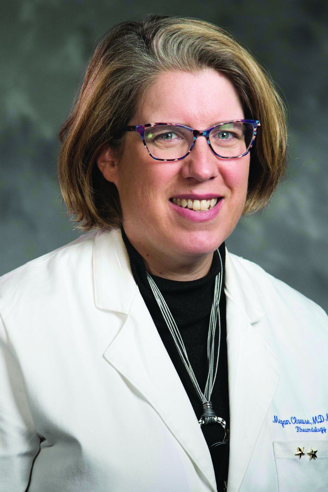
In an interview, coauthor Megan E. B. Clowse, MD, MPH, associate professor of medicine and director of the division of rheumatology and immunology at Duke University, Durham, N.C., said that “the Annals of the Rheumatic Diseases article provides some reassurance that belimumab is unlikely to cause very frequent birth defects. It is clearly not in the risk-range for thalidomide or mycophenolate. However, due to the complexity of collecting these data, this manuscript can’t explore the full extent of possible risks. It also did not provide information about B cells, immune function, or infection risks in exposed offspring.”
A separate case report by Helle Bitter of the department of rheumatology at Sorlandet Hospital Kristiansand (Norway) in Annals of the Rheumatic Diseases is the first to show transplacental passage of belimumab in humans. Other prior reports have shown such transplacental passage for monoclonal IgG antibodies (tumor necrosis factor inhibitors and rituximab). Even though the last infusion was given late in the second trimester, belimumab was present in cord serum at birth, suggesting much higher concentrations before treatment was stopped. While B-cell numbers were reduced at birth, they returned to normal ranges by 4 months post partum when they were undetectable. In the mother, B-cell numbers remained low throughout the study period extending to 7 months after delivery. The authors stated that the child had a normal vaccination response, and except for the reduced B-cell levels at birth, had no adverse effects of prenatal exposure to maternal medication through age 6 years.
“The belimumab transfer in the case report is the level that we would anticipate based on similar studies in infant/mother pairs on other IgG1 antibody biologics like adalimumab – about 60% higher than the maternal level at birth,” Dr. Clowse said. “That the baby has very low B cells at birth is worrisome to me, demonstrating the lasting effect of maternal belimumab on the infant’s immune system, even when the drug was stopped 14 weeks prior to delivery. While this single infant did not have problems with infections, with more widespread use it seems possible that infants would be found to have higher rates of infections after in utero belimumab exposure.”
The field of lupus research greatly needs controlled studies of newer biologics in pregnancy, Dr. Clowse said. “Women with active lupus in pregnancy – particularly with active lupus nephritis – continue to suffer tragic outcomes at an alarming rate. Newer treatments for lupus nephritis provide some hope that we might be able to control lupus nephritis in pregnancy more effectively. The available data suggests the risks of these medications are not so large as to make studies unreasonable. Our current data doesn’t allow us to sufficiently balance the potential risks and benefits in a way that provides clinically useful guidance. Trials of these medications, however, would enable us to identify improved treatment strategies that could result in healthier women, pregnancies, and babies.”
GlaxoSmithKline funded the study. Dr. Clowse reported receiving consulting fees and grants from UCB and GlaxoSmithKline that relate to pregnancy in women with lupus.
FROM ANNALS OF THE RHEUMATIC DISEASES
Abnormal bleeding common among youth with joint hypermobility
A small cohort study of pediatric rheumatology patients with generalized joint hypermobility (GJH) who presented to a specialized rheumatology* clinic suggests that many such patients have abnormal bleeding symptoms, in comparison with health control patients.
The study of 81 patients with GJH found that about three quarters had significantly elevated median bleeding scores, but only 12% had been assessed by hematology for bleeding.
“We propose that screening for bleeding symptoms should be integrated into the routine care for all patients with GJH, with hematology referrals for patients with increased bleeding concerns,” wrote a research team led by Nicole E. Kendel, MD, a pediatric hematologist-oncologist at Akron Children’s Hospital in Ohio, in a study published online in Arthritis Care and Research.
“Further studies are needed to understand the mechanism of bleeding, evaluate comorbidities associated with these bleeding symptoms, and potentially allow for tailored pharmacologic therapy,” the authors stated.
Background
Dr. Kendel’s team had reported moderate menstruation-associated limitations in school, social, and physical activities among female adolescents with GJH. “This cohort also experienced nonreproductive bleeding symptoms and demonstrated minimal hemostatic laboratory abnormalities, indicating that this population may be underdiagnosed and subsequently poorly managed,” she said in an interview. “As excessive bleeding symptoms could have a significant impact on overall health and quality of life, we thought it was important to define the incidence and natural course of bleeding symptoms in a more generalized subset of this population.”
Although the investigators hypothesized that there would be a statistically significant increase in bleeding scores, “we were still impressed by the frequency of abnormal scores, particularly when looking at the low percentage of patients [12%] who had previously been referred to hematology,” she said.
Study results
The median age of the study cohort was 13 years (interquartile range, 10-16 years), and 72.8% were female. The mean Beighton score, which measures joint flexibility, was 6.2 (range, 4-9). All participants were seen by rheumatologists and were diagnosed for conditions on the hypermobility spectrum. Those conditions ranged from GJH to hypermobile Ehlers-Danlos syndrome (hEDS).
Abnormal bleeding, as measured by the International Society on Thrombosis and Haemostasis Bleeding Assessment Tool, was found in 75% (95% confidence interval [CI], 64%-84%). Overall mean and median bleeding scores were 5.2 and 4, respectively; scores ranged from 0 to 16. Abnormal scores of ≥ 3 were observed for patients < 8 years of age, ≥ 4 for men ≥ 18 years of age, and ≥ 6 for women ≥ 18 years of age. These measures were significantly elevated compared with those reported for historical healthy pediatric control persons (P < .001).
The most common hemorrhagic symptom was oral bleeding (74.1%) that occurred with tooth brushing, flossing, tooth loss, or eruption. Others reported easy bruising (59.3%) and bleeding from minor wounds (42%). In terms of procedures, tooth extraction requiring additional packing was reported by 25.9%, and 22.2% reported significant bleeding after otolaryngologic procedures, such as tonsillectomy/adenoidectomy, septoplasty, and nasal turbinate reduction.
Prolonged or heavy menstrual periods were reported by 37.3% of female patients.
Bleeding scores did not differ by biological sex or NSAID use, nor did any correlation emerge between patients’ bleeding and Beighton scores. However, there was a positive correlation with increasing age, a phenomenon observed with other bleeding disorders and in the healthy population, the authors noted.
Of the 10 study participants who had previously undergone hematologic assessment, one had been diagnosed with acquired, heart disease–related von Willebrand disease, and another with mild bleeding disorder.
Severe connective tissue disorders are associated with increased bleeding symptoms in the adult population, Dr. Kendel said, but few studies have assessed bleeding across the GJH spectrum, particularly in children.
Bleeding is thought to be due to modifications of collagen in the blood vessels. “These modifications create mechanical weakness of the vessel wall, as well as defective subendothelial connective tissue supporting those blood vessels,” Dr. Kendel explained. She noted that altered collagen creates defective interactions between collagen and other coagulation factors.
“Even in the presence of a normal laboratory evaluation, GJH can lead to symptoms consistent with a mild bleeding disorder,” she continued. “These symptoms are both preventable and treatable. I’m hopeful more centers will start routinely evaluating for increased bleeding symptoms, with referral to hematology for those with increased bleeding concerns.”
Commenting on the study’s recommendation, Beth S. Gottlieb, MD, chief of the division of pediatric rheumatology at Northwell Health in New Hyde Park, N.Y., who was not involved in the investigation, said a brief questionnaire on bleeding risk is a reasonable addition to a rheumatology office visit.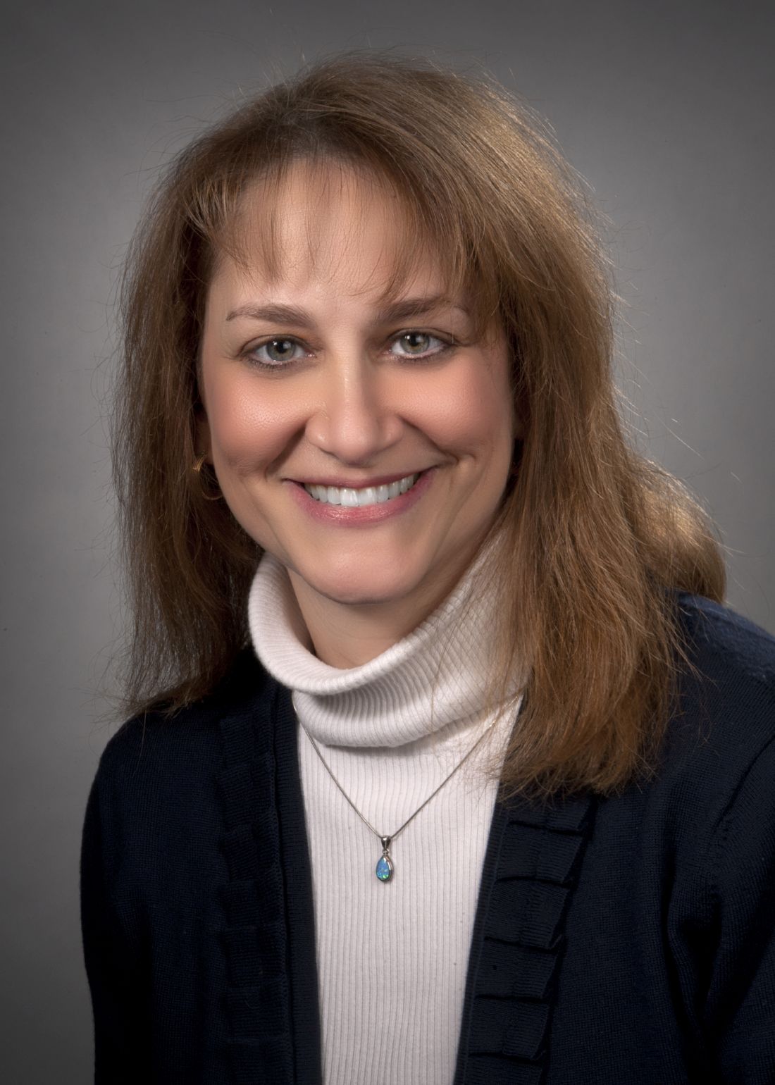
“Joint hypermobility is very common, but not all affected children meet the criteria for the hypermobile form of hEDS,” she told this news organization. “Screening for bleeding tendency is often done as routine medical history questions. Once a child is identified as hypermobile, these screening questions are usually asked, but utilizing one of the formal bleeding risk questionnaires is not currently routine.”
According to Dr. Gottlieb, it remains unclear whether screening would have a significant impact on children who have been diagnosed with hypermobility. “Most of these children are young and may not yet have a significant history for bleeding tendency,” she said. “Education of families is always important, and it will be essential to educate without adding unnecessary stress. Screening guidelines may be an important tool that is easy to incorporate into routine clinical practice.”
Limitation
The study was limited by selection bias, as patients had all been referred to a specialized rheumatology clinic.
The study was supported by the Clinical and Translational Intramural Funding Program of the Abigail Wexner Research Institute. The authors and Dr. Gottlieb have disclosed no relevant financial relationships.
*Correction, 1/11/2023: An earlier version of this story misstated the type of specialty clinic where patients were first seen.
A version of this article first appeared on Medscape.com.
FROM ARTHRITIS CARE AND RESEARCH
Cardiovascular risk score multipliers suggested for rheumatic diseases
A re-evaluation of cardiovascular risk management guidelines intended for use by rheumatologists may be warranted based on findings from a recently published population-based study of the risks for 12 different cardiovascular disease outcomes in patients with autoimmune diseases.
“The notion that patients with rheumatic diseases are at increased risk of developing cardiovascular diseases has been ongoing for many years,” Nathalie Conrad, PhD, and coauthors wrote in a viewpoint article in Annals of the Rheumatic Diseases.
This has “sparked much debate concerning whether and when to initiate cardiovascular prevention therapies,” they said.
Dr. Conrad was first author on the population-based study published in The Lancet in August 2022 that used linked primary and secondary care records from datasets in the U.K. Clinical Practice Research Datalink involving individuals who were recently diagnosed with any of 19 different autoimmune diseases during an 18-year period stretching from 2000 to 2017 but free of cardiovascular disease until at least 12 months after incident autoimmune disease. “Every single autoimmune disorder we looked at was associated with increased cardiovascular risk,” Dr. Conrad, of the department of public health and primary care at Catholic University Leuven (Belgium), said in an interview.
Not only was the risk for cardiovascular disease increased for people with rheumatic diseases by an average of 68%, compared with people without rheumatic diseases, but also the whole spectrum of cardiovascular disorders was seen.
“We saw increases in thromboembolic diseases, degenerative heart diseases, and heart inflammation,” Dr. Conrad said.
Large datasets examined
The idea for the epidemiologic study came from mounting evidence for cardiovascular disease risk among people with autoimmune diseases but not enough to support the design of specific prevention measures.
Dr. Conrad’s Lancet study examined electronic health records of 446,449 individuals with autoimmune diseases and matched them to 2,102,830 individuals without autoimmune disease. This included 160,217 individuals with seven rheumatic diseases: rheumatoid arthritis, polymyalgia rheumatica, vasculitis, systemic lupus erythematosus, Sjögren’s syndrome, ankylosing spondylitis, and systemic sclerosis.
In addition to looking for any evidence of cardiovascular disease, Dr. Conrad and coauthors looked at 12 specific outcomes: atherosclerotic diseases, peripheral arterial disease, stroke or transient ischemic attack, heart failure, valve disorders, thromboembolic disease, atrial fibrillation or flutter, conduction system disease, supraventricular arrhythmias, aortic aneurysm, myocarditis and pericarditis, and infective endocarditis.
CV risk in rheumatic diseases
As might be expected, “greater magnitudes of risk” were seen for individuals with systemic lupus erythematosus and systemic sclerosis than for people in the general population, with the chances of cardiovascular disease being two to four times higher. But what perhaps wasn’t expected was that all rheumatic diseases carried an increased risk for heart or vascular-related problems.
Furthermore, the increased risk could not solely be accounted for by the presence of traditional risk factors, such as blood pressure, smoking, or obesity.
“The background here is that any context of systemic inflammation would be predicted to lead to an increased vascular risk,” Iain McInnes, MD, PhD, professor of medicine and rheumatology at the University of Glasgow, said in an interview. Dr. McInnes was a coauthor of the viewpoint article in Annals of the Rheumatic Diseases.
“The implication is that there may well be increased vascular risk across the whole range of immune-mediated inflammatory diseases,” he added. “We should not, however, infer the magnitude of risk will be the same for each disease.”
What is more intriguing, Dr. McInnes said, is that “we don’t know yet whether there’s one final common pathway that leads to the blood vessel being damaged or whether different diseases might contribute different pathways.”
He added: “A question for the future is to see what are those mechanisms that drive risk across different diseases? And the reason that matters, of course, is that we might want to think about the effectiveness of different therapeutic interventions.”
Determining cardiovascular risk
Dr. Conrad and associates in their viewpoint article suggested that an update to the European Alliance of Associations for Rheumatology guidelines for cardiovascular risk management of rheumatic and musculoskeletal diseases (RMDs) could tailor cardiovascular risk scores to certain diseases.
They suggested that the guidelines could consider a risk multiplier of 2.5 for systemic sclerosis, 2.0 for lupus, and 1.5 for any other rheumatic disease.
“We argue that [EULAR] recommendations should consider this new evidence of poorer cardiovascular health in numerous RMDs and envisage cardiovascular screening and associated prevention measures,” Dr. Conrad said.
While they recognize that risk multipliers aren’t perfect, “they are the best available option until personalized risk prediction tools are developed specifically for patients with RMDs.”
Addressing cardiovascular risk
As a former president of EULAR, Dr. McInnes was keen to point out that “EULAR’s recommendations are evidence based and are rigorously built on [standard operating procedures] that work and have stood the test of time. I’m quite sure that the members of relevant EULAR task forces will be looking at these data, but they’ll be looking at the whole range of literature to see whether change is necessary.”
Good-quality inflammatory disease control will certainly contribute to reducing vascular risk, “but we should not make the assumption that it will be sufficient,” he cautioned. “We still have to be very careful in addressing so called conventional risk factors, but in particular thinking about obesity and cardiometabolic syndrome to be sure that when those are present, that we detect them and we treat them appropriately.”
As to who is best placed to manage a patient’s cardiovascular risk profile, Dr. McInnes said: “I think the rheumatologist has a responsibility to make sure that as much of the patient’s disease spectrum is being treated as possible.”
“As a rheumatologist, I would like to know that those elements of a patient’s disease presentation are being addressed,” whether that is by a primary care physician, cardiologist, diabetologist, or other specialist involved in the optimal management of the patient.
Dr. Conrad acknowledged receiving support from the European Union’s Horizon 2020 Program, the European Society for Cardiology, and grant funding paid to her institution from the Belgian-based Research Foundation Flounders. She also acknowledged receipt of royalties in regard to the intellectual property of a home-monitoring system for heart failure paid to Oxford University Innovation. Dr. McInnes acknowledged financial relationships with many pharmaceutical companies.
*This article was updated 12/30/2022.
FROM ANNALS OF THE RHEUMATIC DISEASES
FDA approves Idacio as eighth adalimumab biosimilar in U.S.
A biosimilar drug to the tumor necrosis factor inhibitor adalimumab, marketed as Idacio (adalimumab-aacf), has been approved by the Food and Drug Administration for use in the United States, according to a press release from manufacturer Fresenius Kabi.
Idacio is a citrate-free, low-concentration formulation of adalimumab and is now approved for use for all but three of the indications that currently apply to the reference adalimumab product (Humira): rheumatoid arthritis, polyarticular juvenile idiopathic arthritis, psoriatic arthritis in adults, ankylosing spondylitis, Crohn’s disease in adults and children aged 6 years or older, ulcerative colitis in adults, and plaque psoriasis in adults. It does not apply to Humira’s indications for hidradenitis suppurativa, uveitis, or ulcerative colitis in pediatric patients aged 5 years and older.
Idacio is the eighth adalimumab biosimilar to be approved in the United States. Its approval was based on evidence of a similar profile of pharmacokinetics, safety, efficacy, and immunogenicity to Humira.
Idacio was first launched in 2019 and has been marketed in more than 37 countries worldwide, according to Fresenius Kabi. The U.S. launch is scheduled for July, and Idacio will be available as a self-administered prefilled syringe or prefilled pen.
A version of this article first appeared on Medscape.com.
Rheumatology workforce shortage demands multipronged approach
PHILADELPHIA – New rheumatology fellowships in underserved areas. Rheumatology training for nonspecialists. Telemedicine training, tools, and resources. These are a few of the remedies the American College of Rheumatology’s new Workforce Solutions Committee has concocted to address a shrinking U.S. rheumatology workforce in the face of a rising population of patients with rheumatologic diseases.
In two sessions at the ACR’s annual meeting, committee members provided insights on current workforce projections since the most recent ACR workforce study in 2015. They also outlined the 1-year-old committee’s response to bridge the looming gap in patient care. Some projects are already underway, while others have yet to be implemented and require sustained monetary and project coordination commitments to make their impacts felt across the targeted regions.
“At the current pace of physician departures from the workforce, even with fellows graduating from adult and pediatric rheumatology training programs, the subspecialty cannot be sustained,” said Daniel Battafarano, DO, chair of the Workforce Solution Committee, professor of medicine at the Uniformed Services University of the Health Sciences, Bethesda, Md., and adjunct professor at the University of Texas Health Science Center, San Antonio.
The Northwest is an example of the problem at hand. “For an adult, there’s 1.65 rheumatologists per 100,000 in 2015. In 2025, there’s going to be 0.5 [adult] rheumatologists per 100,000. That’s a problem. If you look at every region in the United States, every single region is decrementing significantly,” he said.
Even the Northeast, which boasted 3.07 adult rheumatologists per 100,000 in 2015, will drop to 1.61 in 2025. Other regions are much worse off. The Southwest will drop from 1.28 to 0.64, and North Central will drop from 1.64 to 0.69, projections show.
The situation for pediatrics “has been in a crisis since 2015,” Dr. Battafarano said. It is much worse off than the situation for adult rheumatology, with 2015 figures going from 0.17 to 0.20 in the Southwest and from 0.03 to 0.04 in the South Central region by 2025, and in the Northwest from 0.67 to 0.13 and Northeast from 0.83 to 0.16.
“The ill effects of the pandemic are immeasurable to this point, but anecdotally we know we lost colleagues to part-time employment, we saw early retirements, and that happened across the full spectrum of the United States,” he said. It’s possible that up to 10% of full-time equivalents of physicians left practice in the United States since the pandemic began.
Rheumatologists largely practice in the 392 U.S. metropolitan statistical areas (MSAs), which are defined as a “core area containing a substantial population nucleus, together with adjacent communities having a high degree of economic and social integration with that core.” The smallest MSA has nearly 59,000 people. This distribution leaves many people in the Northwest, Southwest, South Central, and North Central regions with few rheumatologists. Over 86% of locations where rheumatologists train and work are in MSAs.
“By definition, if you get into a region where there’s 60,000 people or less, you can’t support a rheumatologist,” Dr. Battafarano said. He also noted that all pediatric hospitals tend to be in dense MSAs where rheumatologists might be.
South Central region serves as an example
People in certain MSAs of the South Central region (Oklahoma, Texas, Arkansas) such as Laredo, Tex., population 281,805 – where there is no adult rheumatologist – have to drive nearly 2.5 hours to the nearest well-served MSA (San Antonio) where there is a rheumatology fellowship program and medical school.
“Texas may have rheumatologists, but it depends on where you live,” Dr. Battafarano said. “The same applies to Waco, Texas; Texarkana, Arkansas; Lawton, Oklahoma; Pine Bluff, Arkansas; and Enid, Oklahoma, and you can see the range of traveling is between 45 minutes and 2.5 hours. That’s adult rheumatology.”
People in Laredo have an even longer drive to find a pediatric rheumatologist. In pediatrics, he said, “we don’t even look at towns, we look at pediatrics by state. You can see there’s a pediatric fellowship program in Dallas, and there’s one in Houston. There’s one or two [pediatric] rheumatologists in Austin. It just depends on how quickly they burn out and how long they stay. I’m in San Antonio. We don’t have a pediatric rheumatologist in San Antonio, the seventh-largest city in the United States.”
Pediatric rheumatologists are very often found where fellowship programs are located. “Ninety-five percent or greater of pediatric rheumatologists are housed in a pediatric hospital,” Dr. Battafarano said. “There are very few adult rheumatologists who see children, for a variety of reasons.”
In a panel discussion and question-and-answer session that took place after Dr. Battafarano outlined concerns with the numbers and distribution of rheumatologists across the United States, committee member Beth Jonas, MD, professor of medicine and chief of the division of rheumatology, allergy, and immunology at the University of North Carolina at Chapel Hill, noted that the rheumatology specialty match is competitive and typically leaves close to 100 residents who had rheumatology as their first choice without a match because there are not enough fellowship positions for them to fill.
The Workforce Solutions Committee hopes to reduce this “bottleneck,” she said, by identifying institutions that are ripe for opening new fellowship programs or have existing programs that can expand their number of positions, as well as expanding musculoskeletal training among nonrheumatologists, such as primary care physicians, advanced practice providers, and sports medicine physicians.
“I think that we’re going to find ourselves needing to be more creative as we become overcome with events with shortages of physicians,” Dr. Battafarano said. He gave an example of a recent pediatric rheumatology fellow in Seattle who wanted to go back home to Montana to practice. At the same time, the pediatric program wanted her to become a faculty member, so they said they would bridge their program to her, rotating fellows and covering her while on vacation.
“That’s an example of a pediatric program spreading its wings,” he said. “I think that we need to think that way, whether we talk about urban, suburban, rural. It’s how can we stage rheumatologists in areas, how can we embrace our primary care providers, and have rheumatology health care teams so that we have rheumatology expertise located within arm’s length but also within telemedicine length.”
Interventions for 2023
As part of a report on the activities of the Workforce Solutions Committee, Dr. Jonas outlined a number of interventions that the ACR is set to launch in 2023. The committee created five intervention teams to support fellowship positions and training providers, recruitment opportunities, fostering patient-centered communities, virtual training programs, and grants for research and training. The ACR’s initial pilot interventions are focused on, but not limited to, the Northwest, Southwest, and South Central regions.
The committee identified 10 potential partner institutions to develop new fellowship programs in the Northwest, Southwest, and South Central regions: University of Nevada, Las Vegas; Texas Tech University Permian Basin Program; Baylor Scott and White Medical Center Round Rock (Tex.) Program; Texas Tech University Health Science Center El Paso Program; University of Texas at Austin Dell Medical School Program; Abrazo Health Network Program in Arizona; University of Arizona Phoenix Program; University of Arkansas (pediatric program); Oklahoma State University; and University of Texas, San Antonio (pediatric program).
Dr. Jonas explained that opening new programs in these underserved areas should help establish new rheumatologists in these areas, because studies have shown that fellows tend to stay within 100-200 miles of where they trained.
In addition to starting new programs, “if you want to grow your program, if you want to add another fellow, we can support that,” Dr. Jonas said.
To help convince institutions of the value of rheumatology care, a position paper will soon be released by the ACR that outlines the benefits of the specialty to a health care system. The paper will describe an annual preventive cost savings of $2,762 per patient who is in the care of a rheumatologist and an annual direct or downstream income of $3.5 million per rheumatologist in a community, committee members said.
To help with recruitment and retention efforts, the ACR will revise its CareerConnection website to make it more useful.
To help in developing patient-centered communities of care, there will be a discussion series with payers on topics such as access, care delivery, and financing, in which 12 payers (including Blue Cross Blue Shield, Aetna, UnitedHealthcare, and Kaiser) will discuss patient access, cost, and quality measures. An in-person payer summit is also scheduled for 2023.
Interventions to enhance rheumatology care will also keep an ongoing focus on virtual training programs for primary care physicians and advanced practice providers, including a Project ECHO (Extension for Community Healthcare Outcomes) virtual lecture and case-based training and mentoring series for nonrheumatologists, an online library of searchable topics targeted to nonrheumatologists, as well as a grand rounds podcast series on certain rheumatic diseases for residents in family medicine, internal medicine, and pediatric programs. For rheumatology educators in 2023, there also will be an online portal curated by the ACR and other sources to use as a resource for developing curriculum.
A newly revised Fundamentals of Rheumatology course started in June 2022, which can help many medical students, residents, primary care physicians, and students in rehabilitation professions to learn about rheumatologic diseases, said committee member Janet Poole, PhD, division chief, professor, and director of the occupational therapy graduate program at the University of New Mexico, Albuquerque. Grants from the Rheumatology Research Foundation are available to offset the costs of the course for attendees.
An “onboarding toolkit” for nurse practitioners and physician assistants is now available to provide advice to practices for hiring, mentoring, and conducting performance evaluations in rheumatology care teams, Dr. Poole said. And fact sheets about the roles of interprofessional team members are being revised to give nonspecialty providers a sense of what other disciplines are available to care for patients with rheumatic and musculoskeletal disorders. The Advanced Rheumatology course is also being revised and will be available in summer 2023.
Other planned interventions aim to optimize telemedicine for the rheumatology community with a telehealth curriculum for rheumatology fellows, tools to improve telehealth implementation and delivery, and educational materials for patients to optimize their participation in telehealth visits.
The committee created targeted outreach to promote all grants focused on workforce expansion. As a result, in the last year the committee noted a three- to fourfold rise in the number of grant applications, including people and programs in underserved areas that had not previously applied. The Rheumatology Research Foundation also created a baseline for measuring success of grant funding in underserved areas and earmarked funds to increase awareness of the grants and facilitate applications.
Early signs of success are evident in the Rheumatology Access Expansion initiative in the ACR’s Collaborative Initiatives (COIN) department, said committee member Rosalind Ramsey-Goldman, MD, DrPH, professor of medicine/rheumatology at Northwestern University, Chicago. A team of rheumatologists, a pharmacist, and Navajo cultural interpreters developed a 12-week rheumatoid arthritis training program for primary care physicians in the Navajo Nation American Indian reservation, where there’s a high prevalence of RA and a shortage of local rheumatology providers. Results of the pilot study were presented in a plenary session at the meeting.
COIN also worked with the ACR’s Diversity, Equity, and Inclusion task force to sponsor 2022 annual meeting attendance for 11 medical students who identify themselves as underrepresented in medicine to expose them to rheumatology early in their career, said Dr. Ramsey-Goldman. The ACR is starting a program for medical students at historically Black colleges and universities and minority-serving institutions to include mentoring, round tables, and attendance at next year’s annual meeting.
None of the rheumatologists had relevant financial disclosures.
AT ACR 2022
Ustekinumab matches TNF inhibitors for psoriatic arthritis in 3-year, real-world study
The interleukin-12/23 inhibitor ustekinumab (Stelara) is nearly as effective as a tumor necrosis factor (TNF) inhibitor for psoriatic arthritis, and patients are slightly more likely to persist with it and have a lower rate of adverse events, a 3-year, real-world study has found.
In a paper published online in Annals of the Rheumatic Diseases, researchers presented the outcomes of the prospective, observational PsABio study of 895 adults with psoriatic arthritis, who were starting treatment for the first time with either ustekinumab or a TNF inhibitor as first-, second-, or third-line treatment.
At 3 years after starting therapy, 49.9% of the 439 patients prescribed ustekinumab were still on that treatment, compared with 47.8% of the 456 patients prescribed a TNF inhibitor. However, there were differences in persistence based on clinical presentation. Patients who had severe skin involvement who were treated with ustekinumab stayed on the drug for longer than did those with severe skin involvement treated with a TNF inhibitor, and they were more likely to persist with their treatment for the 3 years of the study. However, there were numerically more patients with mild or moderate skin involvement taking a TNF inhibitor who stayed persistent with the treatment, compared with those taking ustekinumab, although the differences were not statistically significant.
“In the ustekinumab group, skin response was an important reason for prolonged persistence, with more patients in the ustekinumab group stopping/switching due to lack of effectiveness,” wrote Laure Gossec, MD, of Pitié-Salpêtrière Hospital and Sorbonne University, Paris, and coauthors. “This is expected, as psoriasis can significantly affect morbidity, and successfully treating skin symptoms improves patients’ health-related quality of life.”
The authors also noted that patients on ustekinumab monotherapy had the highest rate of persistence and stayed on treatment longer than did those on TNF inhibitor monotherapy, or on dual therapy with either drug combined with methotrexate. They suggested this could be because patients on TNF inhibitor monotherapy may be more likely to develop antidrug antibodies than those on ustekinumab monotherapy. It could also be because adding methotrexate may increase the risk of adverse events, but without necessarily increasing the effectiveness of ustekinumab on skin involvement.
In terms of efficacy, researchers saw that 69.8% of patients in the TNF inhibitor group had achieved low disease activity and 45% had achieved remission, compared with 58.6% of patients in the ustekinumab group who achieved low disease activity and 31.4% who achieved remission.
A similar pattern was seen for minimal disease activity and very low disease activity, which were achieved by 54.2% and 26.9% respectively of those in the TNF inhibitor group, and 41.4% and 19.2% respectively of those in the ustekinumab group.
Because the study was observational and real-world, the choice of therapy was made by the treating rheumatologist rather than patients being randomized. There were some baseline differences between the ustekinumab and TNF inhibitor groups; for example, patients in ustekinumab group were generally older and with more comorbidities, and were more likely to have previous been treated with biologics. However, they were also less likely to be concurrently treated with methotrexate and NSAIDs, and more likely to have severe skin involvement.
The study saw a higher rate of adverse events in the TNF inhibitor group, compared with the ustekinumab, with 39.7% of patients treated with TNF inhibitor and 34.6% of those treated with ustekinumab reporting at least one adverse event. The rates of serious adverse events and malignancies were similar for the two groups, but overall the ustekinumab group had a lower rate of clinically-relevant adverse events including infections.
The study was sponsored by Janssen, which markets ustekinumab. Ten authors declared personal fees, grants, and nonfinancial support from the pharmaceutical sector, including Janssen. One author was an employee of Janssen, one an employee of Johnson & Johnson, and two are editorial board members of Annals of the Rheumatic Diseases.
FROM ANNALS OF THE RHEUMATIC DISEASES
Metabolic syndrome may promote gout in young men
Metabolic syndrome is associated with a significantly increased risk for gout in young men, but the risk can be mitigated by improvement in individual components of the syndrome, based on data from a pair of population-based studies totaling more than 4 million individuals.
Gout remains the most common type of inflammatory arthritis in men, and the rate has been rising among younger adults, Yeonghee Eun, MD, PhD, of Sungkyunkwan University, Seoul, South Korea, and colleagues wrote. An increasing body of evidence suggests a link between gout and metabolic syndrome (MetS), but large studies have been lacking, especially in younger adults.
In a study published in Frontiers in Medicine, the researchers reviewed data from 3,569,104 men aged 20-39 years who underwent a health checkup between 2009 and 2012 in South Korea, based on the Korean National Health Insurance Service. The primary outcome of incident gout was identified using claims data. The mean age of the participants was 31.5 years.
Over a mean follow-up of 7.4 years, the incidence of gout was 3.36 per 1,000 person-years. The risk of developing gout was more than twice as high among individuals who met MetS criteria than in those who did not (adjusted hazard ratio, 2.44).
MetS was defined as the presence of at least two of the following components: hypertriglyceridemia, abdominal obesity, reduced HDL cholesterol, elevated blood pressure, and elevated fasting glucose.
Overall, individuals with all five MetS components had a fivefold increase in gout risk, compared with people who did not have MetS (aHR, 5.24). In an analysis of each component of MetS, hypertriglyceridemia and abdominal obesity showed the strongest association with gout (aHRs of 2.08 and 2.33, respectively).
The impact of MetS on risk of incident gout was greater in younger participants, which suggests that the management of MetS in young people should be emphasized, the researchers said.
In a further analysis of body mass index subgroups, MetS had the greatest impact on gout risk for individuals who were underweight (aHR, 3.82). “In particular, in the underweight group, the risk of gout increased 10-fold when abdominal obesity was present,” the researchers said.
The study was limited by several factors including potential selection bias and potential overestimation of gout incidence because of the use of diagnostic codes, the researchers noted. Other limitations included lack of control for nutritional or dietary risk factors and the inability to include cases that occurred after the study period.
However, findings were strengthened by the large number of participants with MetS who were underweight or normal weight, the researchers wrote. More research on the mechanism of action is needed, but the data suggest that MetS is a key risk factor in the development of gout in young men.
In a second study, published in Arthritis & Rheumatology, Dr. Eun and colleagues examined associations between MetS changes and incident gout in young men. Although previous studies have shown that changes in MetS status can alter the risk of cardiovascular events, atrial fibrillation, end-stage renal disease, and all-cause mortality, the impact of these changes on gout has not been well studied, they said. The researchers used the same study cohort, the National Health Insurance Service database in South Korea. They reviewed data from 1,293,166 individuals aged 20-39 years. Of these, 18,473 were diagnosed with gout for an incidence rate 3.36/1,000 person-years. The researchers compared gout incidence for men who met criteria for MetS at three health checkups and those without MetS.
Overall, patients with MetS at all three checkups had a nearly fourfold higher risk of gout than those who never had MetS, with an adjusted hazard ratio of 3.82, the researchers wrote. The development of MetS over the study period more than doubled the risk of gout, but recovery from MetS reduced incident gout risk by approximately 50% (aHR, 0.52).
In findings similar to the Frontiers in Medicine study, the greatest associations with gout were noted for changes in elevated triglycerides and changes in abdominal obesity; aHRs for development and recovery for elevated triglycerides were 1.74 and 0.56, respectively, and for abdominal obesity, 1.94 and 0.69, respectively.
More research is needed to explore the mechanism by which both abdominal obesity and elevated triglycerides drive the development of gout, the researchers wrote in their discussion.
Also similar to the Frontiers study, the associations among changes in MetS and incident gout were greater for the youngest participants (in their 20s) and in the underweight or normal weight BMI groups.
Limitations of the second study included possible selection bias because of the study population of workplace employees who participated in regular health checks and the lack of data on women or on men aged 40 years and older, the researchers noted. Other limitations included possible misclassification of MetS because of varying health checkup results and drug claims, and lack of data on serum urate, which prevented assessment of hyperuricemia as a cause of gout.
However, the results were strengthened by the large sample size and suggest that MetS is a modifiable risk factor for gout, the researchers concluded.
Neither of the studies received outside funding. The researchers had no financial conflicts to disclose.
FROM FRONTIERS IN MEDICINE AND ARTHRITIS & RHEUMATOLOGY
RA risk raised by work-related inhaled agents
Exposure to inhaled agents in the workplace could be putting people at risk of developing rheumatoid arthritis, according to research published in Annals of the Rheumatic Diseases.
In an analysis of data from the long-running Swedish Epidemiological Investigation of RA (EIRA) population-based cohort study, there was a 21% increased risk of RA and a 25% increased risk of anti–citrullinated protein antibody (ACPA)–positive RA associated with exposure to any occupationally inhaled agent.
“We have investigated a number of occupational airborne exposures and found that exposure for those agents infer a high risk for RA,” Lars Klareskog, MD, PhD, senior professor of rheumatology at the Karolinska Institute and Karolinska University Hospital (Solna) in Stockholm, said in an interview.
Dr. Klareskog, who is one of the lead authors of the published work, added that the risk is particularly high in individuals who had a genetic susceptibility and in those who smoked.
“The importance of this is that it further demonstrates that exposures to the lung may trigger immune reactions associated with the major subset of rheumatoid arthritis,” Dr. Klareskog said. “Second, it shows that those exposed to these agents should be very keen to not smoke.” “These findings further implicate the respiratory tract mucosa in ACPA-positive RA pathogenesis,” agreed Vanessa L. Kronzer, MD, of the Mayo Clinic in Rochester, Minn., and Jeffrey A. Sparks, MD, MMSc, of Brigham and Women’s Hospital and Harvard Medical School, both in Boston.
They also “impress the need for public policy initiatives related to occupational inhalants to prevent RA,” they suggested in an editorial.
Multiple occupational inhalable exposures assessed
In the analysis, the researchers assessed exposure to 32 inhalable agents in people with (n = 4,033) and without RA (n = 6,485). The list of agents considered included detergents, diesel engine exhaust, fine particulate matter, solvents, and agricultural chemicals.
A total of 17 agents showed a positive association with an increased risk of ACPA-positive RA. Dr. Kronzer and Dr. Sparks noted that breathing in insecticides and fungicides at work was associated with the highest odds ratios for having ACPA-positive RA (both 2.38).
“Importantly, both the number and duration of exposures exhibited a dose-response effect on RA risk,” the editorialists said.
They also picked out that there was “a gene-environment interaction for RA risk for certain inhalants,” including diesel engine exhaust, asbestos, carbon monoxide, and quartz dust.
Smoking amplified the risk for ACPA-positive RA associated with certain agents, such as detergents, and adding in genetic susceptibility for a third exposure increased the risk still further.
A key message is that there are many agents that can affect the airways and increase the risk of RA rather than there being a specific one, Dr. Klareskog said.
“On one hand, it’s a message of public health,” he said. Many public health authorities are aware of the potential risks of inhaled agents on the lung, “but this just adds another dimension that it’s bad also for rheumatoid arthritis.” Thus, greater efforts to help protect people from being exposed at work may be needed.
From the individual’s perspective, “if you have RA or other immune diseases in your family, then you may know that you’re at increased risk,” Dr. Klareskog said. The message here is perhaps to “be aware, [protect yourself], and stop smoking.”
The EIRA study was supported by funding from the Swedish Research Foundation for Health, Working Life, and Welfare, the Swedish Research Council, the AFA foundation, Region Stockholm, King Gustaf V’s 80-year foundation, and the Swedish Rheumatic Foundation. Dr. Klareskog and coauthors had no competing interests to disclose. Dr. Kronzer and Dr. Sparks had no disclosures of relevance to their comments.
FROM ANNALS OF THE RHEUMATIC DISEASES