User login
Survey Highlights Trends in Pediatric Cosmetic Dermatology Procedures
TOPLINE:
METHODOLOGY:
- An anonymous online survey conducted with SurveyMonkey targeted healthcare providers who routinely used lasers to treat cutaneous conditions in pediatric patients.
- The survey included members of the Society for Pediatric Dermatology and the American Society for Laser Medicine and Surgery and Surgery, as well as fellowship directors and current fellows of the American Society for Dermatologic Surgery.
- A total of 85 practitioners responded to the survey, with 86% answering all questions; respondents primarily included pediatric dermatologists (77.65%), general dermatologists (18.82%), cosmetic dermatologists (8.24%), and dermatologic/Mohs surgeons (1.18%).
TAKEAWAY:
- Hypertrophic or traumatic scars ranked as the most frequently treated pediatric cosmetic condition (95.29%), followed by acne (89.41%), axillary and facial hyperhidrosis (77.65%), hypertrichosis/hirsutism (67.06%), and pigmented lesion removal (64.71%).
- The most common procedures performed were vascular lasers (77.65%), laser hair removal (50.59%), lasers for pigmentation (28.24%), neuromodulators (25.88%), and laser skin resurfacing (22.35%).
- Additional treatments respondents performed included chemical peels (20.00%), radiofrequency microneedling (16.47%), soft tissue fillers (4.71%), and cryolipolysis/body contouring (4.17%).
- About 50% of respondents said they would start cosmetic treatment of acne, and about 66% said they would start laser hair removal treatment between the ages of 12 and 15 years.
IN PRACTICE:
Noting that the survey results provided insight into the types of cosmetic procedures being performed for pediatric patients, the authors wrote, “These interventions can play a significant role in addressing the emotional and social challenges faced by pediatric patients with cosmetic concerns, allowing them to navigate social interactions more confidently and positively.” Before any procedure, they added, “It is important that any comorbid conditions be addressed,” they added, and “ethical considerations regarding informed consent, patient autonomy, and long-term consequences should be carefully weighed, given the vulnerable nature of pediatric patients.”
SOURCE:
The study was led by Lauren Hoffman, MD, who practices dermatology in Great Neck, New York. It was published online in December 2024 in Dermatologic Surgery.
LIMITATIONS:
The study was subjective in nature and had a small sample size, and the exact number of survey recipients was unclear, hindering an accurate calculation of the response rate. The absolute number of responses accounted for a small portion of the total memberships of the participating societies. Also, the data collection periods varied among the three academic societies, and dermatologists’ practice types may have influenced the range and nature of treated conditions.
DISCLOSURES:
The authors did not disclose funding information. They declared no conflicts of interest.
This article was created using several editorial tools, including artificial intelligence, as part of the process. Human editors reviewed this content before publication. A version of this article first appeared on Medscape.com.
Smoldering MM: Big Prevention Benefits With Daratumumab?
Among 390 patients with SMM (194 assigned to daratumumab and 196 to active monitoring), progression to active MM or death over a follow-up of 65.2 (0-76.6) months was 51% lower in the daratumumab group vs active monitoring (34.5% vs 50.5%, hazard ratio [HR], 0.49; 95% CI, 0.36-0.67; P < .0001), researchers reported at the American Society of Hematology (ASH) 2024 Annual Meeting and in a simultaneous publication in the New England Journal of Medicine.
Rahul Banerjee, MD, an assistant professor with the University of Washington and Fred Hutchinson Cancer Center, both in Seattle, who wasn’t involved with the research, said the study “is a big deal, and I suspect this will ultimately lead to an FDA [Food and Drug Administration] approval for daratumumab in this setting. If using daratumumab up-front can prevent further myeloma and therefore make patients live longer, this would be immediately adopted at many practices.”
As study first author Meletios Athanasios Dimopoulos, MD, of National and Kapodistrian University of Athens and Alexandra General Hospital in Greece, noted at a news briefing, SMM is common, affecting 0.5% of the population aged over 40, per a 2023 Iceland study.
“Standard practice is close follow-up without immediate intervention. However, this oftentimes ends in organ tissue damage, and hypercalcemia, bone lesions, renal impairment, and anemia,” Dimopoulos said.
According to him, researchers launched the AQUILA study in light of indications that daratumumab may benefit patients with intermediate- and high-risk SMM.
For the study, researchers recruited patients from 2017 to 2019 in 23 countries with confirmed high-risk SMM for ≤ 5 years (median age, 64 [31-86] years; 47%-49% men; 83% White).
In the daratumumab group, the drug was administered in 28-day cycles until cycle 39, 36 months, or disease progression, whichever came first (median treatment duration, 38 months [35 months]).
At 5 years, progression-free survival (PFS) — the primary endpoint — was 63.1% (daratumumab) and 40.8% (active monitoring). Researchers estimated 60-month PFS rates at 63.1% and 40.8%, respectively, and overall response rates were 63.4% vs 2.0% (P < .0001), respectively.
The 60-month overall survival rates were 93.0% and 86.9% (HR, 0.52; 95% CI, 0.27-0.98) with 15 deaths in the daratumumab and 26 in the active monitoring group.
“During the follow-up period, there was continuous improvement in favor of the daratumumab arm,” Dimopoulos said. “Even after treatment was discontinued at 3 years, or even at 5 or 6 years, there was a continuous benefit from treatment with daratumumab.”
By clinical cutoff in May 2024, 65% of patients taking daratumumab had finished 39 cycles/3 years of treatment vs 40.8% in the active monitoring group. Progressive disease was the most common reason that patients stopped treatment (21.8% and 41.8% of patients in the groups, respectively).
Grade 3/4 treatment-emergent adverse events (TEAEs) occurred in 40.4% (daratumumab) and 30.1% (active monitoring) of patients. The most common was hypertension (5.7% and 4.6%, respectively).
In the daratumumab group, 5.7% discontinued therapy because of TEAEs, which the researchers described as a “low” number, and fatal TEAEs were similar in both groups (1.0% and 2.0%, respectively).
Banerjee said that “one theoretical risk of using daratumumab monotherapy to treat perceived high-risk SMM is that if the patient actually has active multiple myeloma, you are undertreating them. For anyone with HR-SMM, active multiple myeloma must be completely ruled out. I always insist on both a PET-CT and a whole-body MRI to evaluate the bone marrow comprehensively.”
For now, Banerjee said, clinicians should wait for the US Food and Drug Administration approval before prescribing daratumumab for high-risk SMM.
Are there alternatives to reduce the risk for SMM turning into MM? “Generally, I advise close observation in most cases, but we do have clinical trials in this space,” Banerjee said. “Technically, it is possible to consider lenalidomide monotherapy in SMM based on the results of a large phase 3 study. But lenalidomide is expensive and has many side effects. Insurance companies often won’t cover it fully, and patients almost always have at least one side effect.”
Also, he added, “only half of patients saw their high-risk SMM disease burden drop. Lenalidomide also has a clear link to rare, delayed toxicities such as second primary malignancies, which makes us nervous.”
Janssen Pharmaceuticals, the maker of daratumumab, funded the study. Dimopoulos disclosed ties with Sanofi, Regeneron, Menarini, Takeda, GSK, BMS, Janssen Pharmaceuticals, BeiGene, Swixx, AstraZeneca, and Amgen. Banerjee disclosed ties with AbbVie, Adaptive, BMS, Caribou, Genentech/Roche, GSK, Karyopharm Therapeutics, Legend, Johnson & Johnson, Novartis, Pack, Pfizer, Prothena, Sanofi Pasteur, and SparkCures. Some other authors reported various and multiple disclosures, including ties with Janssen Pharmaceuticals.
A version of this article first appeared on Medscape.com.
FROM ASH 2024
FDA Approves IL-31 Inhibitor for Atopic Dermatitis
according to a press release from the manufacturer, Galderma.
Nemolizumab (Nemluvio), a monoclonal antibody administered subcutaneously, targets the interleukin (IL)–31 receptor. IL-31 is known to promote itching and inflammation in atopic dermatitis, according to the company.
Approval was based on data from the phase 3 ARCADIA 1 and ARCADIA 2 clinical trials, recently published in The Lancet, which included 1728 patients aged 12 years and older with moderate to severe atopic dermatitis and pruritus who had an inadequate response to topical steroids.
At week 16, significantly more patients randomized to nemolizumab every 4 weeks met the co-primary endpoints, compared with those taking placebo. The co-primary endpoints were an Investigator Global Assessment (IGA) score of 0 (clear skin) or 1 (almost clear skin), with an improvement of at least 2 points from baseline to 16 weeks, and an improvement of at least 75% on the Eczema Area and Severity Index score from baseline to 16 weeks (EASI-75 response). All patients in both trials also received background treatment with topical corticosteroids and/or topical calcineurin inhibitors.
At 16 weeks, 36% and 38% of patients taking nemolizumab met the IGA criteria in ARCADIA 1 and ARCADIA 2, respectively, compared with 25% and 26% of those taking placebo. Similarly, 44% and 42% of those taking nemolizumab in ARCADIA 1 and ARCADIA 2, respectively, achieved EASI-75, compared with 29% and 30% of those taking placebo. Differences between treatment and placebo groups were significant in both studies.
In addition, patients reported significant improvement in all key secondary endpoints, including itch, as early as week 1, and improvement in sleep by week 16, according to the study findings.
Safety profiles were similar between the treatment and placebo groups in both studies; the most common adverse reactions (reported by at least 1% of patients in each group) were headache (5% vs 4%), followed by arthralgia, urticaria, and myalgia (2% or less). In ARCADIA 1 and ARCADIA 2, 50% and 41% of patients taking nemolizumab reported at least one treatment-emergent adverse event, similar to the placebo groups (45% and 44%, respectively).
Serious treatment-emergent adverse events occurred in 1% and 3% of those taking nemolizumab in ARCADIA 1 and ARCADIA 2, respectively, and 1% in the placebo groups in both studies. Ten serious treatment-emergent adverse events potentially related to nemolizumab were reported in five patients in ARCADIA 2. No deaths were reported in either study.
According to the prescribing information, safety profiles were similar between treatment and placebo groups in the subset of adolescents aged 12-17 years.
In August 2024, the FDA approved nemolizumab for the treatment of prurigo nodularis in adults. Authorization applications for nemolizumab for atopic dermatitis and prurigo nodularis are under review by regulatory authorities in Australia, Singapore, Switzerland, Canada, Brazil, and South Korea, according to Galderma.
ARCADIA is funded by Galderma.
A version of this article first appeared on Medscape.com.
Some Antihypertensives Linked to HCC Risk in Patients With MASLD and Cirrhosis
SAN DIEGO — according to new research.
In particular, the use of calcium channel blockers (CCBs), angiotensin-converting enzyme (ACE) inhibitors, and angiotensin receptor blockers (ARBs) was associated with a higher risk of developing HCC, compared with not using these medications.
About half of patients with MASLD have hypertension, and the use of antihypertensives in these patients is beneficial to reduce the risk for cardiovascular disease and complications related to MASLD, said lead author Ahmed Elhariri, MD, a research fellow at the University of Texas MD Anderson Cancer Center, Houston, who conducted the study as a research assistant in gastroenterology and hepatology at the Baylor College of Medicine, also in Houston.
However, previous studies have suggested a possible link between these medications and cancer development, “especially CCBs and breast and lung cancer,” said Elhariri, who presented the findings at The Liver Meeting 2024: American Association for the Study of Liver Diseases (AASLD).
Analyzing Potential Risks
In a case-control study, Elhariri and colleagues analyzed antihypertensive medication use among patients with MASLD-induced HCC, as defined by histology or radiology based on the Liver Imaging Reporting & Data System, and control patients with MASLD but without HCC.
Between 2020 and 2024, the research team recruited 153 newly diagnosed HCC cases with different etiologies and 170 patients with MASLD but without HCC from Baylor College of Medicine’s outpatient clinics. For this study, they selected 47 age- and sex-matched pairs, all of whom had cirrhosis. Only those with a history of hypertension were included, however. Data on risk factors of metabolic syndrome (including diabetes) and HCC were collected, along with details about medication use such as metformin and statins.
A total of 42 patients with MASLD and HCC and 39 MASLD control individuals had a history of hypertension and were treated with antihypertensive medications. The mean age was 66.5 years for the HCC group and 63.5 years for the control group, and the mean body mass index (BMI) was 31.1 for the HCC group and 31.7 for the control group.
After adjusting for age, sex, BMI, Hispanic ethnicity, and use of other medications, patients taking CCBs had an increased HCC risk (odds ratio [OR], 2.76), compared with those not taking CCBs. Patients taking ACE inhibitors or ARBs also had an increased HCC risk (OR, 2.54), compared with those not taking ACE inhibitors or ARBs.
However, there wasn’t a statistically significant difference in HCC risk among patients taking beta-blockers (OR, 0.87).
“Patients with fatty liver in the presence of metabolic syndrome, especially in the presence of cirrhosis and antihypertensives, need to have stricter surveillance for liver cancer,” Elhariri said.
“We need to carefully review blood pressure medications in patients with MASLD and cirrhosis,” he said. CCBs, ACE inhibitors, and ARBs can be replaced with beta-blockers, “which have been shown to reduce progression of cirrhosis-related complications.”
Considering Clinical Implications
“Although our study showed some association between the use of some commonly used antihypertensives and the risk for HCC in this high-risk population, it is based on data collected retrospectively on a small number of selected patients with advanced liver disease,” Elhariri noted.
The associations and underlying mechanisms should be studied in larger populations and prospective trials, he said. “Until we have more data with a significantly larger sample size, it’s premature to raise the concern in the general population.”
“The cardiovascular benefits of controlling blood pressure far outweigh the risk of liver cancer in patients with metabolic syndrome,” Elhariri added.
In ongoing studies, researchers are investigating ways to improve patient outcomes and reduce the negative effects of cirrhosis-associated complications among patients with MASLD and metabolic dysfunction–associated steatohepatitis (MASH), Muhammad Ali Butt, MD, a hepatology fellow at Beth Israel Lahey Hospital & Medical Center in Burlington, Massachusetts, said in an interview.
Butt, who wasn’t involved with this study, presented separate research on statins in MASH patients with cirrhosis, which indicated statistically significant decreases in portal hypertension, thrombosis, hepatorenal syndrome, hepatic encephalopathy, and mortality.
“We know patients with MASLD- and MASH-associated cirrhosis commonly have other comorbidities, including high cardiovascular risks, diabetes, and hyperlipidemia,” he said. “All of these conditions indicate patients to be on other medications such as antihypertensives or statins. It’s important to know the role these medications play, especially given the high-risk profile of these patients.”
Elhariri and Butt reported no relevant disclosures.
A version of this article first appeared on Medscape.com.
FROM AASLD 2024
Wound Healing: Dermatologist’s Toolbox Requires Frequent Updates
NEW YORK CITY — Instructions on wound healing often involve disturbing photographs of severe diabetic ulcers, angry autoimmune blistering, and oozing lesions produced by uncommon genetic disorders, but whether or not they are dramatic, day-to-day dermatologic wound care relies on both the basics as well as novel approaches, according to a well-known wound treatment expert.
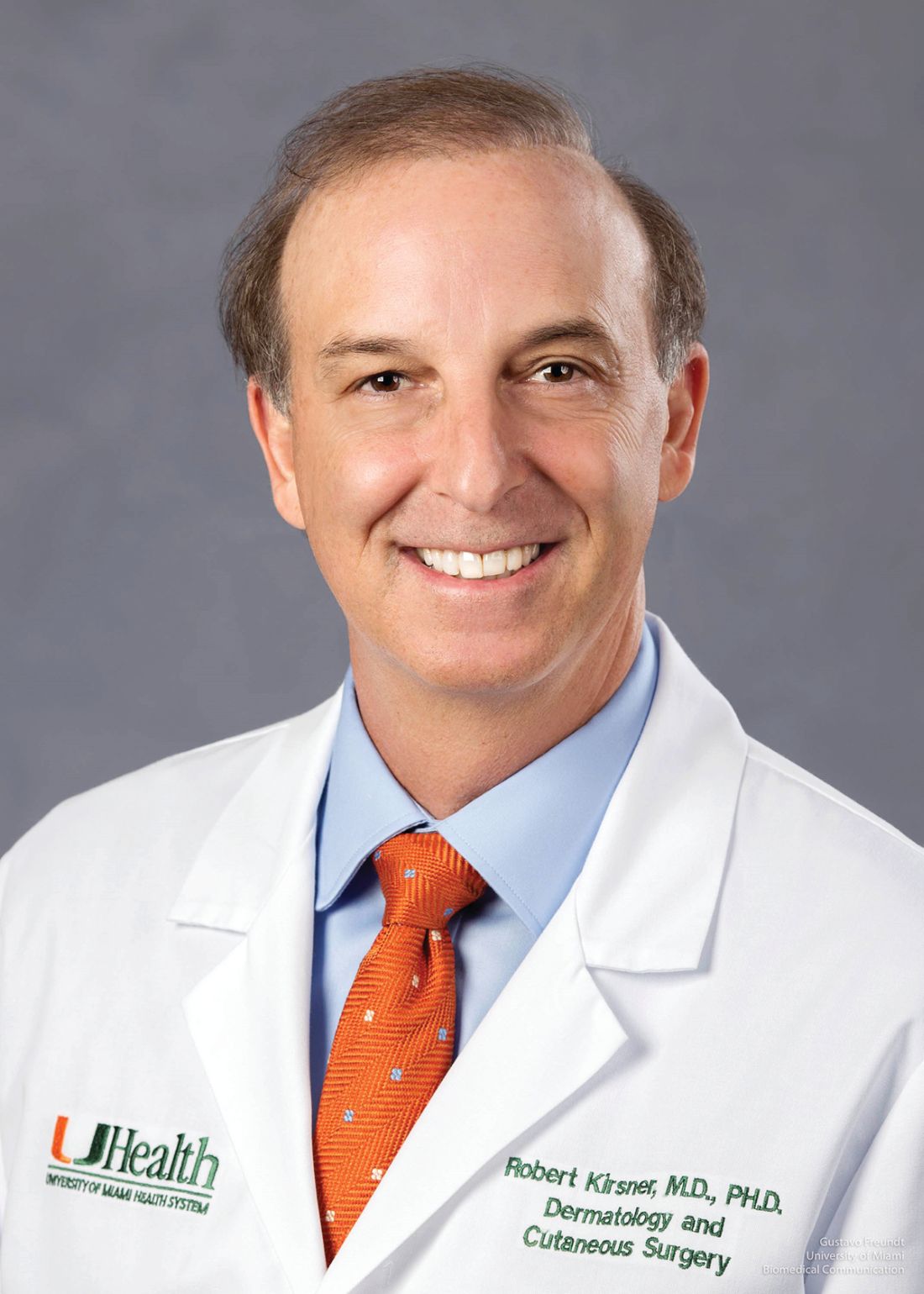
, director of the Wound Clinic at Jackson Memorial Hospital and chair of the Department of Dermatology and Cutaneous Surgery at the University of Miami, Florida.
“We as a specialty make and repair more wounds than any other specialty,” said Kirsner, who provided data to make his point. In a table he showed, the number of wound repairs made annually by dermatologists was several-fold higher than surgeons, the next highest group, and the numbers declined rapidly from there.
Speaking at the 27th Annual Winter Symposium – Advances in Medical and Surgical Dermatology (MSWS) 2024, Kirsner offered an array of clinical pearls, reinforced some basics, and pointed to well-supported strategies he believes are too often overlooked.
Drugs Repurposed for Wound Healing
Of the clinical pearls, he spoke of the repurposing of several agents for wound care. His first example was the monoclonal antibody dupilumab, which inhibits interleukin-4 (IL-4) and IL-13 signaling, to heal selected patients with leg ulcers. The potential of this drug for wound healing was based on a patient with a leg ulcer who presented with concomitant prurigo nodularis and biliary cirrhosis. When offered for the comorbidities, dupilumab provided a “dramatic” benefit with regard to the wound, according to Kirsner.
The explanation for the response is that IL-4 and IL-13 have been found to be upregulated in some patients with leg ulcers. Based on numerous cases, Kirsner spoke of a phenotype of nonhealing leg ulcers from which elevated IL-4 and IL-13 can be isolated; these are the candidates for adding dupilumab to wound care, he said.
Topical beta-blockade is another example of a therapy repurposed for wound healing, according to Kirsner. He said beta-blockers are already a standard of care for burn wounds, but the mechanism is relevant in other wound types.
Several studies have looked at this phenomenon, with experimental studies showing that skin healing is impaired when beta-2 receptors are agonized but accelerated when blocked.
Beta-Blockade Accelerates Wound Healing
A recent review of these mechanisms in soft-tissue wound healing pointed to an anti-inflammatory effect, acceleration of keratinocyte migration, pro-reepithelization effects, and inhibition of bacterial virulence. Beta-blockers were first implicated as mediators of wound healing more than a decade ago, but Kirsner indicated that there is now more attention to this therapy within a comprehensive approach in difficult cases.
Although not specific to wound healing, the potential for teprotumumab to improve control of pretibial myxedema is another example of a repurposed therapy for a challenging skin disease. Teprotumumab, a monoclonal antibody that targets the insulin-like growth factor-1 (IGF-1) receptor, is approved for active thyroid eye disease, but Kirsner cited data showing compelling evidence of benefit in this cutaneous complication of Graves disease.
As for basics, Kirsner devoted some time to emphasizing the importance of compression therapy for improving leg vascularization. This is not something to just consider; rather, he thinks it is part of standard practice. “Compress all leg ulcers,” was Kirsner’s simple message.
Citing encouraging work in identifying targetable molecular events in wound healing, Kirsner suggested that treatment might be increasingly guided by biomarkers. He pointed to ongoing work to characterize wound exudate as a source of biomarkers.
“The discarded dressing contains a wealth of information,” he said, referring to cell types and proteins, such as growth factors. He thinks that the ongoing studies of exudate, which have shown that molecular processes detected at the periphery are often different than those at the focal site of injury, have substantial promise for identifying new treatment targets.
Virtual Reality to Address Pain
From a practical standpoint, Kirsner looked to a well-studied but still underused adjunct to wound debridement and surgical repair: the distraction offered by relatively low-priced virtual reality systems. He described it as a simple way to help patients keep their minds off the pain. It is not a new idea and has been studied for this use numerous times, and the evidence of benefit is essentially uniform, according to Kirsner.
He said effective and sophisticated systems can now be purchased for just hundreds of dollars, and no training is needed. Indeed, he said pediatric patients can typically explain how the system works if the clinician does not know.
“If you can enhance their experience [during wound repair], you can make their lives and your life better,” he said.
Joshua Zeichner, MD, associate professor of dermatology at Mount Sinai Hospital in New York City, concurred that the evidence supports this approach and is easy to do. “I am in favor of anything that improves the experience of the patient,” said Zeichner, who chaired the portion of the meeting during which Kirsner spoke.
Kirsner said he practices what he preaches. “I routinely employ virtual reality for simple surgical procedures or processes that patients might find unpleasant,” he said. He acknowledged that clinicians might have heard this message before, but he believes those who have not yet introduced this into their practice should consider it.
Kirsner has reported no relevant financial relationships. Zeichner has reported serving as a consultant for Beiersdorf.
A version of this article first appeared on Medscape.com.
NEW YORK CITY — Instructions on wound healing often involve disturbing photographs of severe diabetic ulcers, angry autoimmune blistering, and oozing lesions produced by uncommon genetic disorders, but whether or not they are dramatic, day-to-day dermatologic wound care relies on both the basics as well as novel approaches, according to a well-known wound treatment expert.
, director of the Wound Clinic at Jackson Memorial Hospital and chair of the Department of Dermatology and Cutaneous Surgery at the University of Miami, Florida.
“We as a specialty make and repair more wounds than any other specialty,” said Kirsner, who provided data to make his point. In a table he showed, the number of wound repairs made annually by dermatologists was several-fold higher than surgeons, the next highest group, and the numbers declined rapidly from there.
Speaking at the 27th Annual Winter Symposium – Advances in Medical and Surgical Dermatology (MSWS) 2024, Kirsner offered an array of clinical pearls, reinforced some basics, and pointed to well-supported strategies he believes are too often overlooked.
Drugs Repurposed for Wound Healing
Of the clinical pearls, he spoke of the repurposing of several agents for wound care. His first example was the monoclonal antibody dupilumab, which inhibits interleukin-4 (IL-4) and IL-13 signaling, to heal selected patients with leg ulcers. The potential of this drug for wound healing was based on a patient with a leg ulcer who presented with concomitant prurigo nodularis and biliary cirrhosis. When offered for the comorbidities, dupilumab provided a “dramatic” benefit with regard to the wound, according to Kirsner.
The explanation for the response is that IL-4 and IL-13 have been found to be upregulated in some patients with leg ulcers. Based on numerous cases, Kirsner spoke of a phenotype of nonhealing leg ulcers from which elevated IL-4 and IL-13 can be isolated; these are the candidates for adding dupilumab to wound care, he said.
Topical beta-blockade is another example of a therapy repurposed for wound healing, according to Kirsner. He said beta-blockers are already a standard of care for burn wounds, but the mechanism is relevant in other wound types.
Several studies have looked at this phenomenon, with experimental studies showing that skin healing is impaired when beta-2 receptors are agonized but accelerated when blocked.
Beta-Blockade Accelerates Wound Healing
A recent review of these mechanisms in soft-tissue wound healing pointed to an anti-inflammatory effect, acceleration of keratinocyte migration, pro-reepithelization effects, and inhibition of bacterial virulence. Beta-blockers were first implicated as mediators of wound healing more than a decade ago, but Kirsner indicated that there is now more attention to this therapy within a comprehensive approach in difficult cases.
Although not specific to wound healing, the potential for teprotumumab to improve control of pretibial myxedema is another example of a repurposed therapy for a challenging skin disease. Teprotumumab, a monoclonal antibody that targets the insulin-like growth factor-1 (IGF-1) receptor, is approved for active thyroid eye disease, but Kirsner cited data showing compelling evidence of benefit in this cutaneous complication of Graves disease.
As for basics, Kirsner devoted some time to emphasizing the importance of compression therapy for improving leg vascularization. This is not something to just consider; rather, he thinks it is part of standard practice. “Compress all leg ulcers,” was Kirsner’s simple message.
Citing encouraging work in identifying targetable molecular events in wound healing, Kirsner suggested that treatment might be increasingly guided by biomarkers. He pointed to ongoing work to characterize wound exudate as a source of biomarkers.
“The discarded dressing contains a wealth of information,” he said, referring to cell types and proteins, such as growth factors. He thinks that the ongoing studies of exudate, which have shown that molecular processes detected at the periphery are often different than those at the focal site of injury, have substantial promise for identifying new treatment targets.
Virtual Reality to Address Pain
From a practical standpoint, Kirsner looked to a well-studied but still underused adjunct to wound debridement and surgical repair: the distraction offered by relatively low-priced virtual reality systems. He described it as a simple way to help patients keep their minds off the pain. It is not a new idea and has been studied for this use numerous times, and the evidence of benefit is essentially uniform, according to Kirsner.
He said effective and sophisticated systems can now be purchased for just hundreds of dollars, and no training is needed. Indeed, he said pediatric patients can typically explain how the system works if the clinician does not know.
“If you can enhance their experience [during wound repair], you can make their lives and your life better,” he said.
Joshua Zeichner, MD, associate professor of dermatology at Mount Sinai Hospital in New York City, concurred that the evidence supports this approach and is easy to do. “I am in favor of anything that improves the experience of the patient,” said Zeichner, who chaired the portion of the meeting during which Kirsner spoke.
Kirsner said he practices what he preaches. “I routinely employ virtual reality for simple surgical procedures or processes that patients might find unpleasant,” he said. He acknowledged that clinicians might have heard this message before, but he believes those who have not yet introduced this into their practice should consider it.
Kirsner has reported no relevant financial relationships. Zeichner has reported serving as a consultant for Beiersdorf.
A version of this article first appeared on Medscape.com.
FROM MSWS 2024
Tapering Corticosteroids in Severe Alcohol-Associated Hepatitis Appears Safe
SAN DIEGO — , according to new research.
“Although several drugs have been evaluated for severe alcohol-associated hepatitis, none have succeeded in practice. Corticosteroids remain the mainstay of treatment; however, infections remain a major concern in 25%-40% of cases,” said Anand Kulkarni, MD, senior consultant and director of critical care hepatology at the Asian Institute of Gastroenterology in Hyderabad, India.
“There are no standard society guidelines for steroid dosing, and our current practices stem from studies in the 1970s, so there’s a major knowledge gap around optimal dosing and if stepwise tapering helps,” said Kulkarni, who presented the findings at The Liver Meeting 2024: American Association for the Study of Liver Diseases (AASLD).
Assessing Tapered Doses
In a multicenter, open-label randomized controlled trial, 254 patients with SAH from four Indian centers and one Canadian center were randomized to receive either a fixed or tapering dose of 40 mg prednisolone daily for 4 weeks. The patients in the tapering group received a starting dose of 40 mg, which was reduced by 10 mg weekly over 4 weeks.
While taking corticosteroids, 66% of those in the fixed dose group and 55% of those in the tapering group also received prophylactic antibiotics.
The mean age of participants was 41.1 years, the median Model For End-Stage Liver Disease score was 25.6, and 98.4% were men.
The primary objective was to compare the incidence of drug-related adverse events, infections, hospitalization, and mortality through day 90.
The duration of corticosteroid therapy was 22 days in the fixed dose group and 23 days in the tapering dose group.
Overall, the proportion of steroid responders was similar in both groups, at 80.3% in the fixed dose group and 82.5% in the tapering dose group.
However, the incidence of drug-related adverse events was significantly higher in the fixed dose group (52%) than in the tapering dose group (36.2%). The most common adverse events in both groups were infection, hyperglycemia, and hematochezia.
At 90 days, the incidence of infection was significantly lower in the tapering group (19.7%) than in the fixed dose group (33.1%). In both groups, the most common infection sites were the lungs (28.3%) and urinary tract (22.4%).
In terms of liver-related outcomes, some patients developed hepatic encephalopathy (11.8% in fixed dose vs 6.3% in tapering dose) and acute variceal bleed (3.1% in each group), as well as acute kidney injury (26.8% in fixed dose vs 18.9% in tapering dose).
Hospitalization within 90 days was required in 44.1% of the fixed dose group and 33.1% of the tapering dose group.
Survival at day 90 was 83.5% in the fixed dose group and 86.6% in the tapering dose group. Four patients in the fixed dose group and three patients in the tapering dose group underwent living donor liver transplantation by day 90.
Relapse of alcohol use by day 90 occurred in 13.4% of the fixed dose group and 12.6% of the tapering dose group.
“Rapid tapering in severe alcohol-associated hepatitis reduces infections and hospitalizations but doesn’t have a significant impact on survival,” Kulkarni concluded.
Considering Alternative Therapies
Given the high risk for infection in patients with SAH and limited certainty around benefits, the data may also call into question whether to give steroids to these patients at all, said session co-moderator Aleksander Krag, MD, professor of clinical medicine at the University of Southern Denmark, Odense, Denmark, and secretary general of the European Association for the Study of Liver 2023-2025.
“Since there are no other treatments available as of now, we’ll still continue to give steroids,” Kulkarni noted. But “tapering the dose should be beneficial.”
Although steroid therapy has been considered the “mainstay treatment” for SAH for 50 years, it doesn’t always lead to long-term improvement in liver values or survival, said Prasun Jalal, MD, the Stan and Sue Partee Endowed Chair in Hepatology at Baylor College of Medicine, Houston, who wasn’t involved with the study.
Researchers are looking to other connections, such as the gut microbiome, to find treatments for advanced alcoholic liver disease, Jalal said in an interview. In a small pilot study, he and colleagues found that intestinal microbiota transplantation (IMT) appears to be safe and effective for these patients.
“Early analyses suggest that IMT has a favorable outcome on the prognosis of patients with severe alcohol-associated hepatitis and is safe,” Jalal said. “A longer follow-up study with a larger sample size is in progress.”
Kulkarni and Krag reported no relevant disclosures. Jalal has speaking and teaching relationships with AbbVie and Madrigal.
A version of this article appeared on Medscape.com.
FROM AASLD 2024
Diabetes Drugs and Eye Disease: These Protect, These Don’t
TOPLINE:
METHODOLOGY:
- Researchers conducted a retrospective analysis of electronic medical records from the TriNetX health research network to evaluate how systemic medications, such as GLP-1 RAs, fenofibrates, thiazolidinediones, and calcium channel blockers, influence the risk of developing DME in patients with type 2 diabetes.
- They included patients with a 5-year history of type 2 diabetes and an absence of DME at baseline.
- The treatment group included patients who initiated treatment with calcium channel blockers (n = 107,193), GLP-1 RAs (n = 76,583), thiazolidinediones (n = 25,657), or fenofibrates (n = 18,606) after a diagnosis of diabetes. The control group received none of these medications within 1 year of being diagnosed with the condition.
- The researchers used propensity score matching to balance baseline characteristics and comorbidities between both groups.
- The primary outcome was the incidence of diagnoses of DME within a 2-year follow-up period after the initiation of systemic medications.
TAKEAWAY:
- Patients treated with calcium channel blockers showed an increased risk for incident DME (hazard ratio [HR], 1.66; 95% CI, 1.54-1.78) compared with control individuals.
- Treatment with GLP-1 RAs was associated with a reduced risk for DME (HR, 0.77; 95% CI, 0.70-0.85), as was treatment with fenofibrates (HR, 0.83; 95% CI, 0.68-0.98).
- No significant difference in risk for DME was observed between patients taking thiazolidinediones and control individuals.
IN PRACTICE:
“We found a possible protective effect for GLP-1 RA medications and fenofibrate for DME and an adverse effect for calcium channel blockers with regard to the development of DME in patients” with type 2 diabetes, the authors wrote.
“Our preliminary data suggests a protective effect with regard to GLP-1 RA drugs and the development of DME. Clinical studies examining a potential therapeutic effect of GLP-1 RA drugs on DME do seem warranted. A single orally administered drug could conceivably lower blood sugar, reduce weight, offer cardiovascular protection, and treat DME” in patients with type 2 diabetes, they added.
SOURCE:
The study was led by Jawad Muayad, BS, of the School of Medicine at Texas A&M University, in Houston. It was published online on December 5, 2024, in Ophthalmology Retina.
LIMITATIONS:
The study was retrospective in nature. It relied on electronic medical records for the diagnosis of DME instead of directly assessing retinal images or measuring retinal thickness. Moreover, patients on certain medications may have been monitored more closely, potentially influencing the likelihood of a diagnosis of DME being recorded.
DISCLOSURES:
The study did not receive any funding support. One author disclosed receiving consulting fees from various institutions and pharmaceutical companies. The other authors reported no relevant conflicts of interest.
This article was created using several editorial tools, including AI, as part of the process. Human editors reviewed this content before publication. A version of this article first appeared on Medscape.com.
Psychologists and Psychotropic Prescribing: An Old Debate Heats Up
Earlier in 2024, Utah became the seventh state to allow psychologists with the proper training to prescribe psychotropic medications, giving supporters reason to hope that more states might support expanding this scope of practice.
However, the American Psychiatric Association — and some psychologists — oppose granting psychologists this privilege, arguing that the training offered is insufficient and could jeopardize patient safety.
The controversy over whether psychologists should be allowed to prescribe is as old as the so-called RxP movement itself, which began in the early 1990s.
Psychologists have not rushed to become licensed prescribers. After three decades, an estimated 226 psychologists — representing just 0.14% of all those licensed in the United States — have been authorized to prescribe in the six states and one territory where it has been legalized, according to a just-published study in Clinical Psychology.
These are Colorado, Idaho, Illinois, Iowa, Louisiana, New Mexico, and Guam. Data from the study show that only 73 psychologists are prescribing in New Mexico, which authorized it in 2002.
Less is known about the number of psychologists who are prescribing under allowances in the Department of Defense, Indian Health Service and US Public Health Service.
Some psychologists — and the American Psychological Association (APA) — believe that the persistence of the opioid epidemic coupled with a continued lack of access to mental health care for millions of Americans will bring more legislators on-side.
“I feel like we’re on an upswing again,” Deborah Baker, director of legal and regulatory policy for the APA, told Medscape Medical News. “The access issue continues to be a perennial kind of driver.” She noted that at least six states pursued expanding privileges this year.
Robert L. Trestman, MD, chair of the American Psychiatric Association’s Council on Healthcare Systems and Financing, said he doesn’t see new momentum. The interest in having psychologists prescribe “continues to trickle based on just the frustration that people have about not getting adequate access to psychiatry,” he told Medscape Medical News.
While states may be trying to increase access to care, granting psychologists privileges is “not a very effective way of doing it,” said Trestman, Chair of Psychiatry and Behavioral Medicine at Virginia Tech Carilion School of Medicine in Roanoke. Psychologists are needed to deliver psychotherapy, he said. “It makes almost no sense to try to make them into pseudo medical professionals,” said Trestman. “It just exposes people to risks.”
William Robiner, PhD — author of Clinical Psychology study — is a long-time opponent of RxP. The psychologist told Medscape Medical News he’s concerned about patient safety and “about some of the disingenuous reasons” that psychologists want to prescribe. Among these are the ability to increase status and income, said Robiner, a professor of medicine at the University of Minnesota Medical School, Minneapolis, and a board member of Psychologists Opposed to Prescription Privileges for Psychologists.
Adequate Training?
Only PhD and PsyD psychologists are eligible for RxP training, which entails a master’s in clinical psychopharmacology. After receiving the master’s, they must pass the Psychopharmacology Examination for Psychologists and then are only permitted to prescribe medications for mental health disorders.
They must also obtain a Drug Enforcement Administration license but can’t write prescriptions for schedule 2 medications. In some states, psychologists can prescribe buprenorphine and other opioid use disorder medications.
The APA has developed guidance for master’s programs, which currently number just over a handful in the United States.
At Fairleigh Dickinson University in New Jersey, students enrolled in the master’s program — a distance-learning format — complete 10 courses over five 15-week semesters. The curriculum spans a range of topics, from foundational sciences and legal and ethical considerations to strategies for treating specific disorders.
Derek Phillips, PhD, the program’s executive director, said that when he took the position in 2020, enrollment was capped at 45 students, but “we were not routinely enrolling the maximum.” Now, even with class size increased to 60 “we are consistently full and have a waitlist,” he told Medscape Medical News.
Interest is being driven in part by new laws in Colorado (2023) and Utah, said Phillips. But many are enrolling without intending to write a prescription, he said. The degree gives graduates the ability to better collaborate with other clinicians, teach clinical psychopharmacology, and be expert witnesses in medico-legal cases, he said.
In addition, the training gives students “a balanced and thorough biopsychosocial understanding of our patients,” he said. Students also see the “potential of being able to be a ‘one-stop-stop’ of mental health services,” said Phillips.
The American Board of Professional Psychology is developing a board certification in clinical psychopharmacology.
The APA states on its website that prescribing psychologists have “more training in diagnosing and treating (including prescribing) mental health disorders than primary care physicians.”
However, critics argue that the training falls short. Most psychologists, said Robiner, have not completed the undergraduate prerequisites — such as anatomy, physiology, and chemistry — that are required for other prescribing professionals.
In a 2019 article comparing the training of prescribing professionals, Robiner and colleagues reported that psychiatrists undergo 4- to 6-week rotations during medical school and accumulate 8000 clinical hours focused on psychiatric conditions over the course of their 4-year residencies.
States set requirements for clinical hours for prescribing psychologists, but they are generally elective and completed after individuals receive a master’s degree.
Robiner said psychologists aren’t trained in evaluating drug-related adverse events. “If you show a psychologist a rash, they have no idea whether that rash might be a medication adverse effect or poison ivy,” he said.
Trestman pointed out that many psychotropic medications have black box warnings. “The risk of toxicity is by no means trivial, and the majority of people who are seeking care in psychiatry have multiple comorbidities,” he said. “Giving people the equivalent of more or less 10 weeks of training is just woefully inappropriate,” Trestman said.
Increase in Access?
Psychology’s main argument for expanding its scope of practice is that it will increase the number of clinicians available to provide behavioral and mental health care.
Critics said that is a failed experiment, in part because so few psychologists have become prescribers, but also because most psychologists practice in the same areas as psychiatrists. Both specialists tend to cluster in urban regions, which already have high clinician density, said Trestman.
Psychologists are not practicing in underserved rural areas, as even APA data show. A 2018 APA snapshot of the workforce found that the highest density of psychologists was in Washington, DC, Massachusetts, and New York. South Carolina, West Virginia and Mississippi had the fewest number of psychologists per 100,000 people.
The University of Washington Rural Health Research Center reported in 2022 that in 2021, almost half of rural counties did not have a psychologist compared with 15.7% of urban counties.
Psychiatrists also are concentrated on the coasts and New England, according to a study by Ohio State researchers. The highest densities were in Washington, DC (79 per 100,000), Massachusetts (45.3), Rhode Island (42.6), Connecticut (38.6) and Vermont (37.7), whereas the lowest densities were in Idaho (11.8), Mississippi (11.8), Wyoming (12.4), Alabama (13.1), and Indiana (13.5). The study estimated that there were 57,163 psychiatrists responsible for the care of 333,287,557 Americans. “Clinical psychologists, psychotherapists, and counselors can provide alternative forms of intervention, though access to such services is also poor in rural areas,” wrote the authors.
The APA counters with data it says shows that RxP may have increased access. Using the number of psychology practices as a proxy for supply, the authors reported that practices grew in New Mexico, Illinois, Iowa, and Idaho — states that have implemented prescription privileges. Overall, there was an increase of 0.8047 practices per 100,000 residents per county.
However, the access argument “is seriously challenged by the reality of the limited number of psychologists who complete the pathway to prescribing,” Robiner and his colleague Tanya Tompkins countered in Clinical Psychology. They note that in Idaho — a state with shortages of psychologists and psychiatrists — just 10 of the state’s 615 psychologists had prescriptive authority. An estimated 5131 nonpsychologists are prescribers.
Robiner and Tompkins noted that it’s not clear why so few psychologists are pursuing RxP but that many seem to be unaware of the possibility.
Do Benefits Outweigh the Harms?
There is not a large body of literature assessing the harms or benefits of prescribing privileges for psychologists.
Baker shared several studies by Phillip Hughes, PhD, an outcomes researcher at the University of North Carolina Eshelman School of Pharmacy, Chapel Hill, North Carolina. In one study, Hughes found that patients of prescribing psychologists had a 24% lower rate of adverse drug events than patients of psychiatrists. Psychologists’ patients had lower rates of psychotropic polypharmacy but similar rates of emergency room use.
In another paper Hughes suggested that deaths attributable to mental illness had declined in New Mexico after it passed its law. There was no change in Louisiana.
With little evidence of harm — and ongoing provider shortages — making use of nonphysician prescribers is gaining traction with policymakers, claims the psychology association’s Baker, adding that in Utah, the Republican governor was the biggest supporter.
But psychiatrists argue that it’s more important to increase their numbers. Congress agreed in 2021 and 2023 to add 1200 new residency slots — in every specialty — to ease physician shortages. The Centers for Medicare & Medicaid Services recently announced that 70% of the new slots for July 2025 will go to primary care and psychiatry.
“Once those positions are in place, it will be four more years before the first crop of new psychiatrists come out,” noted Trestman. “None of these fixes are quick,” he said.
Baker, Robiner, and Trestman reported no relevant financial relationships.
A version of this article first appeared on Medscape.com.
Multiple Myeloma: Dexamethasone-Sparing Approach Benefits Frail Older Adults
The study “is the first randomized phase 3 study dedicated to frail patients,” said first author Salomon Manier, MD, PhD, an associate professor of hematology at Lille University Hospital, Lille, France. He presented the findings this week at the American Society of Hematology (ASH) 2024 Annual Meeting in San Diego.
“It shows that daratumumab-lenalidomide [with limited dexamethasone] led to a significant reduced risk of progression or death by 49% in frail patients, with a favorable safety profile and an improved health-related quality of life,” Manier said.
Older adult patients who are newly diagnosed with MM have varying levels of fitness or frailty and have been shown to have shorter survival, with higher rates of nonhematologic adverse events and treatment discontinuation.
While the regimen of daratumumab, lenalidomide, and dexamethasone has become a standard of care, with efficacy and tolerance, including for patients with frailty, the infection and pneumonia rates with the approach can be high, particularly for patients with frailty.
To evaluate if an alternative, dexamethasone-sparing approach could improve outcomes while limiting toxicity for older adults, Manier and his colleagues conducted the prospective, open-label phase 3 IFM2017-03 trial, involving 295 patients (age, 65 years) with newly diagnosed MM at 90 centers in France.
The patients had a median age of 81, with 84% older than 75 years and 61% older than 80 years. All had an Eastern Cooperative Oncology Group proxy frailty score ≥ 2.
The patients were randomized 1:2 to treatment either with the Rd regimen of 28-day cycles of lenalidomide (25 mg/d, 21/28) and dexamethasone (20 mg once weekly) or with the dexamethasone-sparing regimen (DR) of daratumumab (1800 mg subcutaneous once weekly for 8 weeks, once every 2 weeks for 16 weeks, and once every 4 weeks thereafter), lenalidomide (25 mg/d, 21/28), and two cycles of dexamethasone (20 mg once weekly for 8 weeks then stopping).
The randomization included stratification based on age and cancer stage. Baseline characteristics were well balanced in the two groups.
Both regimens were administered until disease progression or unacceptable toxicity.
As of the data cut-off in November 2024, with a median follow-up of 46.3 months, 81 of the original 200 patients in the DR arm remained on treatment, and 11 of 95 continued in the Rd arm.
The overall median treatment duration among the 200 original patients in the DR arm was 31.6 months and 14.3 months in the Rd arm.
The study met its primary endpoint of progression-free survival (PFS), with a median rate of 53.4 months in the DR arm vs 22.5 months in the Rd arm (hazard ratio [HR], 0.51; P < .0001).
The improved PFS in the DR arm was observed across all subgroups based on age, Charlson comorbidity index, cancer stage, cytogenetics, and creatinine clearance; however, those with lower frailty scores had better outcomes in both groups.
A median overall survival was not reached in the DR arm vs 47.2 months in the Rd arm (HR, 0.52; P = .0001).
The DR arm also showed a higher overall best response rate of 94% vs 86% (P = .005), respectively, with deeper responses in the DR arm at all time points, including the earliest points at 4 months.
In terms of adverse events (AEs), at least one AE of grade ≥ 3 occurred in 89% in the DR arm and 79% in the Rd arm.
Those in the DR arm had significantly more grade 3 or higher hematologic AEs with neutropenia (62% vs 34%); however, grade ≥ 3 infections were similar, with 19% in the DR arm and 21% in the Rd arm. Infections involving pneumonia occurred in 6% and 8%, respectively.
There were no significant differences between the two arms in discontinuations due to AEs (30% in the DR arm and 34% in the Rd arm).
While health-related quality of life measures at baseline were well balanced between the two groups, those in the DR arm reported significantly shorter times to clinically meaningful improvement in all domains of the EORTC Core Quality of Life questionnaire (EORTC QLQ-C30).
“The safety profile was significantly improved and was favorable, without increased infection or pneumonia rates, with similar rates of treatment discontinuation,” Manier said.
Based on the findings, “we believe that the dexamethasone-sparing strategy is effective and safe for treating these frail patients with multiple myeloma,” he added.
Commenting on the study, Manni Mohyuddin, MD, an assistant professor in the multiple myeloma program at the Huntsman Cancer Institute, University of Utah, Salt Lake City, noted that, with the popularity of triplet regimens such as daratumumab, lenalidomide, and dexamethasone, the Rd regimen in the study is not as commonly used.
“But what this trial tells us is that efficacy outcomes in such a group of patients are encouraging despite getting rid of dexamethasone so quickly,” he said in an interview.
However, “while the tide is changing, dexamethasone is still given long-term, week after week after week in most myeloma protocols and in the community,” he noted. “Hopefully, this trial, and some of the other work, will lead to a change in this space.”
Mohyuddin suggests one approach can involve starting off with just daratumumab and a low dose of dexamethasone, then adding lenalidomide at a lower dose later in a sequential/graded approach.
“Other approaches that should be studied should be bispecific antibodies used sparingly, with less frequent dosing, or finite period,” he added. “There is lot to refine.”
Mohyuddin added a critique that the study’s endpoint of PFS may not have been ideal for the population of elderly and frail patients.
“Older people generally prioritize functional independence and quality of life, and perhaps a more patient-centered endpoint could have led to an even more informative study,” he said.
Manier’s disclosures included consultancy with Takeda Pharmaceuticals, Sanofi, Novartis, Janssen, GlaxoSmithKline, Celgene/BMS, Amgen, Adaptive Biotechnologies, Roche, Regeneron, and AbbVie. Mohyuddin had no disclosures.
A version of this article appeared on Medscape.com.
FROM ASH 2024
New Investigation Casts Doubt on Landmark Ticagrelor Trial
New questions about the landmark trial that launched the antiplatelet drug ticagrelor worldwide are being raised after an investigation uncovered more information about how the PLATO study was conducted.
Peter Doshi, PhD, senior editor at The BMJ, obtained primary records for the trial and unpublished data through a Freedom of Information Act request, and has detailed inconsistencies and omissions in data reporting from the 2009 trial originally published in The New England Journal of Medicine (NEJM). The new investigation into the Platelet Inhibition and Patient Outcomes (PLATO) trial is published in The BMJ.
The findings come as generic versions of ticagrelor (Brilinta) are expected to become available soon in the United States. Ticagrelor is the only P2Y12 inhibitor still under patent, and in 2022, the United States spent more than $750 million on it, according to the report.
PLATO, sponsored by ticagrelor manufacturer AstraZeneca, included more than 18,000 patients in 43 countries. Investigators reported that ticagrelor reduced deaths from vascular causes, heart attack, or stroke compared with clopidogrel (Plavix). However, in a subgroup analysis, among US patients, there were more deaths in the ticagrelor group, and AstraZeneca failed its first bid for approval from the US Food and Drug Administration (FDA).
Failed First Bid for FDA Approval
AstraZeneca resubmitted its application, which was met with objections by some FDA staff members, including medical officer Thomas Marciniak, who called the resubmission “the worst in my experience regarding completeness of the submissions and the sponsor responding completely and accurately to requests,” Doshi reports.
Despite the objections, the FDA in 2011 approved ticagrelor for acute coronary syndrome, kicking off intense controversy over the trial, as several other studies have failed to replicate PLATO’s positive results.
Doubts have grown about its apparent advantage over cheaper, off-patent P2Y12 inhibitors such as clopidogrel and prasugrel.
“Critics said it was noteworthy that ticagrelor failed in the US,” Doshi writes, “the only high enrolling country where sites were not monitored by the sponsor itself.” Doshi’s report points out that critics of the trial “highlight that AstraZeneca itself carried out the data monitoring for PLATO except for sites that were monitored by third party contract research organizations. In the four countries exclusively monitored by non-sponsor personnel—Georgia, Israel, Russia, and the US—ticagrelor fared worse.”
Victor Serebruany, MD, from Johns Hopkins University, said he was initially impressed by the trial results but became skeptical after noticing inconsistencies and anomalies in the data. He filed a complaint with the US District Court in the District of Columbia, suggesting that the cardiovascular events in the study “may have been manipulated.”
US Department of Justice Investigation
The US Department of Justice (DOJ) opened an investigation in 2013 and closed it in 2014 with no further action. Serebruany continues to publish critiques of the trial 15 years later but told The BMJ he has little hope that the questions will be resolved unless the DOJ re-engages with an investigation.
Doshi also points out discrepancies in the data reported. In the 2009 paper, published as an intent-to-treat analysis, investigators said there were 905 total deaths from any cause among all randomized patients. “An internal company report states, however, that 983 patients had died at this point. While 33 deaths occurred after the follow-up period, the NEJM tally still leaves out 45 deaths ‘discovered after withdrawal of consent,’” he reports.
The NEJM responded to Doshi that while it didn’t dispute the error in the number of deaths, it was uncertain about publishing a correction, citing new — not yet published — guidelines from the International Committee of Medical Journal Editors. NEJM Editor-in-Chief Eric Rubin told The BMJ that “for older manuscripts, correction is not necessarily appropriate unless there would be an effect on clinical practice.”
Doshi’s investigation includes an interview with Eric Bates, MD, professor of internal medicine at the University of Michigan in Ann Arbor, and a co-author of the US guidelines that recommend ticagrelor, who said he was “increasingly disturbed by how trial after trial came out as being not dramatically positive in any way.” Bates is now calling for a review of ticagrelor’s recommendation in guidelines, according to the report.
AstraZeneca declined to be interviewed for the BMJ investigation, according to Doshi, and a spokesperson from the company told the journal by email that they have “nothing to add,” directing editors to its 2014 public statement after the DOJ’s investigation into PLATO. The BMJ said PLATO trial co-chairs Robert A. Harrington, MD, and Lars Wallentin, MD, did not respond to The BMJ’s requests for comment.
Will the Guidelines Be Changed Now?
“I know and have worked with Drs Wallentin and Harrington,” Bates told Medscape Medical News, “and find them to be honest, intelligent clinical scientists with the highest ethical standards who manage conflicts of interest as well as can be done in the clinical research arena, where industry support is required to develop new knowledge,” he said.
“If there is a concern that AstraZeneca was manipulating the dataset and FDA submission, that is an important issue,” Bates said. “The US paradox and the failure of any other antiplatelet trial to find a comparative mortality advantage are two unexplained issues with PLATO that provide good fodder for conspiracy theories. I agree with the NEJM that this trial is 15 years old and may not be worth readjudicating in the current treatment era.”
Other calls for revisiting guidelines have come after disappointing postlicensure studies have repeatedly demonstrated that ticagrelor has “similar efficacy to clopidogrel but with increased bleeding and [dyspnea],” Doshi reports.
“My concern is the marketing spin by AstraZeneca and the promotion of ticagrelor by six to eight ‘thought leaders’ consistently funded by AstraZeneca over the past 10 years,” said Bates. “They have flooded the literature with supportive subset and post hoc analyses, review articles, and ‘meta-analyses’ flawed by selection and intellectual bias, and public interviews that consistently discount the findings of the many subsequent randomized controlled trials that have not supported the superiority of ticagrelor over clopidogrel or prasugrel.”
A version of this article first appeared on Medscape.com.
FROM THE BMJ