User login
News and Views that Matter to Physicians
Initial outcomes of PERT at Cleveland Clinic
LOS ANGELES – Initial outcomes measures are beginning to emerge from Pulmonary Embolism Response Teams.
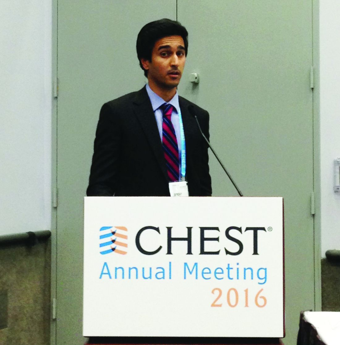
Members of the Cleveland Clinic’s PERT, which was established in 2014, presented some of their preliminary data during a presentation at the CHEST annual meeting.
The concept behind the PERT is to rapidly mobilize a team with varied expertise helpful for treating patients with pulmonary embolisms (PEs). While the PERT “can be activated by any (clinician) for any patient, even low-risk patients ... those with submassive and massive PEs [intermediate- and high-risk patients]” are the target patients, said Dr. Mahar of the Cleveland Clinic.
The first PERT was created at Massachusetts General Hospital in Boston in 2012, according to the National Consortium of Pulmonary Embolism Response Team’s website. As of May 2015, the PERT model has been adopted by physicians and health care professionals from more than 40 institutions.
Dr. Mahar reported that the Cleveland Clinic’s PERT is activated through a single pager that resides with a vascular medicine fellow during the day and a critical care fellow at night. When paged, the fellow promptly evaluates the patient and ensures a complete basic work-up, which includes an ECG, cardiac enzymes, N-terminal pro b-type natriuretic peptide, lower-extremity deep vein thrombosis scans, transthoracic echocardiogram, and confirmatory CT/PE protocol or ventilation/perfusion scan.
Based on the simplified Pulmonary Embolism Severity Index and Bova scores, the patient is risk stratified and the patient’s indications, and relative and absolute contraindications to advanced therapies are reviewed. The fellow next sends a group notification to the PERT via email and text message. The team then convenes online for a virtual meeting and case presentation that includes sharing of lab and test results and images.
The process sounds complex, but the surgeon, interventional radiologist, vascular medicine specialist, and cardiologist are on call and simultaneously get the message and respond, Dr. Mahar said. With a team approach, the decision to use advanced therapies – systemic lytics, surgery, catheter-directed lysis and extracorporeal membrane oxygenation – is expedited. “For example, over the last 2 years, four out of four patients who underwent surgical embolectomies had good outcomes without any deaths,” he said.
Based on a retrospective chart review from October 2014 through August 2016, Cleveland Clinic’s PERT had been activated for 134 patients, 112 of whom were found to have PEs, Dr. Mahar said during his presentation at the annual meeting of the American College of Chest Physicians (CHEST).
The number of low risk, submassive, and massive PEs were 14 (12%), 76 (68%), and 22 (20%), respectively. Just over half of the PE patients, 55% (60 patients), were treated with anticoagulation therapy alone. Inferior vena cava filters were placed in 32 patients (29%); 14 patients received catheter-directed thrombolysis, 3 received a suction thrombectomy, and 4 received a surgical embolectomy.
The 30-day all-cause mortality rate was 9%; the deaths occurred in six patients who had massive PEs, three patients with submassive PEs, and one patient with a low-risk PE. Six of the patients who died had been treated with anticoagulation, two had received catheter-directed thrombolysis, and one had received a full dose of systemic thrombolysis.
Bleeding complications occurred in 10 patients, 6 of whom were treated with anticoagulation alone and 4 of whom underwent catheter-directed thrombolysis.
Cleveland Clinic is a large entity with multiple resources, but the principles of PERT can be applied in smaller facilities, as well, according to Gustavo A. Heresi-Davila, MD, medical director of the Cleveland Clinic’s pulmonary thromboendarterectomy program and the lead researcher for the PERT project at the clinic. “I would emphasize the notion that a PERT has to be multidisciplinary, as people with different backgrounds and expertise bring complementary talent to the discussion of each case. I would not minimize the challenges of assembling such a team,” he said during an interview following the meeting.
The moderator of the meeting session, Robert Schilz, DO, PhD, noted, that the goal of PERT is to determine the best approach for an individual patient based on available resources. To establish a PERT, “you don’t have to be able to put a patient on ECMO [extracorporeal membrane oxygenation] in 15 minutes, and you don’t have to be able to do endarterectomies, embolectomies, and all the catheter-drive techniques emergently. But you do need to have the disposition to have efficient and standardized care, and the solutions may need to be very geographic. What hospital A may do may be very different from hospital B.”
Small hospitals can draw on their available resources, added Dr. Schilz, director of pulmonary vascular disease and lung transplantation at Case Western Reserve University, Cleveland. “Most hospitals have cardiologists on call 24/7, and many have some flavor of interventional radiology; others have clear referral and transfer schemes. Emergency department personnel at small rural hospitals can rapidly identify patients appropriate for transfer.”
Dr. Mahar added that PERTs are already being utilized in smaller hospitals and that he thinks that, in the next 5 years, having a PERT will be the standard protocol.
Dr. Mahar reported no disclosures.
Mary Jo Dales contributed to this report.
FROM CHEST 2016
HCT survivors experience high rates of late respiratory and infectious complications
Cancer survivors who underwent hematopoietic cell transplantation (HCT) face a greater risk for hospitalizations and mortality, compared with survivors who did not have HCT.
New findings show that disparities in infectious and respiratory complications were marked between the two groups, but differences in circulatory disease, mental health diagnoses, and second cancers were insignificant.
“Clinicians who care for long-term survivors of HCT should be aware of comprehensive surveillance guidelines available for this high-risk population,” wrote Eric J. Chow, MD, of the Fred Hutchinson Cancer Research Center, Seattle, and his colleagues (J Clin Oncol. 2016 Nov 21. doi: 10.1200/JCO.2016.68.8457).
There have only been a few comprehensive analyses that have compared HCT with non-HCT cancer survivors. Thus, the authors noted that it is unclear if HCT survivors are at a greater risk of late complications, compared with other cancer survivors.
To address this issue, Dr. Chow and his team matched 2-year cancer survivors who had undergone HCT (n = 1,792; 52% allogeneic and 90% hematologic malignancies) to non-HCT 2-year cancer survivors, using a state cancer registry (n = 5,455), and the general population (n = 16,340), using driver’s license files.
The investigators found that the 10-year cumulative incidence of any hospitalization or death related to all major organ-system outcomes was significantly different (P less than .05) between the HCT survivors and general population.
Patients with a history of HCT had a 30.6% cumulative incidence of infectious complications (difference vs. non-HCT: 8.7%) and a 26.8% incidence of any respiratory complications (difference vs. non-HCT: 6.9%), the investigators reported.
In contrast, the 10-year cumulative incidences of nervous system, circulatory, and genitourinary complications; mental health outcomes; and the development of new cancers did not differ between the HCT and non-HCT groups.
The incidence of pregnancy-related hospitalization among women of childbearing age was lower in the HCT group, compared with non-HCT patients (group difference, 24.4%).
At the 2-year endpoint, Dr. Chow and his associates noted that certain risks were “notably higher” in patients who had undergone HCT, including primary infections (hazard ratio, 1.4), respiratory complications (HR, 1.3), and death from any cause (HR, 1.1).
A significantly greater hospitalization rate also was observed in the HCT group versus the non-HCT group (280 episodes per 1,000 person-years vs. 173 episodes per 1,000 person years; P less than .001).
“Future work to identify more specific risk factors associated with late infections and respiratory complications may help to further refine these guidelines and identify new prevention strategies,” the authors concluded.
The study was funded by grants from the National Institutes of Health. Dr. Chow had no disclosures and several coauthors report relationships with industry.
FROM THE JOURNAL OF CLINICAL ONCOLOGY
Key clinical point: Clinicians who care for HCT survivors should be aware of their high rates of late respiratory and infectious complications.
Major finding: Patients with a history of HCT had a 30.6% cumulative incidence of infectious complications (difference vs. non-HCT: 8.7%) and a 26.8% incidence of any respiratory complications (difference vs. non-HCT: 6.9%).
Data source: Retrospective population study using databases to match outcomes between two patient groups and the general population.
Disclosures: The study was funded by grants from the National Institutes of Health. Dr. Chow has no disclosures and several coauthors report relationships with industry.
Surgeon general’s addiction report calls for better integrated care
Primary and emergency care providers must step up prevention efforts and the use of state-of-the-art medicine when treating patients with addiction, according to the first-ever U.S. Surgeon General’s report on alcohol, drugs, and health.
In the report, “Facing Addiction in America,” Surgeon General Vivek H. Murthy, MD, called on health care providers to increase access to care and approach addiction and substance use disorders as they would any other chronic health condition.
“We must help everyone see that addiction is not a character flaw – it is a chronic illness that we must approach with the same skill and compassion with which we approach heart disease, diabetes, and cancer,” Dr. Murthy explained.
In addition to offering an action plan to address substance use of all kinds, the surgeon general’s report updates the public on the state of the art of addiction science, and includes chapters on neurobiology, prevention, treatment, recovery, health systems integration, and recommendations for the future.
Data from 2015 show that more than 27 million people in the United States used illicit drugs or misuse prescription medications, while a quarter of the entire adult and adolescent population reported binge drinking in the past month. However, only 10% of those with a substance use disorder received relevant specialty care.
For the more than 40% of those with substance use disorders who have a comorbid mental health condition, less than half were treated for either, according to the federal Substance Abuse and Mental Health Services Administration.
That treatment gap is the direct result of lack of access to affordable care, shame, discrimination, and the lack of screening for substance misuse and substance use disorders in the primary care setting, Dr. Murthy wrote.
To address the gap, the surgeon general laid out a plan that starts with increasing community-wide substance use prevention efforts, such as enforcement of underage drinking laws and DUI laws, and offering needle exchange programs.
Dr. Murthy also called for a coordinated public health response to addiction, including an overhaul of criminal justice where substance use is concerned, and an emphasis on preventing known risk factors for substance misuse.
The report underscores the need for a better trained and integrated health care workforce equipped to treat addiction as a chronic disease in the general health care setting, enforcement of addiction and mental health parity laws, and the delivery of services based on the latest research into the psychosocial and biological underpinnings of substance use.
Surgeon General Murthy also used the report to urge professional medical associations to advocate for more access to medication-assisted treatment and prescription drug monitoring programs, and to create evidence-based guidelines for integrating substance use disorder treatment.
The American Medical Association responded positively to the report. In a statement, AMA President Andrew W. Gurman, MD, called it a “crucial starting point” and praised its “important guidance for the nation to see that addiction is a chronic disease and must be treated as such.”
While also supporting the report, others in the addiction medicine field pointed out that such starting points had come and gone before.
“As a profession, we saw what was happening 10 years ago, but there was no strong [push] to respond, for a variety of factors,” Ako Jacintho, MD, director of addiction medicine for HealthRIGHT 360, a California community health network, said in an interview.
“The professional medical societies such as the AMA and the American Academy of Family Physicians should have put more pressure on the American Board of Medical Specialties a decade ago to create an addiction medicine subspecialty,” Dr. Jacintho observed.
Access to addiction specialty care would be wider by now had the ABMS not continued to allow psychiatrists to maintain their hold on the specialty, according to Dr. Jacintho, a family physician who treats patients with addictions.
Earlier this year, the ABMS announced it will certify the subspecialty of addiction medicine through the American Board of Preventive Medicine. A date for the first examination is pending.
With the surgeon general’s imprimatur, Dr. Jacintho expects physicians will be more aware that they should at least screen for substance use. He also predicted the report will lead to more patients demanding that addiction treatment services be made available in the primary care setting – and that practitioners will respond, either by subspecializing in addiction, or by otherwise integrating it into their practices.
That should be easier to do with the recently passed Comprehensive Addiction and Recovery Act of 2016, which expands access to medication-assisted treatment for addiction, Dr. Jacintho said.
Dr. Murthy also urged pharmaceutical companies to continue developing abuse-deterrent formulations of opioids, and to prioritize development of nonopioid alternatives for pain relief.
But the surgeon general’s report does not go far enough, given that five times more Americans suffer from chronic pain than have an opioid use disorder, according to William Maixner, DDS, PhD, professor of anesthesiology and director of Duke University’s Center for Translational Pain Medicine, Durham, N.C.
“There is an interrelationship between this very overt substance abuse epidemic and the subtler and larger covert epidemic of chronic pain,” Dr. Maixner said in a statement. “What it has in common with a huge portion of the substance abuse epidemic is opioids.”
Dr. Maixner said he hoped the report would put greater emphasis on developing alternatives to opioids for pain management, “which would eliminate this key pathway to abuse.
“We have a fundamental problem when we are trying to manage pain for the 100 million people who have some form of chronic pain, and opioids are among the few therapies available that work,” he noted.
The surgeon general’s report also urges researchers to become activists to ensure their findings are not “misrepresented” in public policy debates.
“It’s time to change how we view addiction,” said Dr. Murthy. “Not as a moral failing, but as a chronic illness that must be treated with skill, urgency, and compassion. The way we address this crisis is a test for America.”
Should surgeons change gloves during total laparoscopic hysterectomy?
ORLANDO – Many gynecologic surgeons change gloves, gowns, and even surgical drapes during total laparoscopic hysterectomy to prevent bacterial infections, but little data support the practice.
In a small study of women undergoing total laparoscopic hysterectomy, investigators found that the overall risk of infection from contaminated gowns, gloves, and instruments was very low, with bacterial growth below the infection threshold in 98.9% of samples and no surgical site infections reported during 6 weeks of follow-up after surgery.
“Tradition dictates that even after both fields have been prepped, we refer to the perineum and vagina as ‘dirty,’ and the abdomen as ‘clean,’ ” Dr. Shockley said, “And surgeons habitually change their gown and gloves when inadvertent contact with the perineum or vagina occurs.”
To elucidate the true pathogen picture, Dr. Shockley and her colleagues assessed 31 women undergoing total laparoscopic hysterectomy for a benign indication during 2016. They evaluated the type and quantity of bacteria found intraoperatively on the abdomen, vagina, surgical gloves, instrument tips, and uterus.
All patients received perioperative antibiotic prophylaxis and standard, separate perineovaginal and abdominal prep with chlorhexidine. Investigators swabbed the vaginal fornices and abdomen at six sites, as well as the surgeon’s gloves following placement of the uterine manipulator, tips of instruments used to close the vaginal cuff, uterine fundus after extraction, and surgeon’s gloves following removal of the uterus.
They detected no anaerobic bacterial growth from samples taken from the abdomen, in the vagina, or on the tips of instruments used for cuff closure. Similarly, there was no aerobic growth observed in the vagina of any patient. However, they did detect aerobic bacterial growth in the abdomen, which in all cases was consistent with skin flora.
Three patients demonstrated some growth with the surgeon’s gloves following manipulator placement. Nearly one-third – 32% – of surgeon’s gloves cultured bacteria after removal of the uterus. One sample yielded cumulative growth for a bacterial count considered high enough to potentially cause infection, defined as more than 5,000 colony-forming units (CFU) per milliliter. This was the highest growth sample out of the 180 samples collected.
Additionally, 39% of samples from the uterine fundus were positive, a higher percentage than at any other site, Dr. Shockley reported. “And the one sample with growth exceeding 5,000 CFU/mL – you guessed it – was from the same patient.”
Bacterial growth was scant on the instrument tips used to close the vaginal cuffs.
Overall, bacterial growth in 98.9% of samples was below the infection threshold. “We did not identify any post–surgical site infections during 6 weeks of follow-up,” Dr. Shockley said at the meeting sponsored by AAGL.
“This study does provide a good description and count of the bacteria encountered during total laparoscopic hysterectomy. They are unlikely to cause surgical site infections … but based on concentration and frequency of bacterial growth on the surgeon’s gloves after specimen extraction, we would recommend if you are going to change gloves, do it after this step, before turning your attention back to the abdomen for vaginal cuff closure,” she said.
But changing gloves after placing the Foley and uterine manipulator “seems to be a wasted exercise,” Dr. Shockley said. “There was no growth on the vaginal fornices of any patient.”
The bacteria on the gloves in those three cases developed very low colony counts. “Yes, there was growth after the removal of the specimen, but with the exception of one patient, the colony counts were all below 5,000,” she said. “I think we need more data to reassure ourselves [attire changes are] unnecessary at every step of the [total laparoscopic hysterectomy].”
The study was supported by an educational grant from the Foundation of the AAGL Jerome J. Hoffman Endowment. Dr. Shockley reported having no relevant financial disclosures.
AT THE AAGL GLOBAL CONGRESS
Key clinical point:
Major finding: Bacterial concentrations did not exceed thresholds required to trigger potential infection in almost 99% of cultures.
Data source: A study of 31 women undergoing total laparoscopic hysterectomy for benign indications in 2016.
Disclosures: The study was supported by an educational grant from the Foundation of the AAGL Jerome J. Hoffman Endowment. Dr. Shockley reported having no relevant financial disclosures.
Adjustment for fluid balance improved detection of AKI in critically ill children
CHICAGO – Adjustment for fluid balance increased the detection rate of acute kidney injury, more accurately staged the kidney damage, and distinguished false-positive cases in critically ill children, based on a secondary analysis of the Study of the Prediction of Acute Kidney Injury in Children Using Risk Stratification and Biomarkers (AKI-CHERUB).
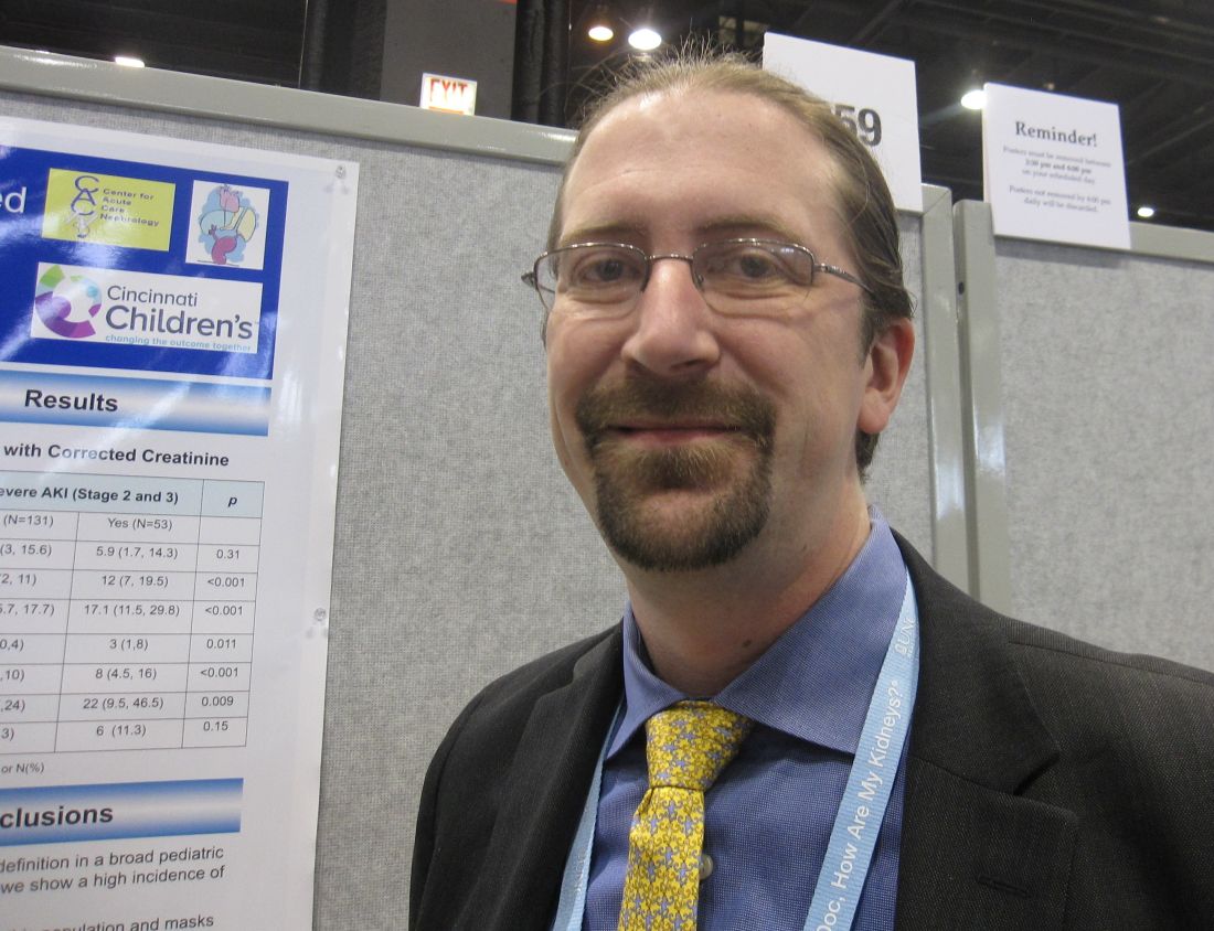
Fluid overload can mask acute kidney injury (AKI) in critically ill children. “The failure to correct serum creatinine measure for fluid overload dilutes the impact of AKI on outcomes,” David T. Selewski, MD, of the University of Michigan, Ann Arbor, said at the annual meeting sponsored by the American Society for Nephrology.
The primary outcome was ICU mortality. Secondary outcomes were length of mechanical ventilation, and length of stay in the ICU and the hospital.
The original study documented an ICU mortality rate of 7.1%. AKI was identified in 77 (41.8%) of the 184 patients. The median peak fluid overload during ICU admission was 12.9 (interquartile range, 7.4-20.8).
The serum creatinine data were corrected for fluid balance and these rates were reassessed. Following the adjustment, the rate of AKI increased from 41.8% to 53.4%, with 30 new cases identified according to standard defined criteria. The mean fluid overload was now 11.2 (interquartile range, 5.7-17.7).
In the original cohort, there were 40 cases of severe AKI (stage 2 and 3). Following the creatinine correction, 13 more cases were judged to be severe. Of these, five cases were associated with a worse outcome in terms of ICU mortality. Additionally, 10 cases that had been diagnosed as AKI were found to be false positives.
The results need to be studied in larger studies and in other populations, such as neonates, Dr. Selewski said.
AT KIDNEY WEEK 2016
Key clinical point: Acute kidney injury can be detected more accurately in critically ill children by correcting for fluid overload.
Major finding: Fluid overload masked diagnosis of over 40% of patients with acute kidney injury in an observational study.
Data source: Secondary analysis of AKI-CHERUB single-center observational study involving 181 critically ill children.
Disclosures: The AKI-CHERUB study was funded by NIH. Dr. Selewski reported having no financial disclosures.
TRUE-AHF: Urgent vasodilator therapy in acute HF provides no long-term benefit
NEW ORLEANS – An investigational synthetic natriuretic peptide given early during hospitalization for acute decompensated heart failure didn’t produce any of the hoped-for intermediate- or long-term clinical benefits in the phase III TRUE-AHF study, Milton Packer, MD, reported at the American Heart Association scientific sessions.
The failure of this investigational vasodilator, ularitide, to influence downstream cardiovascular mortality or early readmission for heart failure closes the door on the once-promising hypothesis that myocardial microinjury occurring during ADHF is due to ventricular distension, observed Dr. Packer, the Distinguished Scholar in Cardiovascular Science at Baylor University Medical Center, Dallas. “Ularitide did exactly what we expected it to do while we were giving it: we caused intravascular decompression, we reduced cardiac wall stress, but we did not affect cardiac microinjury, and we didn’t change long-term cardiovascular mortality or any of our secondary endpoints, including and in particular the 30-day risk of rehospitalization for heart failure,” he said.
During a median follow-up of 15 months there were 236 cardiovascular deaths in the ularitide group and 225 in the control group, a nonsignificant difference. Nor were there any differences between the two groups in secondary endpoints including length of stay in the ICU during the index hospitalization, rehospitalization for ADHF within 30 days of discharge, or the composite of all-cause mortality or cardiovascular hospitalization within 6 months, which occurred in 40.7% of the ularitide group and 37.2% of controls.
The explanation for this lack of long-term benefit lies in the finding that myocardial microinjury wasn’t prevented by the rapid reduction of cardiac distension produced by ularitide. This was evident in the therapy’s inability to dampen the rise in high-sensitivity cardiac troponin T which occurred in the initial 48 hours of the study.
“The trial demonstrated the effects and safety of ularitide. However, to gain long-term benefits on hospitalizations and death in patients following a hospital admission for heart failure, physicians must focus on the drugs that patients take as an outpatient rather than the drugs they receive as an inpatient,” Dr. Packer concluded.
“Readmission rates remain stubbornly at 20% within 30 days and near 50% at 6 months. Acute decompensation is an inflection point in the natural history of heart failure with subsequent 1-year mortality rates consistently approximating 25%. Clearly there is something about the hospitalization that is a herald event which speaks to much worse outcomes, compared with chronic ambulatory heart failure,” said Dr. Yancy, professor of medicine and chief of cardiology at Northwestern University in Chicago.
He agreed with Dr. Packer that in light of the TRUE-AHF results, what Dr. Yancy termed “the early injury hypothesis” isn’t worth further pursuit.
Ularitide thus joins a long list of failed therapies for ADHF. Treatments that have convincingly been shown to have no significant impact on mortality and at best only modest impact on morbidity include continuous IV infusion of loop diuretics in the DOSE trial; the arginine vasopressin antagonists, which failed to impress in EVEREST, SECRET, and TACTICS; nesiritide (Natrecor) in the ASCEND-HF trial; and levosimendan (Simdax), which proved disappointing in the SURVIVE and REVIVE II studies, according to Dr. Yancy.
The jury is still out on serelaxin, he added. The drug showed a favorable signal in the RELAX-AHF trial. The results of RELAX II are awaited with interest.
“Today we still don’t have an effective single intervention for acute decompensated heart failure other than process-of-care improvement,” the cardiologist noted.
What holds promise for improved long-term outcomes in ADHF at this point? Dr. Yancy said sacubitril/valsartan (Entresto) is intriguing based upon the results of the PARADIGM-HF trial (N Engl J Med. 2014;371:993-1004). But the drug needs to be studied prospectively in patients in the throes of ADHF before it can appropriately be recommended for the purpose of changing the natural history of this disorder, Dr. Yancy stressed.
Devices such as the implantable pulmonary catheter are under study as a promising means of altering the natural history of ADHF by identifying actionable signals of impending decompensation weeks beforehand, he added.
The TRUE-AHF trial was sponsored by Cardiorentis. Dr. Packer reported serving as a consultant to that company and more than a dozen other pharmaceutical and medical device companies.
bjancin@frontlinemedcom.com
Key clinical point:
Major finding: Early administration of ularitide during hospitalization for acute decompensated heart failure failed to achieve any long-term clinical benefits.
Data source: The TRUE-AHF trial was a double-blind, placebo-controlled, randomized trial including 2,157 patients hospitalized for acute decompensated heart failure at 156 centers in 23 countries.
Disclosures: The study was sponsored by Cardiorentis. The presenter reported serving as a consultant to that company and more than a dozen other pharmaceutical and medical device companies.
Cardiac rehab also slashes stroke risk
ROME – Cardiac rehabilitation programs have a previously unappreciated benefit: participants enjoy a 60% reduction in the risk of stroke, Gijs van Halewijn, MD, reported at the annual congress of the European Society of Cardiology.
“I think cardiologists are really focused on cardiovascular deaths, especially from MI. But we’ve shown that cardiac rehabilitation also has an effect on cerebrovascular events,” said Dr. van Halewijn of Erasmus University in Rotterdam (the Netherlands).
He presented a meta-analysis of randomized controlled trials of cardiac rehab conducted during 2010-2015. The purpose was to assess the value provided by cardiac rehab in the contemporary era of acute coronary syndrome management featuring primary percutaneous coronary intervention, drug-eluting stents, and potent medications for secondary cardiovascular prevention. That hadn’t previously been looked at systematically.
“The standard meta-analyses cited in the field include randomized trials from as far back as just after World War II,” Dr. van Halewijn noted in an interview.
He employed the same search and analytic methods utilized by the Cochrane Collaboration in evaluating 18 randomized controlled trials of lifestyle- or exercise-based cardiac rehab, compared with usual care, in a total of 7,691 participants.
The results of the meta-analysis indicate cardiac rehab provides powerful secondary prevention benefits above and beyond those obtained through contemporary interventional procedures and preventive medications.
Cardiovascular mortality was reduced by 58% in cardiac rehab participants compared with usual care controls. The risk of acute MI was decreased by 30%. And in a new observation, cerebrovascular events were reduced by 60% in the four randomized trials in which that was an endpoint. All of these differences were highly statistically significant.
“Interestingly, the number needed to treat was 45 for MI, so if you have 45 patients included in your cardiac rehabilitation program, you can prevent one MI. And you can prevent one cerebrovascular event with 82 participants,” according to Dr. van Halewijn.
Cardiac rehab had no effect on all-cause mortality in the overall meta-analysis. However, in the trials involving comprehensive cardiac rehab programs targeting six or more of the components of secondary cardiovascular prevention described by the British Association for Cardiac Prevention and Rehabilitation (Heart. 2013 Aug;99[15]:1069-71), participation was associated with a 37% reduction in the risk of all-cause mortality, compared with usual care.
Those components are smoking, blood pressure, cholesterol, HbA1c, exercise training, counseling about the importance of exercise, stress management, and checking medications.
In addition, Dr. van Halewijn continued, cardiac rehab programs in which a physician or nurse made sure participants were on guideline-directed cardiovascular medications had a 65% reduction in all-cause mortality, compared with usual care.
“Cardiac rehabilitation’s opportunity is to evolve into comprehensive programs addressing all aspects of lifestyle, risk factor management, and prescription of medications to reduce death and nonfatal events,” the physician concluded.
This study was supported by Erasmus University Medical Center, Imperial College London, and the Dutch Heart Foundation. Dr. van Halewijn reported having no financial conflicts of interest.
bjancin@frontlinemedcom.com
AT THE ESC CONGRESS 2016
Key clinical point:
Major finding: The number of patients who need to participate in a cardiac rehabilitation program following an ACS in order to prevent one cerebrovascular event is 85.
Data source: This was a meta-analysis of 18 randomized trials carried out in 2010-2015 comparing contemporary cardiac rehabilitation to usual care in 7,691 patients.
Disclosures: This study was supported by Erasmus University (Rotterdam, the Netherlands) Medical Center, Imperial College London, and the Dutch Heart Foundation. The presenter reported having no financial conflicts of interest.
Discharging select diverticulitis patients from the ED found to be acceptable
CORONADO, CALIF. – Among patients diagnosed with diverticulitis via CT scan in the emergency department who were discharged home, only 13% required a return visit to the hospital, results from a long-term retrospective analysis demonstrated.
“In select patients whose assessment includes a CT scan, discharge to home from the emergency department with treatment for diverticulitis is safe,” study author Anne-Marie Sirany, MD, said at the annual meeting of the Western Surgical Association.
A few years ago, researchers conducted a randomized trial to evaluate the treatment of uncomplicated diverticulitis (Ann Surg. 2014;259[1]:38-44). Patients were diagnosed with diverticulitis in the emergency department and randomized to either hospital admission or outpatient management at home. The investigators found no significant differences between the readmission rates of the inpatient and outpatient groups, but the health care costs were three times lower in the outpatient group. Dr. Sirany and her associates set out to compare the outcomes of patients diagnosed with and treated for diverticulitis in the emergency department who were discharged to home, versus those who were admitted to the hospital. They reviewed the medical records of 240 patients with a primary diagnosis of diverticulitis by CT scan who were evaluated in the emergency department at one of four hospitals and one academic medical center from September 2010 to January 2012. The primary outcome was hospital readmission or return to the emergency department within 30 days, while the secondary outcomes were recurrent diverticulitis or surgical resection for diverticulitis.
The mean age of the 240 patients was 59 years, 45% were men, 22% had a Charlson Comorbidity Index (CCI) of greater than 2, and 7.5% were on steroids or immunosuppressant medications. More than half (62%) were admitted to the hospital, while the remaining 38% were discharged home on oral antibiotics. Compared with patients discharged home, those admitted to the hospital were more likely to be older than age 65 (43% vs. 24%, respectively; P = .003), have a CCI of 2 or greater (28% vs. 13%; P = .007), were more likely to be on immunosuppressant or steroid medications (11% vs. 1%; P = .003), show extraluminal air on CT (30% vs. 7%; P less than .0001), or show abscess on CT (19% vs. 1%; P less than .0001). “Of note: We did not have any patients who had CT scan findings of pneumoperitoneum who were discharged home, and 48% of patients admitted to the hospital had uncomplicated diverticulitis,” she said.
After a median follow-up of 37 months, no significant differences were observed between patients discharged to home and those admitted to the hospital in readmission or return to the emergency department (13% vs. 14%), recurrent diverticulitis (23% in each group), or in colon resection at subsequent encounter (16% vs. 19%). “Among patients discharged to home, only one patient required emergency surgery, and this was 20 months after their index admission,” Dr. Sirany said. “We think that the low rate of readmission in patients discharged home demonstrates that this is a safe approach to management of patients with diverticulitis, when using information from the CT scan.”
Closer analysis of patients who were discharged home revealed that six patients had extraluminal air on CT scan, three of whom returned to the emergency department or were admitted to the hospital. In addition, 11% of those with uncomplicated diverticulitis returned to the emergency department or were admitted to the hospital.
Dr. Sirany acknowledged certain limitations of the study, including its retrospective design, a lack of complete follow-up for all patients, and the fact that it included patients with recurrent diverticulitis. “Despite the limitations, we recommend that young, relatively healthy patients, with uncomplicated findings on CT scan, can be discharged to home and managed as an outpatient,” she said. “In an era where there’s increasing attention to health care costs, we need to think more critically about which patients need to be admitted for management of uncomplicated diverticulitis.” She reported having no financial disclosures.
dbrunk@frontlinemedcom.com
AT WSA 2016
Key clinical point:
Major finding: After a median follow-up of 37 months, no significant differences were observed between patients discharged to home and those admitted to the hospital in readmission or return to the emergency department (13% vs. 14%, respectively).
Data source: A retrospective review of 240 patients with a primary diagnosis of diverticulitis by CT scan who were evaluated in the emergency department at one of four hospitals and one academic medical center from September 2010 to January 2012.
Disclosures: Dr. Sirany reported having no financial disclosures.
Emergent colon cancer resection does not negatively affect patient outcomes
CORONADO, CALIF. – With the exception of patients that present with perforation, emergent resection of colon cancers does not appear to adversely affect operative outcomes or patient survival, a 3-year analysis of data showed.
At the annual meeting of the Western Surgical Association, Jason W. Smith, MD, said that of the estimated 106,100 new cases of colon cancer each year, 6%-30% of patients have symptoms or late complications related to the disease that require an emergency intervention, often leading to dismal outcomes. “The problem with many existing studies of emergent colon cancer resections is that they tend to throw everybody into one large group, making it difficult to compare some of these patients,” said Dr. Smith, a trauma surgeon in the department of surgery at the University of Louisville (Ky.) School of Medicine. “Our thought was, if we provide an appropriate oncologic resection at the time of our initial management in these patients when they come to the emergency department, can we affect the similar rate of overall outcomes for these patients with regard to their cancer prognosis?”
Of the 117 patients in the emergent group, 35 had a perforation and 82 had an emergent resection. In an unmatched analysis comparing perforation, emergent resection, and elective resection, the patients who presented with a perforation had a much higher Charlson Comorbidity Index (CCI) score and a higher American Society of Anesthesiologists (ASA) class. They tended to be on vasopressors or suffering from inflammatory response related to their perforation, they had lower levels of blood pressure and hemoglobin, and they had much higher rates of 30-day mortality and overall 30-day morbidity, compared with their counterparts in the other two groups. Of the eight deaths that occurred in patients with colon perforation, four were related to sepsis and multiple organ failure, one to respiratory failure/acute respiratory distress syndrome, one to acute MI, one to exacerbation of chronic lung disease, and one to transition to palliative care due to cancer diagnosis. “So the overall predominance of the deaths associated in the first 30 days were related to the inflammatory responses associated with that perforation, not specific to the cancer itself,” Dr. Smith said. At the same time, the ASA and CCI scores were not different between those with morbidity/mortality and those who survived. “So it’s difficult to identify these patients out of the gate,” he said.
When the researchers more closely examined data from patients with a perforation, 27 of 35 (77%) survived at 30 days. Survival at 1, 2, and 3 years was 78%, 57%, and 43%, respectively. “This is a mixture of stage II and stage IV patients, so they’re difficult to compare and difficult to standardize across the board,” Dr. Smith noted. “But what you see is that their survival is not significantly different related to their disease if you discount the inflammatory process. Our initial thought was that for these perforated cancers, what we really need to do is provide the appropriate oncologic resection management [in order to] get the same oncologic outcomes.”
Next, the researchers compared the 82 patients who presented without a perforation but required an emergent operation with 82 of the elective surgery patients, matched for age, gender, the CCI, ASA class, oncology stage, and body mass index. There were no differences between the two groups in terms of R0 resection, the number of lymph nodes sampled, or estimated blood loss. However, compared with patients in the elective resection group, those in the emergent resection group had higher rates of ostomy placement (30% vs. 10%, respectively; P = .01), and a longer hospital length of stay (an average of 18 vs. 12 days; P = .0007). “Most of that difference occurred on the front end of hospital stays,” Dr. Smith said. “Their postoperative days were not significantly different.”
As for long-term outcomes, more than 90% of all patients in both groups received chemotherapy within the first year postprocedure, and overall time to initiation of chemotherapy was not significantly different in the emergent vs. elective groups (6.6 vs. 5.5 weeks, respectively; P = .43). However, patients suffering postsurgical complications had an increased risk of delayed chemotherapy.
In a risk-adjusted analysis, overall survival at 3 years was not different between the emergent and elective operation groups (hazard ratio, 1.1; P = .54). Similarly, disease-free survival was not different at 3 years between the two groups (HR, 1.06; P = .84). Independent predictors of poor long-term outcome included age greater than 70 (HR, 1.45; P less than 0.03); elevated ASA class (HR, 2.99 for class III vs. class I-II; P = .08; and HR, 7.45 for ASA class IV vs. I-II; P = .03); presence of residual disease (HR, 3.08; P less than .001), and advanced cancer stage.
He acknowledged certain limitations of the study, including the fact that it was a blinded retrospective cohort with the potential for unrecognized bias, and that it measured 3-year survival instead of 5-year survival data.
“Emergent resection of nonperforated colon cancers does not appear to adversely affect operative outcomes or patient survival when proper oncologic principles are applied to their initial management,” Dr. Smith concluded. “Outcome differences in patients suffering perforation may correlate with the physiologic derangements associated with the perforation rather than the oncologic disease; thus, every effort should be made to provide an appropriate oncologic operation.” He reported having no financial disclosures.
dbrunk@frontlinemedcom.com
AT WSA 2016
Key clinical point:
Major finding: In a risk-adjusted analysis, overall survival at 3 years was not different between the emergent and elective operation groups (HR, 1.1; P = .54).
Data source: A retrospective review of 548 elective and emergent colectomies for colon cancer performed at the University of Louisville (Ky.) from 2011 to 2015.
Disclosures: Dr. Smith reported having no financial disclosures.
Kaiser experience: A helping hand reduces COPD readmissions
LOS ANGELES – With a handful of common-sense steps, the Kaiser Permanente Los Angeles Medical Center reduced 30-day hospital readmissions for chronic obstructive pulmonary disease (COPD) from 17.4/1,000 in Dec. 2013 to 11.9/1,000 in Dec. 2015.
The 57 readmissions avoided in 2015 saved the medical center $700,359, according to a report at the annual meeting of the American College of Chest Physicians.
The quality improvement project – dubbed KP Breath – started in 2013 after staff realized their COPD readmission rates were significantly higher than other area hospitals, and likely to increase. “We knew we had a problem, and that if we did not address it, it was going to be out of control,” Mr. Cam said. There was also the risk of Centers for Medicare & Medicaid Services penalties for COPD readmissions.
Mr. Cam and his colleagues discovered several problems. “Leaving the hospital, [COPD patients] didn’t know what medication was for what, or their medication schedule. They didn’t know how to use their inhalers, and didn’t understand what the disease process was all about, and what it was doing to them,” he said.
There was little continuity of care after discharge; many patients didn’t even have a pulmonologist. Essentially, COPD patients were lost to follow-up until they returned to the emergency department with another exacerbation.
A rapid Plan, Do, Study, Act cycle was the first step; it identified solutions that would work based on COPD management guidelines and published studies. “They were all things that have been shown to reduce rehospitalizations,” said pulmonologist Luis Moreta-Sainz, MD, another key project member.
The team staggered their changes over 2 years. Pulmonary consults for acute exacerbation admissions shot up, and respiratory therapists started to stop by to educate almost every COPD patient about medication use, trigger avoidance, and other matters. Patients began watching educational videos from their bed.
Changes were made after discharge, too. “We felt strongly that pulmonary rehabilitation needed to be an integral part of care, and that patients had to be connected to the pulmonary clinic,” Dr. Moreta-Sainz said.
Patients were booked for a pulmonologist at the clinic soon after they left the hospital, and greeted there by their COPD navigator – a respiratory therapist operating at the top of their license – who bridged the gap between inpatient and outpatient care and oversaw their case, helping with medical, psychosocial, and palliative needs.
Patients were also channeled into pulmonary rehab, three sessions per week for 6-8 weeks, with additional sessions as needed. The outpatient education emphasized and expanded the inpatient lessons, and patients exercised on treadmills and other equipment. They learned how to use resistance bands at home to increase upper body strength and decrease disability. Kaiser increased the number of weekly pulmonary rehab slots from 8 to 64 to make it happen.
After rehab, patients were offered a pedometer to measure how many steps they walked, and a phone number to report it each day. Those who participated got a call from the navigator when they fell below targets.
It has all made a huge difference. Dr. Moreta-Sainz said he’d like to add in-home visits and family support groups, so caregivers know what to do if things head south.
The work was funded by Kaiser; Dr. Moreta-Sainz and Mr. Cam have no disclosures.
aotto@frontlinemedcom.com
On a recent morning, two of my scheduled clinic patients were “no-shows.” Both of them were patients with COPD that I had recently cared for in-hospital for an exacerbation. While I know that snow may have played a role, there are other barriers to care, including lack of access to transportation, poor health literacy, and no effective health insurance.
On a recent morning, two of my scheduled clinic patients were “no-shows.” Both of them were patients with COPD that I had recently cared for in-hospital for an exacerbation. While I know that snow may have played a role, there are other barriers to care, including lack of access to transportation, poor health literacy, and no effective health insurance.
On a recent morning, two of my scheduled clinic patients were “no-shows.” Both of them were patients with COPD that I had recently cared for in-hospital for an exacerbation. While I know that snow may have played a role, there are other barriers to care, including lack of access to transportation, poor health literacy, and no effective health insurance.
LOS ANGELES – With a handful of common-sense steps, the Kaiser Permanente Los Angeles Medical Center reduced 30-day hospital readmissions for chronic obstructive pulmonary disease (COPD) from 17.4/1,000 in Dec. 2013 to 11.9/1,000 in Dec. 2015.
The 57 readmissions avoided in 2015 saved the medical center $700,359, according to a report at the annual meeting of the American College of Chest Physicians.
The quality improvement project – dubbed KP Breath – started in 2013 after staff realized their COPD readmission rates were significantly higher than other area hospitals, and likely to increase. “We knew we had a problem, and that if we did not address it, it was going to be out of control,” Mr. Cam said. There was also the risk of Centers for Medicare & Medicaid Services penalties for COPD readmissions.
Mr. Cam and his colleagues discovered several problems. “Leaving the hospital, [COPD patients] didn’t know what medication was for what, or their medication schedule. They didn’t know how to use their inhalers, and didn’t understand what the disease process was all about, and what it was doing to them,” he said.
There was little continuity of care after discharge; many patients didn’t even have a pulmonologist. Essentially, COPD patients were lost to follow-up until they returned to the emergency department with another exacerbation.
A rapid Plan, Do, Study, Act cycle was the first step; it identified solutions that would work based on COPD management guidelines and published studies. “They were all things that have been shown to reduce rehospitalizations,” said pulmonologist Luis Moreta-Sainz, MD, another key project member.
The team staggered their changes over 2 years. Pulmonary consults for acute exacerbation admissions shot up, and respiratory therapists started to stop by to educate almost every COPD patient about medication use, trigger avoidance, and other matters. Patients began watching educational videos from their bed.
Changes were made after discharge, too. “We felt strongly that pulmonary rehabilitation needed to be an integral part of care, and that patients had to be connected to the pulmonary clinic,” Dr. Moreta-Sainz said.
Patients were booked for a pulmonologist at the clinic soon after they left the hospital, and greeted there by their COPD navigator – a respiratory therapist operating at the top of their license – who bridged the gap between inpatient and outpatient care and oversaw their case, helping with medical, psychosocial, and palliative needs.
Patients were also channeled into pulmonary rehab, three sessions per week for 6-8 weeks, with additional sessions as needed. The outpatient education emphasized and expanded the inpatient lessons, and patients exercised on treadmills and other equipment. They learned how to use resistance bands at home to increase upper body strength and decrease disability. Kaiser increased the number of weekly pulmonary rehab slots from 8 to 64 to make it happen.
After rehab, patients were offered a pedometer to measure how many steps they walked, and a phone number to report it each day. Those who participated got a call from the navigator when they fell below targets.
It has all made a huge difference. Dr. Moreta-Sainz said he’d like to add in-home visits and family support groups, so caregivers know what to do if things head south.
The work was funded by Kaiser; Dr. Moreta-Sainz and Mr. Cam have no disclosures.
aotto@frontlinemedcom.com
AT CHEST 2016
