User login
Certain DMTs in MS linked to more psoriasis
, a new study finds. However, overall rates of reported disease are very low, and there’s no confirmation of a connection.
“People with MS and comorbid psoriasis – or those at a known high-risk for developing psoriasis – may benefit from a careful consideration of disease-modifying therapy (DMT), specifically when B cell-depleting therapies are considered,” study coauthor and Medical College of Wisconsin neurologist Ahmed Obeidat, MD, PhD, said in an interview. The findings were presented at the 2021 Annual Meeting of the Consortium of Multiple Sclerosis Centers (CMSC).
Dr. Obeidat and colleagues launched the study after noticing cases of psoriasis that developed months to years after patients started taking ocrelizumab, a B cell-depleting therapy. “We referred to the published literature and only found very scant reports of MS, psoriasis, and B cell-depleting therapy use,” he said. “Thus we decided to pursue an investigation of a large [Food and Drug Administration] database to examine for possible out-of-proportion reports for psoriasis in patients with MS who were receiving B cell-depleting therapy.”
The researchers tracked case reports of psoriasis in patients with MS on DMTs from 2009 to 2020 via the FDA Adverse Event Reporting System. They found 517 psoriasis reports among 45,547 reports of skin/cutaneous conditions. The reports were linked to interferon beta 1a (136 reports, 26% of total), natalizumab (107, 21%), fingolimod (75, 15%), dimethyl fumarate (64, 12%), ocrelizumab (49, 10%), teriflunomide (28, 5%), interferon beta 1b (22, 4%), glatiramer acetate (12, 2%), rituximab (10, 2%), and alemtuzumab (9, 2%).
The total numbers of cases is low, but this may reflect underreporting due to the assumption that “autoimmunity begets autoimmunity” and therefore cases of psoriasis in MS are not alarming, medical student Mokshal H. Porwal, the study lead author, said in an interview.
The average age of patients – 48-51 – was similar for all of the drugs except alemtuzumab (mean age 41), which had a very small number of cases. The percentage of cases in females was 71%-77% for most of the drugs, with a few exceptions: rituximab (60%), ocrelizumab (63%), and alemtuzumab (33%).
Other drugs – cladribine, siponimod, and ozanimod – had 1, 1, and 0 reports, respectively, and were not included in the age and gender analyses.
The researchers also found that psoriasis made up about 65% of all skin/cutaneous adverse reports for rituximab, the highest number among DMTs. By comparison, that number was about 30% for ocrelizumab and under 1% for dimethyl fumarate and alemtuzumab.
Links between psoriasis and MS are murky, Dr. Obeidat said. “Some studies consider the presence of psoriasis as a possible indicator of increased future risk for MS, but there’s no clear association between the two conditions,” he said.
As for DMTs, “a few case reports of psoriasis in association with interferon-beta and rare case reports in association with ocrelizumab therapy have been published. However, the possible association between certain DMTs and psoriasis remains unclear,” he said.
Going forward, “we advise that patients with psoriasis on B cell-depleting agents are monitored more closely,” Dr. Obeidat said. “If the psoriasis worsens, it may be beneficial to think about potential alternative therapies.”
No study funding is reported. Dr. Obeidat reports various disclosures; the other authors report no disclosures.
FROM CMSC 2021
Challenges in Treating Patients with Tardive Dyskinesia
In this supplement to Current Psychiatry, Greg Mattingly, MD and Manish K. Jha, MBBS review dilemmas and treatment options for patients with tardive dyskinesia.
Click here to read the supplement and earn free CME/CE credits.
In this supplement to Current Psychiatry, Greg Mattingly, MD and Manish K. Jha, MBBS review dilemmas and treatment options for patients with tardive dyskinesia.
Click here to read the supplement and earn free CME/CE credits.
In this supplement to Current Psychiatry, Greg Mattingly, MD and Manish K. Jha, MBBS review dilemmas and treatment options for patients with tardive dyskinesia.
Click here to read the supplement and earn free CME/CE credits.
Boxed warnings: Legal risks that many physicians never see coming
Almost all physicians write prescriptions, and each prescription requires a physician to assess the risks and benefits of the drug. If an adverse drug reaction occurs, physicians may be called on to defend their risk-benefit assessment in court.

The assessment of risk is complicated when there is a boxed warning that describes potentially serious and life-threatening adverse reactions associated with a drug. Some of our most commonly prescribed drugs have boxed warnings, and drugs that were initially approved by the Food and Drug Administration without boxed warnings may have them added years later.
One serious problem with boxed warnings is that there are no reliable mechanisms for making sure that physicians are aware of them. The warnings are typically not seen by physicians as printed product labels, just as physicians often don’t see the pills and capsules that they prescribe. Pharmacists who receive packaged drugs from manufacturers may be the only ones to see an actual printed boxed warning, but even those pharmacists have little reason to read each label and note changes when handling many bulk packages.
This problem is aggravated by misperceptions that many physicians have about boxed warnings and the increasingly intense scrutiny given to them by mass media and the courts. Lawyers can use boxed warnings to make a drug look dangerous, even when it’s not, and to make physicians look reckless when prescribing it. Therefore, it is important for physicians to understand what boxed warnings are, what they are not, the problems they cause, and how to minimize these problems.
What is a ‘boxed warning’?
The marketing and sale of drugs in the United States requires approval by the FDA. Approval requires manufacturers to prepare a document containing “Full Prescribing Information” for the drug and to include a printed copy in every package of the drug that is sold. This document is commonly called a “package insert,” but the FDA designates this document as the manufacturer’s product “label.”
In 1979, the FDA began requiring some labels to appear within thick, black rectangular borders; these have come to be known as boxed warnings. Boxed warnings are usually placed at the beginning of a label. They may be added to the label of a previously approved drug already on the market or included in the product label when first approved and marketed.
The requirement for a boxed warning most often arises when a signal appears during review of postmarketing surveillance data suggesting a possible and plausible association between a drug and an adverse reaction. Warnings may also be initiated in response to petitions from public interest groups, or upon the discovery of serious toxicity in animals. Regardless of their origin, the intent of a boxed warning is to highlight information that may have important therapeutic consequences and warrants heightened awareness among physicians.
What a boxed warning is not
A boxed warning is not “issued” by the FDA; it is merely required by the FDA. Specific wording or a template may be suggested by the FDA, but product labels and boxed warnings are written and issued by the manufacturer. This distinction may seem minor, but extensive litigation has occurred over whether manufacturers have met their duty to warn consumers about possible risks when using their products, and this duty cannot be shifted to the FDA.
A boxed warning may not be added to a product label at the option of a manufacturer. The FDA allows a boxed warning only if it requires the warning, to preserve its impact. It should be noted that some medical information sources (e.g., PDR.net) may include a “BOXED WARNING” in their drug monographs, but monographs not written by a manufacturer are not regulated by the FDA, and the text of their boxed warnings do not always correspond to the boxed warning that was approved by the FDA.
A boxed warning is not an indication that revocation of FDA approval is being considered or that it is likely to be revoked. FDA approval is subject to ongoing review and may be revoked at any time, without a prior boxed warning.
A boxed warning is not the highest level of warning. The FDA may require a manufacturer to send out a “Dear Health Care Provider” (DHCP) letter when an even higher or more urgent level of warning is deemed necessary. DHCP letters are usually accompanied by revisions of the product label, but most label revisions – and even most boxed warnings – are not accompanied by DHCP letters.
A boxed warning is not a statement about causation. Most warnings describe an “association” between a drug and an adverse effect, or “increased risk,” or instances of a particular adverse effect that “have been reported” in persons taking a drug. The words in a boxed warning are carefully chosen and require careful reading; in most cases they refrain from stating that a drug actually causes an adverse effect. The postmarketing surveillance data on which most warnings are based generally cannot provide the kind of evidence required to establish causation, and an association may be nothing more than an uncommon manifestation of the disorder for which the drug has been prescribed.
A boxed warning is not a statement about the probability of an adverse reaction occurring. The requirement for a boxed warning correlates better to the new recognition of a possible association than to the probability of an association. For example, penicillin has long been known to cause fatal anaphylaxis in 1/100,000 first-time administrations, but it does not have a boxed warning. The adverse consequences described in boxed warnings are often far less frequent – so much so that most physicians will never see them.
A boxed warning does not define the standard of care. The warning is a requirement imposed on the manufacturer, not on the practice of medicine. For legal purposes, the “standard of care” for the practice of medicine is defined state by state and is typically cast in terms such as “what most physicians would do in similar circumstances.” Physicians often prescribe drugs in spite of boxed warnings, just as they often prescribe drugs for “off label” indications, always balancing risk versus benefit.
A boxed warning does not constitute a contraindication to the use of a medication. Some warnings state that a drug is contraindicated in some situations, but product labels have another mandated section for listing contraindications, and most boxed warnings have no corresponding entry in that section.
A boxed warning does not necessarily constitute current information, nor is it always updated when new or contrary information becomes available. Revisions to boxed warnings, and to product labels in general, are made only after detailed review at the FDA, and the process of deciding whether an existing boxed warning continues to be appropriate may divert limited regulatory resources from more urgent priorities. Consequently, revisions to a boxed warning may lag behind the data that justify a revision by months or years. Revisions may never occur if softening or eliminating a boxed warning is deemed to be not worth the cost by a manufacturer.
Boxed warning problems for physicians
There is no reliable mechanism for manufacturers or the FDA to communicate boxed warnings directly to physicians, so it’s not clear how physicians are expected to stay informed about the issuance or revision of boxed warnings. They may first learn about new or revised warnings in the mass media, which is paying ever-increasing attention to press releases from the FDA. However, it can be difficult for the media to accurately convey the subtle and complex nature of a boxed warning in nontechnical terms.
Many physicians subscribe to various medical news alerts and attend continuing medical education (CME) programs, which often do an excellent job of highlighting new warnings, while hospitals, clinics, and pharmacies may broadcast news about boxed warnings in newsletters or other notices. But these notifications are ephemeral and may be missed by physicians who are overwhelmed by email, notices, newsletters, and CME programs.
The warnings that pop up in electronic medical records systems are often so numerous that physicians become trained to ignore them. Printed advertisements in professional journals must include mandated boxed warnings, but their visibility is waning as physicians increasingly read journals online.
Another conundrum is how to inform the public about boxed warnings.
Manufacturers are prohibited from direct-to-consumer advertising of drugs with boxed warnings, although the warnings are easily found on the Internet. Some patients expect and welcome detailed information from their physicians, so it’s a good policy to always and repeatedly review this information with them, especially if they are members of an identified risk group. However, that policy may be counterproductive if it dissuades anxious patients from needed therapy despite risk-benefit considerations that strongly favor it. Boxed warnings are well known to have “spillover effects” in which the aspersions cast by a boxed warning for a relatively small subgroup of patients causes use of a drug to decline among all patients.
Compounding this conundrum is that physicians rarely have sufficient information to gauge the magnitude of a risk, given that boxed warnings are often based on information from surveillance systems that cannot accurately quantify the risk or even establish a causal relationship. The text of a boxed warning generally does not provide the information needed for evidence-based clinical practice such as a quantitative estimate of effect, information about source and trustworthiness of the evidence, and guidance on implementation. For these and other reasons, FDA policies about various boxed warnings have been the target of significant criticism.
Medication guides are one mechanism to address the challenge of informing patients about the risks of drugs they are taking. FDA-approved medication guides are available for most drugs dispensed as outpatient prescriptions, they’re written in plain language for the consumer, and they include paraphrased versions of any boxed warning. Ideally, patients review these guides with their physicians or pharmacists, but the guides may be lengthy and raise questions that may not be answerable (e.g., about incidence rates). Patients may decline to review this information when a drug is prescribed or dispensed, and they may discard printed copies given to them without reading.
What can physicians do to minimize boxed warning problems?
Physicians should periodically review the product labels for drugs they commonly prescribe, including drugs they’ve prescribed for a long time. Prescription renewal requests can be used as a prompt to check for changes in a patient’s condition or other medications that might place a patient in the target population of a boxed warning. Physicians can subscribe to newsletters that announce and discuss significant product label changes, including alerts directly from the FDA. Physicians may also enlist their office staff to find and review boxed warnings for drugs being prescribed, noting which ones should require a conversation with any patient who has been or will be receiving this drug. They may want to make explicit mention in their encounter record that a boxed warning, medication guide, or overall risk-benefit assessment has been discussed.
Summary
The nature of boxed warnings, the means by which they are disseminated, and their role in clinical practice are all in great need of improvement. Until that occurs, boxed warnings offer some, but only very limited, help to patients and physicians who struggle to understand the risks of medications.
Dr. Axelsen is professor in the departments of pharmacology, biochemistry, and biophysics, and of medicine, infectious diseases section, University of Pennsylvania, Philadelphia. He disclosed no relevant financial relationships. A version of this article first appeared on Medscape.com.
Almost all physicians write prescriptions, and each prescription requires a physician to assess the risks and benefits of the drug. If an adverse drug reaction occurs, physicians may be called on to defend their risk-benefit assessment in court.
The assessment of risk is complicated when there is a boxed warning that describes potentially serious and life-threatening adverse reactions associated with a drug. Some of our most commonly prescribed drugs have boxed warnings, and drugs that were initially approved by the Food and Drug Administration without boxed warnings may have them added years later.
One serious problem with boxed warnings is that there are no reliable mechanisms for making sure that physicians are aware of them. The warnings are typically not seen by physicians as printed product labels, just as physicians often don’t see the pills and capsules that they prescribe. Pharmacists who receive packaged drugs from manufacturers may be the only ones to see an actual printed boxed warning, but even those pharmacists have little reason to read each label and note changes when handling many bulk packages.
This problem is aggravated by misperceptions that many physicians have about boxed warnings and the increasingly intense scrutiny given to them by mass media and the courts. Lawyers can use boxed warnings to make a drug look dangerous, even when it’s not, and to make physicians look reckless when prescribing it. Therefore, it is important for physicians to understand what boxed warnings are, what they are not, the problems they cause, and how to minimize these problems.
What is a ‘boxed warning’?
The marketing and sale of drugs in the United States requires approval by the FDA. Approval requires manufacturers to prepare a document containing “Full Prescribing Information” for the drug and to include a printed copy in every package of the drug that is sold. This document is commonly called a “package insert,” but the FDA designates this document as the manufacturer’s product “label.”
In 1979, the FDA began requiring some labels to appear within thick, black rectangular borders; these have come to be known as boxed warnings. Boxed warnings are usually placed at the beginning of a label. They may be added to the label of a previously approved drug already on the market or included in the product label when first approved and marketed.
The requirement for a boxed warning most often arises when a signal appears during review of postmarketing surveillance data suggesting a possible and plausible association between a drug and an adverse reaction. Warnings may also be initiated in response to petitions from public interest groups, or upon the discovery of serious toxicity in animals. Regardless of their origin, the intent of a boxed warning is to highlight information that may have important therapeutic consequences and warrants heightened awareness among physicians.
What a boxed warning is not
A boxed warning is not “issued” by the FDA; it is merely required by the FDA. Specific wording or a template may be suggested by the FDA, but product labels and boxed warnings are written and issued by the manufacturer. This distinction may seem minor, but extensive litigation has occurred over whether manufacturers have met their duty to warn consumers about possible risks when using their products, and this duty cannot be shifted to the FDA.
A boxed warning may not be added to a product label at the option of a manufacturer. The FDA allows a boxed warning only if it requires the warning, to preserve its impact. It should be noted that some medical information sources (e.g., PDR.net) may include a “BOXED WARNING” in their drug monographs, but monographs not written by a manufacturer are not regulated by the FDA, and the text of their boxed warnings do not always correspond to the boxed warning that was approved by the FDA.
A boxed warning is not an indication that revocation of FDA approval is being considered or that it is likely to be revoked. FDA approval is subject to ongoing review and may be revoked at any time, without a prior boxed warning.
A boxed warning is not the highest level of warning. The FDA may require a manufacturer to send out a “Dear Health Care Provider” (DHCP) letter when an even higher or more urgent level of warning is deemed necessary. DHCP letters are usually accompanied by revisions of the product label, but most label revisions – and even most boxed warnings – are not accompanied by DHCP letters.
A boxed warning is not a statement about causation. Most warnings describe an “association” between a drug and an adverse effect, or “increased risk,” or instances of a particular adverse effect that “have been reported” in persons taking a drug. The words in a boxed warning are carefully chosen and require careful reading; in most cases they refrain from stating that a drug actually causes an adverse effect. The postmarketing surveillance data on which most warnings are based generally cannot provide the kind of evidence required to establish causation, and an association may be nothing more than an uncommon manifestation of the disorder for which the drug has been prescribed.
A boxed warning is not a statement about the probability of an adverse reaction occurring. The requirement for a boxed warning correlates better to the new recognition of a possible association than to the probability of an association. For example, penicillin has long been known to cause fatal anaphylaxis in 1/100,000 first-time administrations, but it does not have a boxed warning. The adverse consequences described in boxed warnings are often far less frequent – so much so that most physicians will never see them.
A boxed warning does not define the standard of care. The warning is a requirement imposed on the manufacturer, not on the practice of medicine. For legal purposes, the “standard of care” for the practice of medicine is defined state by state and is typically cast in terms such as “what most physicians would do in similar circumstances.” Physicians often prescribe drugs in spite of boxed warnings, just as they often prescribe drugs for “off label” indications, always balancing risk versus benefit.
A boxed warning does not constitute a contraindication to the use of a medication. Some warnings state that a drug is contraindicated in some situations, but product labels have another mandated section for listing contraindications, and most boxed warnings have no corresponding entry in that section.
A boxed warning does not necessarily constitute current information, nor is it always updated when new or contrary information becomes available. Revisions to boxed warnings, and to product labels in general, are made only after detailed review at the FDA, and the process of deciding whether an existing boxed warning continues to be appropriate may divert limited regulatory resources from more urgent priorities. Consequently, revisions to a boxed warning may lag behind the data that justify a revision by months or years. Revisions may never occur if softening or eliminating a boxed warning is deemed to be not worth the cost by a manufacturer.
Boxed warning problems for physicians
There is no reliable mechanism for manufacturers or the FDA to communicate boxed warnings directly to physicians, so it’s not clear how physicians are expected to stay informed about the issuance or revision of boxed warnings. They may first learn about new or revised warnings in the mass media, which is paying ever-increasing attention to press releases from the FDA. However, it can be difficult for the media to accurately convey the subtle and complex nature of a boxed warning in nontechnical terms.
Many physicians subscribe to various medical news alerts and attend continuing medical education (CME) programs, which often do an excellent job of highlighting new warnings, while hospitals, clinics, and pharmacies may broadcast news about boxed warnings in newsletters or other notices. But these notifications are ephemeral and may be missed by physicians who are overwhelmed by email, notices, newsletters, and CME programs.
The warnings that pop up in electronic medical records systems are often so numerous that physicians become trained to ignore them. Printed advertisements in professional journals must include mandated boxed warnings, but their visibility is waning as physicians increasingly read journals online.
Another conundrum is how to inform the public about boxed warnings.
Manufacturers are prohibited from direct-to-consumer advertising of drugs with boxed warnings, although the warnings are easily found on the Internet. Some patients expect and welcome detailed information from their physicians, so it’s a good policy to always and repeatedly review this information with them, especially if they are members of an identified risk group. However, that policy may be counterproductive if it dissuades anxious patients from needed therapy despite risk-benefit considerations that strongly favor it. Boxed warnings are well known to have “spillover effects” in which the aspersions cast by a boxed warning for a relatively small subgroup of patients causes use of a drug to decline among all patients.
Compounding this conundrum is that physicians rarely have sufficient information to gauge the magnitude of a risk, given that boxed warnings are often based on information from surveillance systems that cannot accurately quantify the risk or even establish a causal relationship. The text of a boxed warning generally does not provide the information needed for evidence-based clinical practice such as a quantitative estimate of effect, information about source and trustworthiness of the evidence, and guidance on implementation. For these and other reasons, FDA policies about various boxed warnings have been the target of significant criticism.
Medication guides are one mechanism to address the challenge of informing patients about the risks of drugs they are taking. FDA-approved medication guides are available for most drugs dispensed as outpatient prescriptions, they’re written in plain language for the consumer, and they include paraphrased versions of any boxed warning. Ideally, patients review these guides with their physicians or pharmacists, but the guides may be lengthy and raise questions that may not be answerable (e.g., about incidence rates). Patients may decline to review this information when a drug is prescribed or dispensed, and they may discard printed copies given to them without reading.
What can physicians do to minimize boxed warning problems?
Physicians should periodically review the product labels for drugs they commonly prescribe, including drugs they’ve prescribed for a long time. Prescription renewal requests can be used as a prompt to check for changes in a patient’s condition or other medications that might place a patient in the target population of a boxed warning. Physicians can subscribe to newsletters that announce and discuss significant product label changes, including alerts directly from the FDA. Physicians may also enlist their office staff to find and review boxed warnings for drugs being prescribed, noting which ones should require a conversation with any patient who has been or will be receiving this drug. They may want to make explicit mention in their encounter record that a boxed warning, medication guide, or overall risk-benefit assessment has been discussed.
Summary
The nature of boxed warnings, the means by which they are disseminated, and their role in clinical practice are all in great need of improvement. Until that occurs, boxed warnings offer some, but only very limited, help to patients and physicians who struggle to understand the risks of medications.
Dr. Axelsen is professor in the departments of pharmacology, biochemistry, and biophysics, and of medicine, infectious diseases section, University of Pennsylvania, Philadelphia. He disclosed no relevant financial relationships. A version of this article first appeared on Medscape.com.
A Clinical Review of Eslicarbazepine Acetate
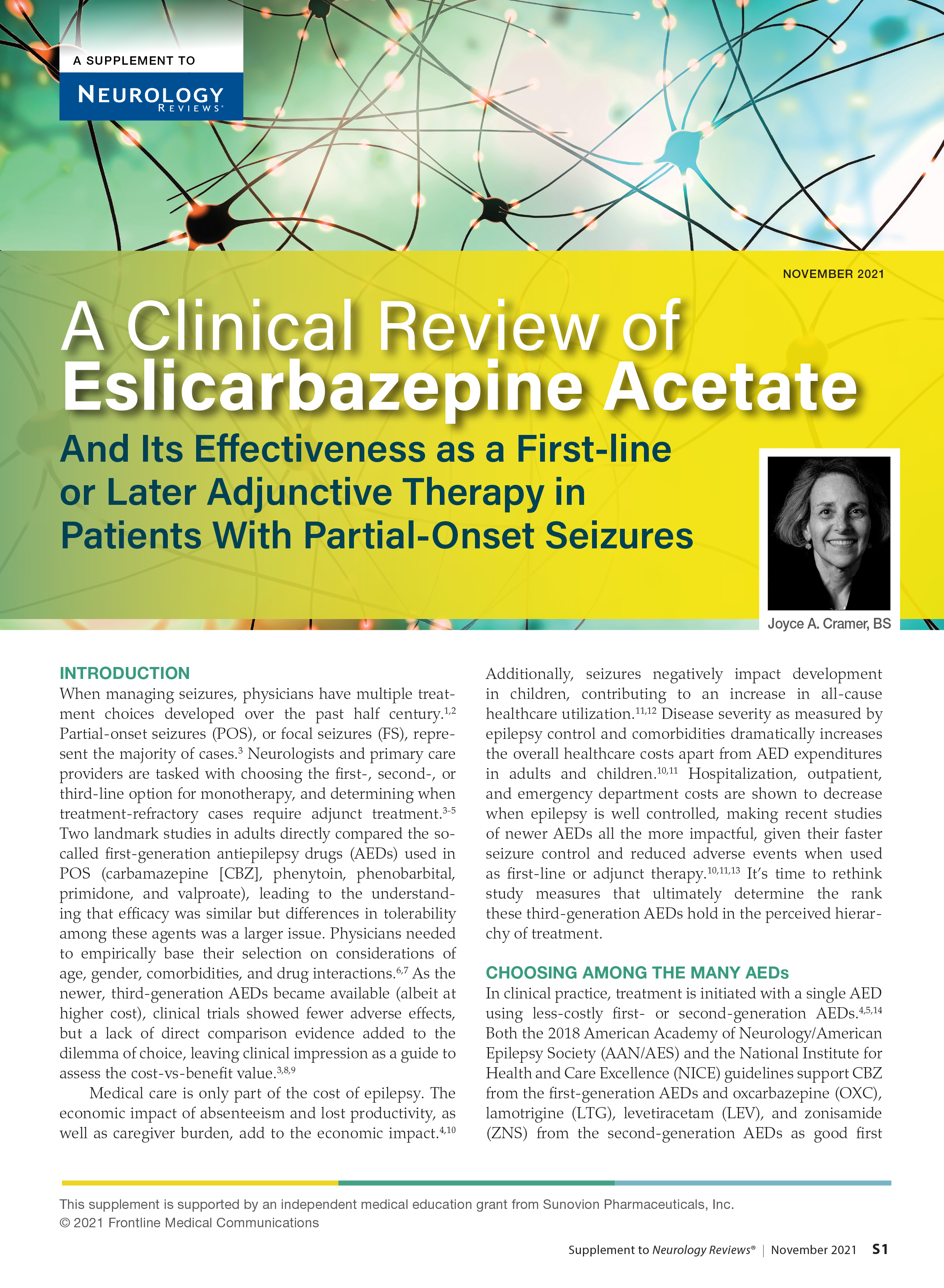
When managing seizures, physicians have multiple treatment choices developed over the past half century. Partial-onset seizures, or focal seizures, represent the majority of cases. Neurologists and primary care providers are tasked with choosing the first-, second-, or third-line option for monotherapy, and determining when treatment-refractory cases require adjunct treatment.
This supplement reviews Eslicarbazepine Acetate and its effectiveness as a first-line or later adjunctive therapy in patients with partial-onset seizures.
With a Captive Audience, a Hospitalist Tries to Reach the Unvaccinated
Cheryl K. Lee, MD, an Assistant Professor Of Medicine at Northwestern Feinberg School of Medicine, practices internal medicine and pediatrics at Northwestern Memorial and the Ann & Robert H. Lurie Children's Hospital, both in Chicago, IL. She also serves on the Northwestern Medicine Covid Quality Committee and as core clinical faculty in the Internal Medicine Residency.
Dr. Lee reported no disclosures.

You have been treating COVID-19 patients since before the US Food and Drug Administration (FDA) granted emergency authorization to 3 pharma vaccine producers. But now you have patients, on oxygen or under observation, who have foregone vaccination. What do you think about that?
This question raises a good point that is often missed: how the unvaccinated are often portrayed. The reasons these patients remain unvaccinated are not necessarily uniform.
What we know based on attitude surveys done by the Kaiser Family Foundation1 is that people are vaccine hesitant for varied reasons. And this finding isn’t unique. The pediatric literature shows that those who are opposed to childhood vaccination do not share the same motivations.2 Yes, some are strident about their beliefs against vaccination, usually in concert with popularized myths. Many unvaccinated people are hesitant based on misconceptions, do not have access to a clinician who can answer their questions, can’t afford to lose a day of work due to the vaccine’s expected side effects, or understandably mistrust the healthcare community based on personal or historical context.
What do the unvaccinated have in common? Education levels, income levels?
We know from surveys3 that generally, more men than women are hesitant. Those who are uninsured or underinsured4 and those of lower socioeconomic status are more hesitant than their counterparts. It's changing a bit, but those who are in minority communities, Black and Latinx communities, are more likely to be unvaccinated compared to other groups. Even in Chicago, where we have a relatively good vaccination rate (59%),5 Black and Latinx communities are under vaccinated as compared to those who are White or Asian. The reasons for this are complex and include historical disinvestment in communities and decreased access to medical care. Some wonderful agencies are pairing up with community leaders in target neighborhoods to address this equity gap.
What do you say to these patients, if anything, about their status?
It’s not what you might expect. At first, I listen. I find that most are well-intentioned people trying to make the right decision for themselves and their family. It is, therefore, helpful to hear what their motivations and fears are first, before delving into facts. Furthermore, although facts are wonderful and necessary, what is more persuasive is a personal anecdote. I will tell folks my personal story about deciding to be vaccinated. I talk about how I found accurate information about the vaccine and what a relief it was afterwards to know that I would be safe, especially as a mom. I even talk about feeling tired and achy after the second shot, which means that the vaccine is working. I joke that it is the only time I’ve felt so relieved to feel sick. Last, I often say that it’s okay to feel scared or apprehensive, and that they deserve to get the best information. What’s important is that these conversations feel genuine.
Can you share an anecdote or two?
A few months ago, I took care of an unvaccinated gentleman who was in the hospital for a chronic medical condition. Before this hospitalization, his personal physicians had tried to convince him to get the vaccine over a period of several months.
It would have been easy to assume that he would remain unvaccinated and that I should put my energy into convincing someone else. However, I found him surprisingly open to discussion, and we were able to have many conversations about what he'd heard from nonmedical sources. We bonded over the sheer volume of available information and how difficult it is to know what is true. We then walked through what was truth vs fiction, and I tailored the discussion to how the vaccine could specifically improve his quality of life and his family's. He confided that what made his decision more difficult was the fact that he hadn’t met anyone who had gotten the vaccine among his friends and family. He ultimately did decide to get vaccinated, along with a family member. We made the appointment for the week after he was discharged. What a feeling it was to get a text message from his clinic physician saying that he got his first shot and that it went great!
I wasn’t the only physician who had spoken to this patient about getting vaccinated; others had done the same before he came to the hospital. It is a good reminder that each conversation can act like a gentle nudge in the right direction.
In terms of the data on the unvaccinated–reasons they stay away, what their backgrounds are and so forth–how close do those data play out in real life?
It is not advisable to assume why someone would be unvaccinated based on first impressions. I find the reasons are highly specific to that individual, ranging from false impressions about fertility to concerns about missing work. In my experience, several patients simply wanted to get more facts from a healthcare worker directly before signing up. Pregnancy is particularly important to talk about, considering how devastating the Delta variant has been to this group of women. One gentleman that I spoke to was worried about affecting his wife’s pregnancy with the vaccine. We know now that vaccines are safe and prevent pregnant patients from getting seriously ill and dying, but that knowledge isn’t widely known to the public. So many kind and well-meaning people have foregone vaccination because they're concerned about doing anything to upset the pregnancy.
How long, generally, does it take for unvaccinated patients to discuss the reasons for their choice?
It takes time, and that's a real barrier for many healthcare professionals, especially in a clinic setting where the luxury of extra time is nonexistent. How much time differs for everyone, and usually a change of heart takes more than one conversation.
Truly, the first conversation is just to listen, to understand their hesitation, and to develop trust. For anyone to really hear what I have to say, they must trust that what I'm saying is solely motivated by caring about what happens to them and their family.
One gentleman said something pointed during our first conversation: Thank you for listening. When I tell people I am not vaccinated I can feel them judging me, that they've already decided what to think of me.
I always tell people that they have good questions because they do. I respect the fact that they're feeling open enough to share what they're hearing or what they're afraid of. It's a privilege for me to be involved in that conversation.
What advice would you give other hospitalists in terms of treating and counseling patients who are unvaccinated?
Every hospitalization, whether it’s COVID-related or not, is an opportunity to speak with those who are still unvaccinated. Every encounter can be used to further the conversation about vaccines, by increasing their trust in the healthcare community, answering their questions, and providing facts in place of confusion. Using those opportunities is the best way to get us out of this pandemic.
That said, it's been a long two years, so it's okay if physicians don't have the emotional bandwidth or the time to have these discussions. Maybe save that conversation for another day. But for some providers, perhaps knowing that those who are unvaccinated can change and that anxiety could be preventing some from getting their shot will motivate them to start these conversations with their patients.
References
1. Does the public want to get a Covid-19 vaccine? When? Kaiser Family Foundation. Sept. 13-22, 2021. Accessed October 26, 2021. https://www.kff.org/coronavirus-covid-19/dashboard/kff-covid-19-vaccine-monitor-dashboard/#concernsorbarriers
2. Report of the SAGE Working Group on Vaccine Hesitancy. World Health Organization. November 12, 2014. Accessed October 25, 2021. https://www.who.int/immunization/sage/meetings/2014/october/SAGE_working_group_revised_report_vaccine_hesitancy.pdf?ua=1
3. Lazarus JV, Ratzan SC, Palayew A, et al. A global survey of potential acceptance of a COVID-19 vaccine. Nat Med. 2021;27:225-228. Erratum in: Nat Med. 2021;27:354.
Cheryl K. Lee, MD, an Assistant Professor Of Medicine at Northwestern Feinberg School of Medicine, practices internal medicine and pediatrics at Northwestern Memorial and the Ann & Robert H. Lurie Children's Hospital, both in Chicago, IL. She also serves on the Northwestern Medicine Covid Quality Committee and as core clinical faculty in the Internal Medicine Residency.
Dr. Lee reported no disclosures.
You have been treating COVID-19 patients since before the US Food and Drug Administration (FDA) granted emergency authorization to 3 pharma vaccine producers. But now you have patients, on oxygen or under observation, who have foregone vaccination. What do you think about that?
This question raises a good point that is often missed: how the unvaccinated are often portrayed. The reasons these patients remain unvaccinated are not necessarily uniform.
What we know based on attitude surveys done by the Kaiser Family Foundation1 is that people are vaccine hesitant for varied reasons. And this finding isn’t unique. The pediatric literature shows that those who are opposed to childhood vaccination do not share the same motivations.2 Yes, some are strident about their beliefs against vaccination, usually in concert with popularized myths. Many unvaccinated people are hesitant based on misconceptions, do not have access to a clinician who can answer their questions, can’t afford to lose a day of work due to the vaccine’s expected side effects, or understandably mistrust the healthcare community based on personal or historical context.
What do the unvaccinated have in common? Education levels, income levels?
We know from surveys3 that generally, more men than women are hesitant. Those who are uninsured or underinsured4 and those of lower socioeconomic status are more hesitant than their counterparts. It's changing a bit, but those who are in minority communities, Black and Latinx communities, are more likely to be unvaccinated compared to other groups. Even in Chicago, where we have a relatively good vaccination rate (59%),5 Black and Latinx communities are under vaccinated as compared to those who are White or Asian. The reasons for this are complex and include historical disinvestment in communities and decreased access to medical care. Some wonderful agencies are pairing up with community leaders in target neighborhoods to address this equity gap.
What do you say to these patients, if anything, about their status?
It’s not what you might expect. At first, I listen. I find that most are well-intentioned people trying to make the right decision for themselves and their family. It is, therefore, helpful to hear what their motivations and fears are first, before delving into facts. Furthermore, although facts are wonderful and necessary, what is more persuasive is a personal anecdote. I will tell folks my personal story about deciding to be vaccinated. I talk about how I found accurate information about the vaccine and what a relief it was afterwards to know that I would be safe, especially as a mom. I even talk about feeling tired and achy after the second shot, which means that the vaccine is working. I joke that it is the only time I’ve felt so relieved to feel sick. Last, I often say that it’s okay to feel scared or apprehensive, and that they deserve to get the best information. What’s important is that these conversations feel genuine.
Can you share an anecdote or two?
A few months ago, I took care of an unvaccinated gentleman who was in the hospital for a chronic medical condition. Before this hospitalization, his personal physicians had tried to convince him to get the vaccine over a period of several months.
It would have been easy to assume that he would remain unvaccinated and that I should put my energy into convincing someone else. However, I found him surprisingly open to discussion, and we were able to have many conversations about what he'd heard from nonmedical sources. We bonded over the sheer volume of available information and how difficult it is to know what is true. We then walked through what was truth vs fiction, and I tailored the discussion to how the vaccine could specifically improve his quality of life and his family's. He confided that what made his decision more difficult was the fact that he hadn’t met anyone who had gotten the vaccine among his friends and family. He ultimately did decide to get vaccinated, along with a family member. We made the appointment for the week after he was discharged. What a feeling it was to get a text message from his clinic physician saying that he got his first shot and that it went great!
I wasn’t the only physician who had spoken to this patient about getting vaccinated; others had done the same before he came to the hospital. It is a good reminder that each conversation can act like a gentle nudge in the right direction.
In terms of the data on the unvaccinated–reasons they stay away, what their backgrounds are and so forth–how close do those data play out in real life?
It is not advisable to assume why someone would be unvaccinated based on first impressions. I find the reasons are highly specific to that individual, ranging from false impressions about fertility to concerns about missing work. In my experience, several patients simply wanted to get more facts from a healthcare worker directly before signing up. Pregnancy is particularly important to talk about, considering how devastating the Delta variant has been to this group of women. One gentleman that I spoke to was worried about affecting his wife’s pregnancy with the vaccine. We know now that vaccines are safe and prevent pregnant patients from getting seriously ill and dying, but that knowledge isn’t widely known to the public. So many kind and well-meaning people have foregone vaccination because they're concerned about doing anything to upset the pregnancy.
How long, generally, does it take for unvaccinated patients to discuss the reasons for their choice?
It takes time, and that's a real barrier for many healthcare professionals, especially in a clinic setting where the luxury of extra time is nonexistent. How much time differs for everyone, and usually a change of heart takes more than one conversation.
Truly, the first conversation is just to listen, to understand their hesitation, and to develop trust. For anyone to really hear what I have to say, they must trust that what I'm saying is solely motivated by caring about what happens to them and their family.
One gentleman said something pointed during our first conversation: Thank you for listening. When I tell people I am not vaccinated I can feel them judging me, that they've already decided what to think of me.
I always tell people that they have good questions because they do. I respect the fact that they're feeling open enough to share what they're hearing or what they're afraid of. It's a privilege for me to be involved in that conversation.
What advice would you give other hospitalists in terms of treating and counseling patients who are unvaccinated?
Every hospitalization, whether it’s COVID-related or not, is an opportunity to speak with those who are still unvaccinated. Every encounter can be used to further the conversation about vaccines, by increasing their trust in the healthcare community, answering their questions, and providing facts in place of confusion. Using those opportunities is the best way to get us out of this pandemic.
That said, it's been a long two years, so it's okay if physicians don't have the emotional bandwidth or the time to have these discussions. Maybe save that conversation for another day. But for some providers, perhaps knowing that those who are unvaccinated can change and that anxiety could be preventing some from getting their shot will motivate them to start these conversations with their patients.
References
1. Does the public want to get a Covid-19 vaccine? When? Kaiser Family Foundation. Sept. 13-22, 2021. Accessed October 26, 2021. https://www.kff.org/coronavirus-covid-19/dashboard/kff-covid-19-vaccine-monitor-dashboard/#concernsorbarriers
2. Report of the SAGE Working Group on Vaccine Hesitancy. World Health Organization. November 12, 2014. Accessed October 25, 2021. https://www.who.int/immunization/sage/meetings/2014/october/SAGE_working_group_revised_report_vaccine_hesitancy.pdf?ua=1
3. Lazarus JV, Ratzan SC, Palayew A, et al. A global survey of potential acceptance of a COVID-19 vaccine. Nat Med. 2021;27:225-228. Erratum in: Nat Med. 2021;27:354.
Giving thanks
Thanksgiving has long been my favorite holiday: a chance to reconnect with family and friends as well as time for reflection, gratitude, and hope. While Thanksgiving 2020 (sadly) was spent eating takeout turkey on the couch due to the pandemic, I am hopeful that Thanksgiving 2021 will for most of us bring a return to the holiday traditions that sustain us.

In this month’s issue of GIHN, we highlight several important studies impacting frontline clinical practice. Relevant to patients with liver disease, we highlight work evaluating the potential supra-additive effects of alcohol intake and obesity in impacting cirrhosis incidence and assessing the comparative performance of non-invasive screening tests in detecting NASH-related fibrosis. Another study of note, relevant to clinical management of GERD, suggests that combinations of abnormal pH-impedance monitoring metrics may predict PPI nonresponders better than individual metrics and could be used to identify patients more likely to respond to invasive GERD management.
We also wish to acknowledge in this issue the outstanding work that AGA and its fellow societies do on behalf of the gastroenterology community in developing and harmonizing ACGME Reporting Milestones for GI and Transplant Hepatology fellowship programs to assist with trainee assessment. Our fellowship trainees represent the future of our profession, and it is of critical importance that we train competent, compassionate professionals who will provide outstanding clinical care to our patients. Kudos to the team, including Dr. Brijen Shah, GI & Hepatology News associate editor Dr. Janice Jou, and others, for their hard work on Milestones 2.0!
Megan A. Adams, MD, JD, MSc
Editor in Chief
Thanksgiving has long been my favorite holiday: a chance to reconnect with family and friends as well as time for reflection, gratitude, and hope. While Thanksgiving 2020 (sadly) was spent eating takeout turkey on the couch due to the pandemic, I am hopeful that Thanksgiving 2021 will for most of us bring a return to the holiday traditions that sustain us.
In this month’s issue of GIHN, we highlight several important studies impacting frontline clinical practice. Relevant to patients with liver disease, we highlight work evaluating the potential supra-additive effects of alcohol intake and obesity in impacting cirrhosis incidence and assessing the comparative performance of non-invasive screening tests in detecting NASH-related fibrosis. Another study of note, relevant to clinical management of GERD, suggests that combinations of abnormal pH-impedance monitoring metrics may predict PPI nonresponders better than individual metrics and could be used to identify patients more likely to respond to invasive GERD management.
We also wish to acknowledge in this issue the outstanding work that AGA and its fellow societies do on behalf of the gastroenterology community in developing and harmonizing ACGME Reporting Milestones for GI and Transplant Hepatology fellowship programs to assist with trainee assessment. Our fellowship trainees represent the future of our profession, and it is of critical importance that we train competent, compassionate professionals who will provide outstanding clinical care to our patients. Kudos to the team, including Dr. Brijen Shah, GI & Hepatology News associate editor Dr. Janice Jou, and others, for their hard work on Milestones 2.0!
Megan A. Adams, MD, JD, MSc
Editor in Chief
Clinical Edge Journal Scan Commentary: RA November 2021

Biosimilar medications have received significant attention recently in the treatment of rheumatoid arthritis (RA), though the effects of switching from the reference drug to a biosimilar is not known. The study by Fleischmann et al1 looks at the safety and efficacy, as well as immunogenicity, of biosimilar adalimumab (ADL-PF) compared to the reference ADL-EU (European Union sourced adalimumab) in a randomized double-blind study. Patients were randomized to start and continue ADL-PF or start ADL-EU and switch to ADL-PF at week 26 or 52. Immunogenicity was measured using anti-drug antibodies (ADA). American College of Rheumatology (ACR20) response rates were similar among all groups, while ACR50 and ACR70 response rates were numerically lower in the week 52 switch group. ADA development was comparable between groups. Data were analyzed using descriptive statistics, but overall, safety, efficacy, and immunogenicity were similar between patients maintained on ADL-PF and those switched from ADL-EU at week 26 or 52.
Statins have long been associated with musculoskeletal symptoms in patients, but have also been postulated to have anti-inflammatory and immunomodulatory effects. Complicating the relationship is the knowledge that inflammation increases cardiovascular risk in RA patients. In a case-control study, Peterson et al2 used an administrative database to examine the influence of statin use on development of RA. Cases were identified based on ICD-9 coding as well as a prescription for methotrexate after diagnosis. They were matched 1:1 with controls based on demographics and year of diagnosis. Statin use was evaluated. Statin use was associated with a small increase in risk of RA, which was diminished after adjusting for hyperlipidemia; a trend towards increase in risk with higher dose and duration of statin use was not statistically significant. Beyond this, the reduced risk after adjustment for hyperlipidemia is hard to interpret as an explanation of cause or effect of autoimmunity, and, given the small magnitude of increases and decreases in risk, may not be clinically meaningful.
In addition to patients with RA having a higher burden of cardiovascular disease necessitating use of statins, they also have a high risk of progressive secondary osteoarthritis requiring joint replacement surgery. Chang et al3 used a national claims-based dataset from China to examine risk of total knee or hip replacement (TKR and THR, respectively) in patients with RA. From 2000-2013 (ie, the onset of the biologic era), TKR and THR rates were examined in a cohort of biologic disease-modifying antirheumatic drug (bDMARD) users compared to conventional synthetic DMARD (csDMARD) users. Adjusted hazard ratios (HR) were lower for both TKD and THR in bDMARD users. Though RA activity was not examined, combined with the knowledge of the association of disease severity with bDMARD use, this study lends evidence to the benefits of more aggressive treatment.
Finally, much is made of the link between bDMARD and targeted synthetic DMARD use and malignancy due to reduced immunosurveillance, but concrete evidence is conflicting. Wetzman et al4 performed a systematic review of studies of patients with inflammatory arthritis (RA, psoriatic arthritis, and Ankylosing spondylitis) looking at cancer relapse or occurrence of new cancer. An increase in skin cancers (HR 1.32) was noted, but reassuringly no other increase in risk of recurrent or new cancer was seen.
References
- Fleischmann R et al. Long-term efficacy, safety, and immunogenicity of the adalimumab biosimilar, PF-06410293, in patients with rheumatoid arthritis after switching from reference adalimumab (Humira®) or continuing biosimilar therapy: week 52–92 data from a randomized, double-blind, phase 3 trialArthritis Res Ther. 2021(Sep 25);23:248.
- Peterson MN et al. Risk of rheumatoid arthritis diagnosis in statin users in a large nationwide US study. Arthritis Res Ther. 2021(Sep 18):23:244.
- Chang YS et al. Effects of biologics on reducing the risks of total knee replacement and total hip replacement in rheumatoid arthritis. Rheumatology (Oxford). 2021(Sep 17):keab671.
- Wetzman A et al. Risk of cancer after initiation of targeted therapies in patients with rheumatoid arthritis and a prior cancer: systematic review with meta-analysis. Arthritis Care Res (Hoboken). 2021(Sep 21): acr.24784.
Clinical Edge Journal Scan Commentary: PsA November 2021

There have been quite a few papers published in October that have provided further insights into psoriatic arthritis (PsA). Understanding risk factors for developing PsA in patients with psoriasis is of ongoing interest, but there is limited data on the relationship between the severity of psoriasis and the risk of developing PsA especially in the USA population. Using the Optum electronic health records (EHR) database, Merola et al1 assessed the incidence, prevalence, and predictors of PsA among 114,868 patients with psoriasis between January 1, 2009, and March 31, 2019. The severity of psoriasis was determined by treatment received during the 1 year after psoriasis diagnosis as follows: mild (89.3%) topicals and phototherapy only; moderate (5.5%) nonbiologic systemic therapies (acitretin, apremilast, cyclosporine, methotrexate), and severe (5.2%) biologic therapies (adalimumab, certolizumab pegol, etanercept, golimumab, guselkumab, infliximab, ixekizumab, secukinumab, ustekinumab). They found that the overall incidence of PsA was 2.9 (95% CI 2.9-3.0) events per 100 patient-years of follow up (PY). The incidence (100 PY, 95% CI) by severity was lowest (2.1 [95% CI 2.1-2.1]) in the mild, higher in the moderate (9.9 [95% CI 9.5-10.4]), and highest (17.6 [95% CI 16.9-18.3]) in the severe psoriasis category. The study thus confirms that patients with more severe psoriasis have higher risk of developing PsA.
The effect of treatment of psoriasis on the development of PsA is also have great interest. Recent studies2 have indicated that biologic treatment of psoriasis may reduce the incidence of PsA. However, Meer et al3 in a retrospective cohort study using of 1,93,709 patients with psoriasis without PsA in the Optum Insights EHR database report that biologic use was associated with the development of PsA among patients with psoriasis. After propensity score matching, the hazard ratio was 2.14 (95% CI 2.00-2.28) for patients on biologics compared to those on oral therapy or phototherapy. Such studies are influenced by confounding factors ,by indication, and protopathic bias and hence prospective studies are warranted.
Better treatment outcomes are likely if patient priorities are taken into account when choosing a therapy. However, there are few studies addressing this issue. Sumpton et al4 conducted a discrete choice experiment in patients with PsA in Sydney, Australia, to assess preferences for different attributes of biologics. They identified the following attributes in order of preference: oral route (compared to subcutaneous and intravenous routes), avoiding severe side effects, increasing ability to attend to normal activities, avoiding infections, improvement in enthesitis pain, improvement in psoriasis, increasing chance of remission and improvement in joint pain. Thus, patients valued ease of administration, avoiding side effects, and physical function more when choosing a therapy. With increased availability of treatment choices, developing decision support systems that facilitate shared decision making between patients and clinicians is required to improve care of PsA patients.
Ultrasound is increasingly being used at the point of care in rheumatology, but until now ultrasound was not used as a primary outcome in a clinical trial. In the first randomized, placebo-controlled, phase III study using power Doppler ultrasound (PDUS) D’Agostino et al5 demonstrated that treatment with secukinumab (dosed according to psoriasis severity) led to statistically significant improvement in synovitis measured using the Global European League Against Rheumatism and Outcome Measures in Rheumatoid Arthritis Clinical Trials Synovitis Score (GLOESS) compared to placebo. Thus, secukinumab, an IL-17A inhibitor, reduces synovitis as detected by ultrasound as well as symptoms and clinical signs of PsA.
References
- Merola JF et al. Incidence and Prevalence of Psoriatic Arthritis in Patients With Psoriasis Stratified by Psoriasis Disease Severity: Retrospective Analysis of a US Electronic Health Records Database. J Am Acad Dermatol. 2021(Sep 18):S0190-9622(21)02494-4.
- Acosta Felquer ML et al. Treating the skin with biologics in patients with psoriasis decreases the incidence of psoriatic arthritis. Ann Rheum Dis. 2021 (Jul 19):annrheumdis-2021-220865.
- Meer E et al. Does biologic therapy impact the development of PsA among patients with psoriasis? Ann Rheum Dis. 2021(Oct 6):annrheumdis-2021-220761.
- Sumpton D et al. Preferences for biologic treatment in patients with psoriatic arthritis: a discrete choice experiment. Arthritis Care Res (Hoboken). 2021(Sep 13):acr.24782
- D'Agostino MA et al. Response to secukinumab on synovitis using power Doppler ultrasound in psoriatic arthritis: 12-week results from a phase III study, ULTIMATE. Rheumatology (Oxford). 2021(Sep 16):keab628.
Clinical Edge Journal Scan Commentary: EPI November 2021

While this month’s journal scan for clinical research reviews in exocrine pancreatic insufficiency (EPI) may not reveal a landmark paper, there certainly are novel findings worthy of note. In particular researchers Halabitska and Babinets1 from Ternopil National Medical University in Ukraine1 looked at the impact of NSAID use on fecal elastase and nutritional parameters in patients with baseline EPI.
The researchers’ primary aim is based on the idea that osteoarthritis (OA) accounts for a significant global disease burden, particularly in advanced age with other concomitant comorbidities, yet most attention is paid to the study of peptic ulcer disease (PUD) and nonsteroidal anti-inflammatory drug (NSAID)-related enteropathies of the gastrointestinal tract after NSAID use. Researchers state, “however, no studies have been found to study the effect of NSAIDs on the progression of EPI and the development of trophological disorders, especially under conditions of primary OA comorbidity with diseases accompanied by EPI.”
This study included 87 adult patients with primary OA along with EPI and 30 healthy controls. The patients in the interventional group received a 14-day course of NSAIDs which are officially recommended for the treatment of pain in primary OA (15 mg/day meloxicam, 200 mg/day nimesulide, 150 mg/day diclofenac sodium). Fecal α-elastase levels were analyzed before and after NSAID treatment in patients with primary OA and concomitant EPI. After NSAID treatment, there was a statistically significant decrease in EPI levels, which was most pronounced in the subgroup of patients with EPI from chronic pancreatitis. Further, the levels of trophologic parameters (magnesium, calcium, iron, zinc, selenium, albumin, and vitamins A, E, and K) decreased significantly after NSAID treatment (all P < .05).
The authors concluded that NSAID use likely worsens EPI and nutritional parameters in patients with primary osteoarthritis with concomitant EPI at baseline. This is potentially practice changing, which might suggest that a closer monitoring of EPI parameters during a course of NSAID treatment is needed. Patients with EPI are of course already at risk of fat soluble vitamin deficiencies, and it may provide only more reason to check and replenish micronutrients and vitamin levels after a course of NSAID therapy.
References
1. Halabitska IM et al. Different consequences of the treatment of osteoarthritis in gastrointestinal comorbidity with exocrine pancreatic insufficiency. Fam Med Prim Care Rev. 2021 (Oct 5);23(4):10.5114/fmpcr.2021.108207.
Is telemedicine here to stay?
Dear colleagues and friends,
I am fortunate to receive the baton from Charles Kahi, MD, in facilitating the fascinating and timely debates that have characterized the AGA Perspective series. Favorable reimbursement changes and the need for social distancing fast-tracked telemedicine, a care delivery model that had been slowly evolving.

In this month’s Perspective column, Dr. Hernaez and Dr. Vaughn discuss the pros and cons of telemedicine in GI. Is it the new office visit? Or simply just good enough for when we really need it? I look forward to hearing your thoughts and experiences on the AGA Community forum as well as by email (ginews@gastro.org).
Gyanprakash A. Ketwaroo, MD, is assistant professor of medicine at Baylor College of Medicine, Houston. He is an associate editor for GI & Hepatology News.
It holds promise
BY RUBEN HERNAEZ, MD, MPH, PHD
It was around January 2020, when COVID-19 was something far, far away, and not particularly worrisome. I was performing a routine care visit of one of my out-of-state patients waitlisted for a liver transplant. All was going fine until he stated, “Hey doc, while I appreciate your time and visits, these travels to Houston are quite inconvenient for my family and me: It is a logistical ordeal, and my wife is always afraid of catching something in the airplane. Could you do the same remotely such as a videoconference?” And just like that, it sparked my interest in how to maintain his liver transplant care from a distance. The Federation of State Medical Boards defines telemedicine as “the practice of medicine using electronic communication, information technology, or other means between a physician in one location, and a patient in another location, with or without an intervening health care provider.1 What my patient was asking was to use a mode of telemedicine – a video visit – to receive the same quality of care. He brought up three critical points that I will discuss further: access to specialty care (such as transplant hepatology), reduction of costs (time and money), and improved patient satisfaction.

Arora and colleagues pioneered the Extension for Community Healthcare Outcomes (ECHO) project, providing complex specialty medical care to underserved populations through a model of team-based interdisciplinary development in hepatitis C infection treatment in underserved communities, with cure rates similar to the university settings.1 The University of Michigan–Veterans Affairs Medical Center used a similar approach, called Specialty Care Access Network–Extension of Community Healthcare Outcome (SCAN‐ECHO). They showed that telemedicine improved survival in 513 patients evaluated in this program compared to regular care (hazard ratio [HR]of 0.54, 95% confidence interval 0.36‐0.81, P = .003).2 So the evidence backs my patient’s request in providing advanced medical care using a telemedicine platform.
An extra benefit of telemedicine in the current climate crisis is reducing the carbon footprint: There’s no need to travel. Telemedicine has been shown to be cost effective: A study of claims data from Jefferson Health reported that patients who received care from an on-demand telemedicine program had net cost savings per telemedicine visit between $19 and $121 per visit compared with traditional in-person visits.3 Using telemonitoring, a form of telemedicine, Bloom et al. showed in 100 simulated patients with cirrhosis and ascites over a 6-month horizon that standard of care was $167,500 more expensive than telemonitoring. The net savings of telemonitoring was always superior in different clinical scenarios.4
Further, our patients significantly decrease travel time (almost instant), improve compliance with medical appointments (more flexibility), and no more headaches related to parking or getting lost around the medical campus. Not surprisingly, these perks from telemedicine are associated with patient-reported outcomes. Reed and colleagues reported patients’ experience with video telemedicine visits in Kaiser Permanente Northern California (n = 1,274) and showed that “67% generally needed to make one or more arrangements to attend an in-person office visit (55% time off from work, 29% coverage for another activity or responsibility, 15% child care or caregiving, and 10% another person to accompany them)”; in contrast, 87% reported a video visit as “more convenient for me,” and 93% stated that “my video visit adequately addressed my needs.”5 In liver transplantation, John et al. showed that, in the Veterans Health Administration, telehealth was associated with a significantly shorter time on the liver transplant waitlist (138.8 vs. 249 days), reduction in the time from referral to evaluation (HR, 0.15; 95% CI, 0.09-0.21; P < .01) and listing (HR, 0.26; 95% CI, 0.12-0.40; P < .01) in a study of 232 patients with advanced cirrhosis.6
So, should I change my approach to patients undergoing care for chronic liver/gastrointestinal diseases? I think so. Telemedicine and its tools provide clear benefits to our patients by increasing access to care, time and money savings, and satisfaction. I am fortunate to work within the largest healthcare network in the Nation – the Veterans Health Administration – and therefore, I can cross state lines to provide medical care/advice using the video-visit tool (VA VideoConnect). One could argue that some patients might find it challenging to access telemedicine appointments, but with adequate coaching or support from our teams, telemedicine visits are a click away.
Going back to my patient, I embraced his request and coached him on using VA VideoConnect. We can continue his waitlist medical care in the following months despite the COVID-19 pandemic using telemedicine. I can assess his asterixis and ascites via his cellphone; his primary care team fills in the vitals and labs to complete a virtual visit. There’s no question in my mind that telemedicine is here to stay and that we will continue to adapt e-health tools into video visits (for example, integrating vitals, measurement of frailty, and remote monitoring). The future of our specialty is here, and I envision we will eventually have home-based hospitalizations with daily virtual rounds.
Dr. Hernaez is with the section of gastroenterology and the Center for Innovations in Quality, Effectiveness, and Safety at the Michael E. DeBakey Veterans Affairs Medical Center and in the section of gastroenterology and hepatology in the department of medicine at Baylor College of Medicine, both in Houston. He has no relevant conflicts of interest.
References
1. Arora S et al. N Engl J Med. 2011 Jun 9;364(23):2199-207.
2. Su GL et al. Hepatology. 2018 Dec;68(6):2317-24.
3. Nord G et al. Am J Emerg Med. 2019 May;37(5):890-4.
4. Bloom PP et al. Dig Dis Sci. 2021. doi: 10.1007/s10620-021-07013-2.
5. Reed ME et al. Ann Intern Med. 2019 Aug;171:222-4.
6. John BV et al. Clin Gastroenterol Hepatol. 2020 Jul;18:1822-30.e4.
It has its limits
BY BYRON P. VAUGHN, MD, MS
The post-pandemic world will include telehealth. Technology disrupts business as usual and often brings positive change. But there are consequences. To employ telehealth into routine care equitably and effectively within a gastroenterology practice, we should consider two general questions: “Was the care I provided the same quality as if the patient was seen in person?” and more broadly, “am I satisfied with my practice’s implementation of telehealth?” This perspective will highlight several areas affecting gastroenterology care: lack of physical exam, disproportionate impact on certain populations, development of a patient-provider relationship, impact on physician well-being, and potential financial ramifications. We will all have to adapt to telemedicine to some extent. Understanding the trade-offs of this technology can help us position effectively in a gastroenterology practice.

Perhaps the most obvious limitation of telemedicine is the lack of vital signs and physical exam. Determining if a patient is “sick or not” is often one of the first lessons trainees learn overnight. Vital signs and physical exam are crucial in the complex triaging that occurs when evaluating for diagnoses with potentially urgent interventions. While most outpatient gastroenterology clinics are not evaluating an acute abdomen, in the correct context, the physical exam provides important nuance and often reassurance. My personal estimate is that 90% of a diagnosis is based on the history. But without physical contact, providers may increase costly downstream diagnostic testing or referrals to the emergency department. Increasing use of at-home or wearable health technology could help but requires system investments in infrastructure to implement.
Telemedicine requires a baseline level of equipment and knowledge to participate. A variety of populations will have either a knowledge gap or technology gap. Lack of rural high-speed Internet can lead to poor video quality, inhibiting effective communication and frustrating both provider and patient. In urban areas, there is a drastic wealth divide, and some groups may have difficulty obtaining sufficient equipment to complete a video visit. Even with adequate infrastructure and equipment, certain groups may be disadvantaged because of a lack of the technological savvy or literacy needed to navigate a virtual visit. The addition of interpreter services adds complexity to communication on top of the virtual interaction. These technology and knowledge gaps can produce confusion and potentially lead to worse care.1 Careful selection of appropriate patients for telemedicine is essential. Is the quality of care over a virtual visit the same for a business executive as that of a non-English speaking refugee?
The term “webside manner” precedes the pandemic but will be important in the lexicon of doctor-patient relationships moving forward. We routinely train physicians about the importance of small actions to improve our bedside manner, such as sitting down, reacting to body language, and making eye contact. First impressions matter in relationship building. For many of my established IBD patients, I can easily hop into a comfortable repertoire in person, virtually, or even on the phone. In addition, I know these people well enough to trust that I am providing the same level of care regardless of visit medium. However, a new patient virtual encounter requires nuance. I have met new patients while they are driving (I requested the patient park!), in public places, and at work. Despite instructions given to patients about the appropriate location for a virtual visit, the patient location is not in our control. For some patients this may increase the comfort of the visit. However, for others, it can lead to distractions or potentially limit the amount of information a patient is willing to share. Forming a patient-provider relationship virtually will require a new set of skills and specific training for many practitioners.
Telehealth can contribute to provider burnout. While a busy in-person clinic can be exhausting, I have found I can be more exhausted after a half-day of virtual clinic. There is an element of human connection that is difficult to replace online.2 On top of that loss, video visits are more psychologically demanding than in-person interactions. I also spend more time in a chair, have fewer coffee breaks, and have fewer professional interactions with the clinic staff and professional colleagues. Several other micro-stressors exist in virtual care that may make “Zoom fatigue” a real occupational hazard.3
Lastly, there are implications on reimbursement with telehealth. In Minnesota, a 2015 telemedicine law required private and state employee health plans to provide the same coverage for telemedicine as in-person visits, although patients had to drive to a clinic or facility to use secure telehealth equipment and have vital signs taken. With the pandemic, this stipulation is waived, and it seems likely to become permanent. However, reimbursement questions will arise, as there is a perception that a 30-minute telephone call should not cost as much as a 30-minute in-person visit, regardless of the content of the conversation.
We will have to learn to move forward with telehealth. The strength of telehealth is likely in patients with chronic, well controlled diseases, who have frequent interactions with health care. Examples of this include (although certainly are not limited to) established patients with well-controlled IBD, non-cirrhotic liver disease, and irritable bowel syndrome. Triaging patients who need in-person evaluation, ensuring patient and provider well-being, and creating a financially sustainable model of care are yet unresolved issues. Providers will likely vary in their personal acceptance of telehealth and will need to advocate within their own systems to obtain a hybrid model of telehealth that maximizes quality of care with job satisfaction.
Dr. Vaughn is associate professor of medicine and codirector of the inflammatory bowel disease program in the division of gastroenterology, hepatology, and nutrition at University of Minnesota, Minneapolis. He has received consulting fees from Prometheus and research support from Roche, Takeda, Celgene, Diasorin, and Crestovo.
References
1. George S et al. Stud Health Technol Inform. 2013;192:946.
2. Blank S. “What’s missing from Zoom reminds us what it means to be human,” 2020 Apr 27, Medium.
3. Williams N. Occup Med (Lond). 2021 Apr. doi: 10.1093/occmed/kqab041.
Dear colleagues and friends,
I am fortunate to receive the baton from Charles Kahi, MD, in facilitating the fascinating and timely debates that have characterized the AGA Perspective series. Favorable reimbursement changes and the need for social distancing fast-tracked telemedicine, a care delivery model that had been slowly evolving.
In this month’s Perspective column, Dr. Hernaez and Dr. Vaughn discuss the pros and cons of telemedicine in GI. Is it the new office visit? Or simply just good enough for when we really need it? I look forward to hearing your thoughts and experiences on the AGA Community forum as well as by email (ginews@gastro.org).
Gyanprakash A. Ketwaroo, MD, is assistant professor of medicine at Baylor College of Medicine, Houston. He is an associate editor for GI & Hepatology News.
It holds promise
BY RUBEN HERNAEZ, MD, MPH, PHD
It was around January 2020, when COVID-19 was something far, far away, and not particularly worrisome. I was performing a routine care visit of one of my out-of-state patients waitlisted for a liver transplant. All was going fine until he stated, “Hey doc, while I appreciate your time and visits, these travels to Houston are quite inconvenient for my family and me: It is a logistical ordeal, and my wife is always afraid of catching something in the airplane. Could you do the same remotely such as a videoconference?” And just like that, it sparked my interest in how to maintain his liver transplant care from a distance. The Federation of State Medical Boards defines telemedicine as “the practice of medicine using electronic communication, information technology, or other means between a physician in one location, and a patient in another location, with or without an intervening health care provider.1 What my patient was asking was to use a mode of telemedicine – a video visit – to receive the same quality of care. He brought up three critical points that I will discuss further: access to specialty care (such as transplant hepatology), reduction of costs (time and money), and improved patient satisfaction.
Arora and colleagues pioneered the Extension for Community Healthcare Outcomes (ECHO) project, providing complex specialty medical care to underserved populations through a model of team-based interdisciplinary development in hepatitis C infection treatment in underserved communities, with cure rates similar to the university settings.1 The University of Michigan–Veterans Affairs Medical Center used a similar approach, called Specialty Care Access Network–Extension of Community Healthcare Outcome (SCAN‐ECHO). They showed that telemedicine improved survival in 513 patients evaluated in this program compared to regular care (hazard ratio [HR]of 0.54, 95% confidence interval 0.36‐0.81, P = .003).2 So the evidence backs my patient’s request in providing advanced medical care using a telemedicine platform.
An extra benefit of telemedicine in the current climate crisis is reducing the carbon footprint: There’s no need to travel. Telemedicine has been shown to be cost effective: A study of claims data from Jefferson Health reported that patients who received care from an on-demand telemedicine program had net cost savings per telemedicine visit between $19 and $121 per visit compared with traditional in-person visits.3 Using telemonitoring, a form of telemedicine, Bloom et al. showed in 100 simulated patients with cirrhosis and ascites over a 6-month horizon that standard of care was $167,500 more expensive than telemonitoring. The net savings of telemonitoring was always superior in different clinical scenarios.4
Further, our patients significantly decrease travel time (almost instant), improve compliance with medical appointments (more flexibility), and no more headaches related to parking or getting lost around the medical campus. Not surprisingly, these perks from telemedicine are associated with patient-reported outcomes. Reed and colleagues reported patients’ experience with video telemedicine visits in Kaiser Permanente Northern California (n = 1,274) and showed that “67% generally needed to make one or more arrangements to attend an in-person office visit (55% time off from work, 29% coverage for another activity or responsibility, 15% child care or caregiving, and 10% another person to accompany them)”; in contrast, 87% reported a video visit as “more convenient for me,” and 93% stated that “my video visit adequately addressed my needs.”5 In liver transplantation, John et al. showed that, in the Veterans Health Administration, telehealth was associated with a significantly shorter time on the liver transplant waitlist (138.8 vs. 249 days), reduction in the time from referral to evaluation (HR, 0.15; 95% CI, 0.09-0.21; P < .01) and listing (HR, 0.26; 95% CI, 0.12-0.40; P < .01) in a study of 232 patients with advanced cirrhosis.6
So, should I change my approach to patients undergoing care for chronic liver/gastrointestinal diseases? I think so. Telemedicine and its tools provide clear benefits to our patients by increasing access to care, time and money savings, and satisfaction. I am fortunate to work within the largest healthcare network in the Nation – the Veterans Health Administration – and therefore, I can cross state lines to provide medical care/advice using the video-visit tool (VA VideoConnect). One could argue that some patients might find it challenging to access telemedicine appointments, but with adequate coaching or support from our teams, telemedicine visits are a click away.
Going back to my patient, I embraced his request and coached him on using VA VideoConnect. We can continue his waitlist medical care in the following months despite the COVID-19 pandemic using telemedicine. I can assess his asterixis and ascites via his cellphone; his primary care team fills in the vitals and labs to complete a virtual visit. There’s no question in my mind that telemedicine is here to stay and that we will continue to adapt e-health tools into video visits (for example, integrating vitals, measurement of frailty, and remote monitoring). The future of our specialty is here, and I envision we will eventually have home-based hospitalizations with daily virtual rounds.
Dr. Hernaez is with the section of gastroenterology and the Center for Innovations in Quality, Effectiveness, and Safety at the Michael E. DeBakey Veterans Affairs Medical Center and in the section of gastroenterology and hepatology in the department of medicine at Baylor College of Medicine, both in Houston. He has no relevant conflicts of interest.
References
1. Arora S et al. N Engl J Med. 2011 Jun 9;364(23):2199-207.
2. Su GL et al. Hepatology. 2018 Dec;68(6):2317-24.
3. Nord G et al. Am J Emerg Med. 2019 May;37(5):890-4.
4. Bloom PP et al. Dig Dis Sci. 2021. doi: 10.1007/s10620-021-07013-2.
5. Reed ME et al. Ann Intern Med. 2019 Aug;171:222-4.
6. John BV et al. Clin Gastroenterol Hepatol. 2020 Jul;18:1822-30.e4.
It has its limits
BY BYRON P. VAUGHN, MD, MS
The post-pandemic world will include telehealth. Technology disrupts business as usual and often brings positive change. But there are consequences. To employ telehealth into routine care equitably and effectively within a gastroenterology practice, we should consider two general questions: “Was the care I provided the same quality as if the patient was seen in person?” and more broadly, “am I satisfied with my practice’s implementation of telehealth?” This perspective will highlight several areas affecting gastroenterology care: lack of physical exam, disproportionate impact on certain populations, development of a patient-provider relationship, impact on physician well-being, and potential financial ramifications. We will all have to adapt to telemedicine to some extent. Understanding the trade-offs of this technology can help us position effectively in a gastroenterology practice.
Perhaps the most obvious limitation of telemedicine is the lack of vital signs and physical exam. Determining if a patient is “sick or not” is often one of the first lessons trainees learn overnight. Vital signs and physical exam are crucial in the complex triaging that occurs when evaluating for diagnoses with potentially urgent interventions. While most outpatient gastroenterology clinics are not evaluating an acute abdomen, in the correct context, the physical exam provides important nuance and often reassurance. My personal estimate is that 90% of a diagnosis is based on the history. But without physical contact, providers may increase costly downstream diagnostic testing or referrals to the emergency department. Increasing use of at-home or wearable health technology could help but requires system investments in infrastructure to implement.
Telemedicine requires a baseline level of equipment and knowledge to participate. A variety of populations will have either a knowledge gap or technology gap. Lack of rural high-speed Internet can lead to poor video quality, inhibiting effective communication and frustrating both provider and patient. In urban areas, there is a drastic wealth divide, and some groups may have difficulty obtaining sufficient equipment to complete a video visit. Even with adequate infrastructure and equipment, certain groups may be disadvantaged because of a lack of the technological savvy or literacy needed to navigate a virtual visit. The addition of interpreter services adds complexity to communication on top of the virtual interaction. These technology and knowledge gaps can produce confusion and potentially lead to worse care.1 Careful selection of appropriate patients for telemedicine is essential. Is the quality of care over a virtual visit the same for a business executive as that of a non-English speaking refugee?
The term “webside manner” precedes the pandemic but will be important in the lexicon of doctor-patient relationships moving forward. We routinely train physicians about the importance of small actions to improve our bedside manner, such as sitting down, reacting to body language, and making eye contact. First impressions matter in relationship building. For many of my established IBD patients, I can easily hop into a comfortable repertoire in person, virtually, or even on the phone. In addition, I know these people well enough to trust that I am providing the same level of care regardless of visit medium. However, a new patient virtual encounter requires nuance. I have met new patients while they are driving (I requested the patient park!), in public places, and at work. Despite instructions given to patients about the appropriate location for a virtual visit, the patient location is not in our control. For some patients this may increase the comfort of the visit. However, for others, it can lead to distractions or potentially limit the amount of information a patient is willing to share. Forming a patient-provider relationship virtually will require a new set of skills and specific training for many practitioners.
Telehealth can contribute to provider burnout. While a busy in-person clinic can be exhausting, I have found I can be more exhausted after a half-day of virtual clinic. There is an element of human connection that is difficult to replace online.2 On top of that loss, video visits are more psychologically demanding than in-person interactions. I also spend more time in a chair, have fewer coffee breaks, and have fewer professional interactions with the clinic staff and professional colleagues. Several other micro-stressors exist in virtual care that may make “Zoom fatigue” a real occupational hazard.3
Lastly, there are implications on reimbursement with telehealth. In Minnesota, a 2015 telemedicine law required private and state employee health plans to provide the same coverage for telemedicine as in-person visits, although patients had to drive to a clinic or facility to use secure telehealth equipment and have vital signs taken. With the pandemic, this stipulation is waived, and it seems likely to become permanent. However, reimbursement questions will arise, as there is a perception that a 30-minute telephone call should not cost as much as a 30-minute in-person visit, regardless of the content of the conversation.
We will have to learn to move forward with telehealth. The strength of telehealth is likely in patients with chronic, well controlled diseases, who have frequent interactions with health care. Examples of this include (although certainly are not limited to) established patients with well-controlled IBD, non-cirrhotic liver disease, and irritable bowel syndrome. Triaging patients who need in-person evaluation, ensuring patient and provider well-being, and creating a financially sustainable model of care are yet unresolved issues. Providers will likely vary in their personal acceptance of telehealth and will need to advocate within their own systems to obtain a hybrid model of telehealth that maximizes quality of care with job satisfaction.
Dr. Vaughn is associate professor of medicine and codirector of the inflammatory bowel disease program in the division of gastroenterology, hepatology, and nutrition at University of Minnesota, Minneapolis. He has received consulting fees from Prometheus and research support from Roche, Takeda, Celgene, Diasorin, and Crestovo.
References
1. George S et al. Stud Health Technol Inform. 2013;192:946.
2. Blank S. “What’s missing from Zoom reminds us what it means to be human,” 2020 Apr 27, Medium.
3. Williams N. Occup Med (Lond). 2021 Apr. doi: 10.1093/occmed/kqab041.
