User login
Consider centralized pain in patients with rheumatic disease
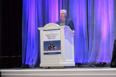
Las Vegas – A fibromyalgia survey may provide important information about the degree to which patients with rheumatic disease experience centralized pain. This information may guide treatment decisions, said Daniel J. Clauw, MD, professor of anesthesiology, rheumatology, and psychiatry and director of the Chronic Pain and Fatigue Research Center at the University of Michigan in Ann Arbor.

The questionnaire that Dr. Clauw uses is a patient self-report survey for the assessment of fibromyalgia based on criteria in the 2011 modification of the American College of Rheumatology preliminary diagnostic criteria for fibromyalgia. In it, he asks patients to report where they experience pain throughout the body and symptoms such as fatigue, sleep problems, and memory problems. The survey predicts outcomes of surgery for osteoarthritis better than x-rays, MRI scans, or psychological factors do, he said.
Physicians should ask every patient with chronic pain, including patients with OA, rheumatoid arthritis, or lupus, to complete the survey, Dr. Clauw said at the annual Perspectives in Rheumatic Diseases held by Global Academy for Medical Education. “This score will tell you the degree to which their central nervous system is augmenting or amplifying what is going on in their body,” he said. “And the higher their score is, the more you should treat them like you would someone with fibromyalgia, even if their underlying disease might be an autoimmune disease.”
Physicians should not use a cutoff of 13 points on the fibromyalgia measure to define whether a patient has the disease, as has been done in the past, he said. The threshold is arbitrary, he said. “We should not think about fibromyalgia as ‘yes’ or ‘no.’ We should think of the degree of fibromyalgia that people have.”
A poor relationship between pain and imaging
Some patients who have severe knee OA on imaging walk without pain. Other patients have normal x-rays, but severe pain. “There is a terrible relationship between what you see on a knee x-ray or an MRI and whether someone has pain,” Dr. Clauw said. Furthermore, the poor relationship between imaging and pain is common across chronic pain conditions, he said.
This phenomenon may occur because pain manifests in different ways, similar to there being multiple ways to adjust the volume of an electric guitar, he said. How hard the strings are strummed affects the volume. But so does the amplifier setting. “In these centralized pain conditions, the problem is an amplifier problem, not a guitar problem,” he said. “The amplifier, i.e., the central nervous system, is set too high.”
Researchers have found that people who have severe OA of the knee on x-ray but do not experience pain “have a very low amplifier setting,” he said. That is, they are nontender and less sensitive to pain. Most of these patients are men. “On average, men have a much lower amplifier setting than women,” he said. “This is also why ... women have 1.5 to 2 times the rate of any type of chronic pain than men, because on average women have a higher amplifier setting. ... In OA, at any given age, men and women have the exact same percentage of radiographic OA. But if you look at the clinical condition of OA, it is always two-thirds women, one-third men.”
Opioid responsiveness
To examine whether fibromyalgia survey results correlate with outcomes after knee and hip arthroplasty, Dr. Clauw and colleagues conducted a prospective, observational cohort study that included approximately 500 people. Patients completed the questionnaire on the day of surgery.
Patients with higher levels of fibromyalgia were less responsive to opioids. “For each 1-point increase in the fibromyalgia score, people needed about one more hydrocodone tablet in the first 24-48 hours to control their pain,” he said (Anesthesiology. 2013 Dec;119[6]:1434-43). In addition, each 1-point increase in the fibromyalgia score made people about 25% less likely to have a 50% improvement in knee pain level after 6 months (Arthritis Rheumatol. 2015 May;67[5]:1386-94). The correlations were independent of psychological factors. In addition, the associations were linear. “There was nothing magical about a fibromyalgia score of 13,” Dr. Clauw said.
Dr. Clauw is a coauthor of a study to be presented at the 2019 American College of Rheumatology/Association of Rheumatology Professionals annual meeting that found pain centralization in patients with RA is associated with poor response to disease-modifying antirheumatic drugs (DMARDs).
Prior studies in patients with RA have found that the degree of fibromyalgia is a better predictor of pain and disability than erythrocyte sedimentation rate or the number of swollen joints.
Diagnosed cases are the “tip of the iceberg”
Researchers at Dr. Clauw’s institution have identified dozens of patients undergoing knee surgery who met criteria for fibromyalgia but had not received the diagnosis. “This is at the University of Michigan, which is the epicenter for fibromyalgia research. If we are not seeing fibromyalgia superimposed on OA in our patients, no one is seeing it,” he said.
Patients with diagnosed fibromyalgia are “the tip of the iceberg,” he said. “There are far greater numbers of individuals whose primary diagnosis is OA, RA, lupus, ankylosing spondylitis, cancer pain, or sickle cell disease that have the same fundamental problem as fibromyalgia patients. But you do not see it because you label them as having an autoimmune disease or osteoarthritis. And that is at your peril and at their peril. Because treating that individual as if all of their pain and other symptoms are due to a problem out on the periphery will not make that person better.”
Patients with high levels of centralized pain may be less responsive to peripherally directed therapies such as surgery or injections, Dr. Clauw said. Pharmacologic options for patients with centralized pain include gabapentinoids (e.g., pregabalin and gabapentin), serotonin-norepinephrine reuptake inhibitors (e.g., duloxetine and milnacipran), and tricyclic compounds (e.g., amitriptyline and cyclobenzaprine), he said. “Opioids are going to be quite unlikely to help these individuals,” he said. “In fact, it is likely that opioids will make this kind of pain worse.”
Dr. Clauw is a consultant for Aptinyx, Daiichi Sankyo, Eli Lilly, Intec Pharma, Pfizer, Samumed, Theravance, Tonix, and Zynerba Pharma. He has received grant or research support from Aptinyx and Pfizer and is an expert witness.
Global Academy for Medical Education and this news organization are owned by the same parent company.
Las Vegas – A fibromyalgia survey may provide important information about the degree to which patients with rheumatic disease experience centralized pain. This information may guide treatment decisions, said Daniel J. Clauw, MD, professor of anesthesiology, rheumatology, and psychiatry and director of the Chronic Pain and Fatigue Research Center at the University of Michigan in Ann Arbor.
The questionnaire that Dr. Clauw uses is a patient self-report survey for the assessment of fibromyalgia based on criteria in the 2011 modification of the American College of Rheumatology preliminary diagnostic criteria for fibromyalgia. In it, he asks patients to report where they experience pain throughout the body and symptoms such as fatigue, sleep problems, and memory problems. The survey predicts outcomes of surgery for osteoarthritis better than x-rays, MRI scans, or psychological factors do, he said.
Physicians should ask every patient with chronic pain, including patients with OA, rheumatoid arthritis, or lupus, to complete the survey, Dr. Clauw said at the annual Perspectives in Rheumatic Diseases held by Global Academy for Medical Education. “This score will tell you the degree to which their central nervous system is augmenting or amplifying what is going on in their body,” he said. “And the higher their score is, the more you should treat them like you would someone with fibromyalgia, even if their underlying disease might be an autoimmune disease.”
Physicians should not use a cutoff of 13 points on the fibromyalgia measure to define whether a patient has the disease, as has been done in the past, he said. The threshold is arbitrary, he said. “We should not think about fibromyalgia as ‘yes’ or ‘no.’ We should think of the degree of fibromyalgia that people have.”
A poor relationship between pain and imaging
Some patients who have severe knee OA on imaging walk without pain. Other patients have normal x-rays, but severe pain. “There is a terrible relationship between what you see on a knee x-ray or an MRI and whether someone has pain,” Dr. Clauw said. Furthermore, the poor relationship between imaging and pain is common across chronic pain conditions, he said.
This phenomenon may occur because pain manifests in different ways, similar to there being multiple ways to adjust the volume of an electric guitar, he said. How hard the strings are strummed affects the volume. But so does the amplifier setting. “In these centralized pain conditions, the problem is an amplifier problem, not a guitar problem,” he said. “The amplifier, i.e., the central nervous system, is set too high.”
Researchers have found that people who have severe OA of the knee on x-ray but do not experience pain “have a very low amplifier setting,” he said. That is, they are nontender and less sensitive to pain. Most of these patients are men. “On average, men have a much lower amplifier setting than women,” he said. “This is also why ... women have 1.5 to 2 times the rate of any type of chronic pain than men, because on average women have a higher amplifier setting. ... In OA, at any given age, men and women have the exact same percentage of radiographic OA. But if you look at the clinical condition of OA, it is always two-thirds women, one-third men.”
Opioid responsiveness
To examine whether fibromyalgia survey results correlate with outcomes after knee and hip arthroplasty, Dr. Clauw and colleagues conducted a prospective, observational cohort study that included approximately 500 people. Patients completed the questionnaire on the day of surgery.
Patients with higher levels of fibromyalgia were less responsive to opioids. “For each 1-point increase in the fibromyalgia score, people needed about one more hydrocodone tablet in the first 24-48 hours to control their pain,” he said (Anesthesiology. 2013 Dec;119[6]:1434-43). In addition, each 1-point increase in the fibromyalgia score made people about 25% less likely to have a 50% improvement in knee pain level after 6 months (Arthritis Rheumatol. 2015 May;67[5]:1386-94). The correlations were independent of psychological factors. In addition, the associations were linear. “There was nothing magical about a fibromyalgia score of 13,” Dr. Clauw said.
Dr. Clauw is a coauthor of a study to be presented at the 2019 American College of Rheumatology/Association of Rheumatology Professionals annual meeting that found pain centralization in patients with RA is associated with poor response to disease-modifying antirheumatic drugs (DMARDs).
Prior studies in patients with RA have found that the degree of fibromyalgia is a better predictor of pain and disability than erythrocyte sedimentation rate or the number of swollen joints.
Diagnosed cases are the “tip of the iceberg”
Researchers at Dr. Clauw’s institution have identified dozens of patients undergoing knee surgery who met criteria for fibromyalgia but had not received the diagnosis. “This is at the University of Michigan, which is the epicenter for fibromyalgia research. If we are not seeing fibromyalgia superimposed on OA in our patients, no one is seeing it,” he said.
Patients with diagnosed fibromyalgia are “the tip of the iceberg,” he said. “There are far greater numbers of individuals whose primary diagnosis is OA, RA, lupus, ankylosing spondylitis, cancer pain, or sickle cell disease that have the same fundamental problem as fibromyalgia patients. But you do not see it because you label them as having an autoimmune disease or osteoarthritis. And that is at your peril and at their peril. Because treating that individual as if all of their pain and other symptoms are due to a problem out on the periphery will not make that person better.”
Patients with high levels of centralized pain may be less responsive to peripherally directed therapies such as surgery or injections, Dr. Clauw said. Pharmacologic options for patients with centralized pain include gabapentinoids (e.g., pregabalin and gabapentin), serotonin-norepinephrine reuptake inhibitors (e.g., duloxetine and milnacipran), and tricyclic compounds (e.g., amitriptyline and cyclobenzaprine), he said. “Opioids are going to be quite unlikely to help these individuals,” he said. “In fact, it is likely that opioids will make this kind of pain worse.”
Dr. Clauw is a consultant for Aptinyx, Daiichi Sankyo, Eli Lilly, Intec Pharma, Pfizer, Samumed, Theravance, Tonix, and Zynerba Pharma. He has received grant or research support from Aptinyx and Pfizer and is an expert witness.
Global Academy for Medical Education and this news organization are owned by the same parent company.
Las Vegas – A fibromyalgia survey may provide important information about the degree to which patients with rheumatic disease experience centralized pain. This information may guide treatment decisions, said Daniel J. Clauw, MD, professor of anesthesiology, rheumatology, and psychiatry and director of the Chronic Pain and Fatigue Research Center at the University of Michigan in Ann Arbor.
The questionnaire that Dr. Clauw uses is a patient self-report survey for the assessment of fibromyalgia based on criteria in the 2011 modification of the American College of Rheumatology preliminary diagnostic criteria for fibromyalgia. In it, he asks patients to report where they experience pain throughout the body and symptoms such as fatigue, sleep problems, and memory problems. The survey predicts outcomes of surgery for osteoarthritis better than x-rays, MRI scans, or psychological factors do, he said.
Physicians should ask every patient with chronic pain, including patients with OA, rheumatoid arthritis, or lupus, to complete the survey, Dr. Clauw said at the annual Perspectives in Rheumatic Diseases held by Global Academy for Medical Education. “This score will tell you the degree to which their central nervous system is augmenting or amplifying what is going on in their body,” he said. “And the higher their score is, the more you should treat them like you would someone with fibromyalgia, even if their underlying disease might be an autoimmune disease.”
Physicians should not use a cutoff of 13 points on the fibromyalgia measure to define whether a patient has the disease, as has been done in the past, he said. The threshold is arbitrary, he said. “We should not think about fibromyalgia as ‘yes’ or ‘no.’ We should think of the degree of fibromyalgia that people have.”
A poor relationship between pain and imaging
Some patients who have severe knee OA on imaging walk without pain. Other patients have normal x-rays, but severe pain. “There is a terrible relationship between what you see on a knee x-ray or an MRI and whether someone has pain,” Dr. Clauw said. Furthermore, the poor relationship between imaging and pain is common across chronic pain conditions, he said.
This phenomenon may occur because pain manifests in different ways, similar to there being multiple ways to adjust the volume of an electric guitar, he said. How hard the strings are strummed affects the volume. But so does the amplifier setting. “In these centralized pain conditions, the problem is an amplifier problem, not a guitar problem,” he said. “The amplifier, i.e., the central nervous system, is set too high.”
Researchers have found that people who have severe OA of the knee on x-ray but do not experience pain “have a very low amplifier setting,” he said. That is, they are nontender and less sensitive to pain. Most of these patients are men. “On average, men have a much lower amplifier setting than women,” he said. “This is also why ... women have 1.5 to 2 times the rate of any type of chronic pain than men, because on average women have a higher amplifier setting. ... In OA, at any given age, men and women have the exact same percentage of radiographic OA. But if you look at the clinical condition of OA, it is always two-thirds women, one-third men.”
Opioid responsiveness
To examine whether fibromyalgia survey results correlate with outcomes after knee and hip arthroplasty, Dr. Clauw and colleagues conducted a prospective, observational cohort study that included approximately 500 people. Patients completed the questionnaire on the day of surgery.
Patients with higher levels of fibromyalgia were less responsive to opioids. “For each 1-point increase in the fibromyalgia score, people needed about one more hydrocodone tablet in the first 24-48 hours to control their pain,” he said (Anesthesiology. 2013 Dec;119[6]:1434-43). In addition, each 1-point increase in the fibromyalgia score made people about 25% less likely to have a 50% improvement in knee pain level after 6 months (Arthritis Rheumatol. 2015 May;67[5]:1386-94). The correlations were independent of psychological factors. In addition, the associations were linear. “There was nothing magical about a fibromyalgia score of 13,” Dr. Clauw said.
Dr. Clauw is a coauthor of a study to be presented at the 2019 American College of Rheumatology/Association of Rheumatology Professionals annual meeting that found pain centralization in patients with RA is associated with poor response to disease-modifying antirheumatic drugs (DMARDs).
Prior studies in patients with RA have found that the degree of fibromyalgia is a better predictor of pain and disability than erythrocyte sedimentation rate or the number of swollen joints.
Diagnosed cases are the “tip of the iceberg”
Researchers at Dr. Clauw’s institution have identified dozens of patients undergoing knee surgery who met criteria for fibromyalgia but had not received the diagnosis. “This is at the University of Michigan, which is the epicenter for fibromyalgia research. If we are not seeing fibromyalgia superimposed on OA in our patients, no one is seeing it,” he said.
Patients with diagnosed fibromyalgia are “the tip of the iceberg,” he said. “There are far greater numbers of individuals whose primary diagnosis is OA, RA, lupus, ankylosing spondylitis, cancer pain, or sickle cell disease that have the same fundamental problem as fibromyalgia patients. But you do not see it because you label them as having an autoimmune disease or osteoarthritis. And that is at your peril and at their peril. Because treating that individual as if all of their pain and other symptoms are due to a problem out on the periphery will not make that person better.”
Patients with high levels of centralized pain may be less responsive to peripherally directed therapies such as surgery or injections, Dr. Clauw said. Pharmacologic options for patients with centralized pain include gabapentinoids (e.g., pregabalin and gabapentin), serotonin-norepinephrine reuptake inhibitors (e.g., duloxetine and milnacipran), and tricyclic compounds (e.g., amitriptyline and cyclobenzaprine), he said. “Opioids are going to be quite unlikely to help these individuals,” he said. “In fact, it is likely that opioids will make this kind of pain worse.”
Dr. Clauw is a consultant for Aptinyx, Daiichi Sankyo, Eli Lilly, Intec Pharma, Pfizer, Samumed, Theravance, Tonix, and Zynerba Pharma. He has received grant or research support from Aptinyx and Pfizer and is an expert witness.
Global Academy for Medical Education and this news organization are owned by the same parent company.
EXPERT ANALYSIS FROM PRD 2019
What is the optimal duration of maintenance in myeloma?
SAN FRANCISCO – Should patients with multiple myeloma receive maintenance therapy until progression?

Yvonne A. Efebera, MD, of The Ohio State University Comprehensive Cancer Center – Arthur G. James Cancer Hospital in Columbus, and Nina Shah, MD, of the University of California San Francisco Health, faced off on this question at the National Comprehensive Cancer Network Hematologic Malignancies Annual Congress.

Dr. Shah said maintenance therapy improves survival in myeloma patients, so it follows that treating them until progression would confer a survival advantage. While Dr. Efebera agreed that maintenance can improve survival, she said the optimal duration of that treatment is unknown.
Treat until progression
Dr. Shah cited studies suggesting that maintenance improves progression-free survival (PFS) and may prolong overall survival (OS) in multiple myeloma.
A meta-analysis of data from the IFM 2005-02, CALGB 100104, and GIMEMA RV-MM-PI-209 trials showed that lenalidomide maintenance prolonged PFS and OS. The median PFS was 52.8 months in patients who received maintenance and 23.5 months in those who received placebo or observation (hazard ratio [HR], 0.48). At a median follow-up of 79.5 months, the median OS was not reached for the maintenance group and was 86.0 months for the no-maintenance group (HR, 0.75; P = .001; J Clin Oncol. 2017 Oct 10;35[29]:3279-89).
In the Myeloma XI trial, maintenance improved PFS, but not OS, in both transplant-eligible and ineligible patients. Overall, the median PFS was 39 months in the lenalidomide maintenance arm and 20 months in the observation arm (P less than .0001). Among transplant-eligible patients, the median PFS was 57 months and 30 months, respectively (P less than .0001). Among transplant-ineligible patients, the median PFS was 26 months and 11 months, respectively (P less than .0001; Lancet Oncol. 2019 Jan;20[1]:57-73).
These data suggest maintenance can improve survival, “but the question is, how long should we have therapy,” Dr. Shah said. “No one has looked at this in a prospective manner, so we really have to look at our retrospective data.”
One study suggested a longer duration of lenalidomide maintenance improves PFS. The HR for progression or death was 0.39 for patients who received maintenance for 12-24 months, compared with those who received maintenance for less than 12 months. The HR was 0.13 for patients who received maintenance for more than 24 months, compared with less than 12 months (Leuk Lymphoma. 2019 Feb;60[2]:511-4).
Dr. Shah also cited a pooled analysis of three phase 3 trials suggesting that continuous therapy is superior to fixed-duration therapy in patients with newly diagnosed myeloma. The median PFS was 32 months with continuous therapy and 16 months with fixed-duration therapy (P less than .001). The 4-year OS was 69% and 60%, respectively (P = .003; J Clin Oncol. 2015 Oct 20;33[30]:3459-66).
These data suggest that “continuous therapy, more therapy, has a survival advantage,” Dr. Shah said.
Don’t treat until progression
Dr. Efebera also discussed data from studies showing that lenalidomide maintenance can prolong survival in multiple myeloma. However, she said, it’s unclear how long maintenance should last.
Different durations of maintenance have proved effective in different trials. In the CALGB 100104 trial, the median duration of maintenance was 31 months (Lancet Haematol. 2017 Sep;4[9]:e431-e442). In the meta-analysis of the CALGB, IFM, and GIMEMA trials, the median duration was 22 months. And in Myeloma XI, the median duration was 18 months.
As there is no randomized trial comparing different durations of maintenance, Dr. Efebera proposed that researchers conduct one. She said this “perfect study” would involve induction with an immunomodulatory agent, a proteasome inhibitor, dexamethasone, and perhaps an anti-CD38 therapy. Transplant-eligible patients would receive four cycles of induction before transplant. Transplant-ineligible patients would receive eight cycles of induction. Then, all patients would be randomized to lenalidomide maintenance for 3 years, 5 years, or 7-10 years.
Until a trial like this reveals the optimal duration of maintenance, we cannot conclude that treating patients until progression is better, Dr. Efebera said.
She added that maintenance has been shown to have detrimental effects, and these should be taken into consideration. For instance, neutropenia, other hematologic adverse events, and second primary malignancies have been shown to be more common among patients who receive lenalidomide maintenance (N Engl J Med. 2012; 366:1782-91).
The cost of maintenance is another factor to consider. Researchers analyzed data from the CALGB 100104 and IFM 2005-02 trials to compare the cost of lenalidomide maintenance with no maintenance. In the CALGB 100104 trial, patients who received lenalidomide maintenance had 5.72 quality-adjusted life years (QALYs), and those who received no maintenance had 4.61 QALYs. The incremental cost-utility ratio (ICUR) was more than 277,000 euros per QALY.
In the IFM2005-02 trial, patients in the lenalidomide group had 5.13 QALYs, and those who didn’t receive maintenance had 4.98 QALYs. The ICUR was more than 1.5 million euros per QALY. The researchers said the high ICURs and budgetary impact add “uncertainty about the maximum prudent duration of the treatment” (Bone Marrow Transplant. 2019 May 31. doi: 10.1038/s41409-019-0574-5).
Dr. Efebera reported relationships with Akcea Therapeutics, Janssen, and Takeda. Dr. Shah reported having no relevant financial relationships.
SAN FRANCISCO – Should patients with multiple myeloma receive maintenance therapy until progression?
Yvonne A. Efebera, MD, of The Ohio State University Comprehensive Cancer Center – Arthur G. James Cancer Hospital in Columbus, and Nina Shah, MD, of the University of California San Francisco Health, faced off on this question at the National Comprehensive Cancer Network Hematologic Malignancies Annual Congress.
Dr. Shah said maintenance therapy improves survival in myeloma patients, so it follows that treating them until progression would confer a survival advantage. While Dr. Efebera agreed that maintenance can improve survival, she said the optimal duration of that treatment is unknown.
Treat until progression
Dr. Shah cited studies suggesting that maintenance improves progression-free survival (PFS) and may prolong overall survival (OS) in multiple myeloma.
A meta-analysis of data from the IFM 2005-02, CALGB 100104, and GIMEMA RV-MM-PI-209 trials showed that lenalidomide maintenance prolonged PFS and OS. The median PFS was 52.8 months in patients who received maintenance and 23.5 months in those who received placebo or observation (hazard ratio [HR], 0.48). At a median follow-up of 79.5 months, the median OS was not reached for the maintenance group and was 86.0 months for the no-maintenance group (HR, 0.75; P = .001; J Clin Oncol. 2017 Oct 10;35[29]:3279-89).
In the Myeloma XI trial, maintenance improved PFS, but not OS, in both transplant-eligible and ineligible patients. Overall, the median PFS was 39 months in the lenalidomide maintenance arm and 20 months in the observation arm (P less than .0001). Among transplant-eligible patients, the median PFS was 57 months and 30 months, respectively (P less than .0001). Among transplant-ineligible patients, the median PFS was 26 months and 11 months, respectively (P less than .0001; Lancet Oncol. 2019 Jan;20[1]:57-73).
These data suggest maintenance can improve survival, “but the question is, how long should we have therapy,” Dr. Shah said. “No one has looked at this in a prospective manner, so we really have to look at our retrospective data.”
One study suggested a longer duration of lenalidomide maintenance improves PFS. The HR for progression or death was 0.39 for patients who received maintenance for 12-24 months, compared with those who received maintenance for less than 12 months. The HR was 0.13 for patients who received maintenance for more than 24 months, compared with less than 12 months (Leuk Lymphoma. 2019 Feb;60[2]:511-4).
Dr. Shah also cited a pooled analysis of three phase 3 trials suggesting that continuous therapy is superior to fixed-duration therapy in patients with newly diagnosed myeloma. The median PFS was 32 months with continuous therapy and 16 months with fixed-duration therapy (P less than .001). The 4-year OS was 69% and 60%, respectively (P = .003; J Clin Oncol. 2015 Oct 20;33[30]:3459-66).
These data suggest that “continuous therapy, more therapy, has a survival advantage,” Dr. Shah said.
Don’t treat until progression
Dr. Efebera also discussed data from studies showing that lenalidomide maintenance can prolong survival in multiple myeloma. However, she said, it’s unclear how long maintenance should last.
Different durations of maintenance have proved effective in different trials. In the CALGB 100104 trial, the median duration of maintenance was 31 months (Lancet Haematol. 2017 Sep;4[9]:e431-e442). In the meta-analysis of the CALGB, IFM, and GIMEMA trials, the median duration was 22 months. And in Myeloma XI, the median duration was 18 months.
As there is no randomized trial comparing different durations of maintenance, Dr. Efebera proposed that researchers conduct one. She said this “perfect study” would involve induction with an immunomodulatory agent, a proteasome inhibitor, dexamethasone, and perhaps an anti-CD38 therapy. Transplant-eligible patients would receive four cycles of induction before transplant. Transplant-ineligible patients would receive eight cycles of induction. Then, all patients would be randomized to lenalidomide maintenance for 3 years, 5 years, or 7-10 years.
Until a trial like this reveals the optimal duration of maintenance, we cannot conclude that treating patients until progression is better, Dr. Efebera said.
She added that maintenance has been shown to have detrimental effects, and these should be taken into consideration. For instance, neutropenia, other hematologic adverse events, and second primary malignancies have been shown to be more common among patients who receive lenalidomide maintenance (N Engl J Med. 2012; 366:1782-91).
The cost of maintenance is another factor to consider. Researchers analyzed data from the CALGB 100104 and IFM 2005-02 trials to compare the cost of lenalidomide maintenance with no maintenance. In the CALGB 100104 trial, patients who received lenalidomide maintenance had 5.72 quality-adjusted life years (QALYs), and those who received no maintenance had 4.61 QALYs. The incremental cost-utility ratio (ICUR) was more than 277,000 euros per QALY.
In the IFM2005-02 trial, patients in the lenalidomide group had 5.13 QALYs, and those who didn’t receive maintenance had 4.98 QALYs. The ICUR was more than 1.5 million euros per QALY. The researchers said the high ICURs and budgetary impact add “uncertainty about the maximum prudent duration of the treatment” (Bone Marrow Transplant. 2019 May 31. doi: 10.1038/s41409-019-0574-5).
Dr. Efebera reported relationships with Akcea Therapeutics, Janssen, and Takeda. Dr. Shah reported having no relevant financial relationships.
SAN FRANCISCO – Should patients with multiple myeloma receive maintenance therapy until progression?
Yvonne A. Efebera, MD, of The Ohio State University Comprehensive Cancer Center – Arthur G. James Cancer Hospital in Columbus, and Nina Shah, MD, of the University of California San Francisco Health, faced off on this question at the National Comprehensive Cancer Network Hematologic Malignancies Annual Congress.
Dr. Shah said maintenance therapy improves survival in myeloma patients, so it follows that treating them until progression would confer a survival advantage. While Dr. Efebera agreed that maintenance can improve survival, she said the optimal duration of that treatment is unknown.
Treat until progression
Dr. Shah cited studies suggesting that maintenance improves progression-free survival (PFS) and may prolong overall survival (OS) in multiple myeloma.
A meta-analysis of data from the IFM 2005-02, CALGB 100104, and GIMEMA RV-MM-PI-209 trials showed that lenalidomide maintenance prolonged PFS and OS. The median PFS was 52.8 months in patients who received maintenance and 23.5 months in those who received placebo or observation (hazard ratio [HR], 0.48). At a median follow-up of 79.5 months, the median OS was not reached for the maintenance group and was 86.0 months for the no-maintenance group (HR, 0.75; P = .001; J Clin Oncol. 2017 Oct 10;35[29]:3279-89).
In the Myeloma XI trial, maintenance improved PFS, but not OS, in both transplant-eligible and ineligible patients. Overall, the median PFS was 39 months in the lenalidomide maintenance arm and 20 months in the observation arm (P less than .0001). Among transplant-eligible patients, the median PFS was 57 months and 30 months, respectively (P less than .0001). Among transplant-ineligible patients, the median PFS was 26 months and 11 months, respectively (P less than .0001; Lancet Oncol. 2019 Jan;20[1]:57-73).
These data suggest maintenance can improve survival, “but the question is, how long should we have therapy,” Dr. Shah said. “No one has looked at this in a prospective manner, so we really have to look at our retrospective data.”
One study suggested a longer duration of lenalidomide maintenance improves PFS. The HR for progression or death was 0.39 for patients who received maintenance for 12-24 months, compared with those who received maintenance for less than 12 months. The HR was 0.13 for patients who received maintenance for more than 24 months, compared with less than 12 months (Leuk Lymphoma. 2019 Feb;60[2]:511-4).
Dr. Shah also cited a pooled analysis of three phase 3 trials suggesting that continuous therapy is superior to fixed-duration therapy in patients with newly diagnosed myeloma. The median PFS was 32 months with continuous therapy and 16 months with fixed-duration therapy (P less than .001). The 4-year OS was 69% and 60%, respectively (P = .003; J Clin Oncol. 2015 Oct 20;33[30]:3459-66).
These data suggest that “continuous therapy, more therapy, has a survival advantage,” Dr. Shah said.
Don’t treat until progression
Dr. Efebera also discussed data from studies showing that lenalidomide maintenance can prolong survival in multiple myeloma. However, she said, it’s unclear how long maintenance should last.
Different durations of maintenance have proved effective in different trials. In the CALGB 100104 trial, the median duration of maintenance was 31 months (Lancet Haematol. 2017 Sep;4[9]:e431-e442). In the meta-analysis of the CALGB, IFM, and GIMEMA trials, the median duration was 22 months. And in Myeloma XI, the median duration was 18 months.
As there is no randomized trial comparing different durations of maintenance, Dr. Efebera proposed that researchers conduct one. She said this “perfect study” would involve induction with an immunomodulatory agent, a proteasome inhibitor, dexamethasone, and perhaps an anti-CD38 therapy. Transplant-eligible patients would receive four cycles of induction before transplant. Transplant-ineligible patients would receive eight cycles of induction. Then, all patients would be randomized to lenalidomide maintenance for 3 years, 5 years, or 7-10 years.
Until a trial like this reveals the optimal duration of maintenance, we cannot conclude that treating patients until progression is better, Dr. Efebera said.
She added that maintenance has been shown to have detrimental effects, and these should be taken into consideration. For instance, neutropenia, other hematologic adverse events, and second primary malignancies have been shown to be more common among patients who receive lenalidomide maintenance (N Engl J Med. 2012; 366:1782-91).
The cost of maintenance is another factor to consider. Researchers analyzed data from the CALGB 100104 and IFM 2005-02 trials to compare the cost of lenalidomide maintenance with no maintenance. In the CALGB 100104 trial, patients who received lenalidomide maintenance had 5.72 quality-adjusted life years (QALYs), and those who received no maintenance had 4.61 QALYs. The incremental cost-utility ratio (ICUR) was more than 277,000 euros per QALY.
In the IFM2005-02 trial, patients in the lenalidomide group had 5.13 QALYs, and those who didn’t receive maintenance had 4.98 QALYs. The ICUR was more than 1.5 million euros per QALY. The researchers said the high ICURs and budgetary impact add “uncertainty about the maximum prudent duration of the treatment” (Bone Marrow Transplant. 2019 May 31. doi: 10.1038/s41409-019-0574-5).
Dr. Efebera reported relationships with Akcea Therapeutics, Janssen, and Takeda. Dr. Shah reported having no relevant financial relationships.
EXPERT ANALYSIS FROM NCCN HEMATOLOGIC MALIGNANCIES

Atezolizumab plus chemo gives slight PFS edge in mUC
BARCELONA – Adding the immune checkpoint inhibitor atezolizumab (Tecentriq) to standard platinum-based chemotherapy was associated with a small but statistically significant progression-free survival benefit for patients with previously untreated metastatic urothelial carcinoma, investigators in the IMvigor130 trial found.

Among 1,213 patients with newly diagnosed metastatic urothelial carcinoma assigned to receive either atezolizumab monotherapy or chemotherapy with a platinum compound and gemcitabine plus either atezolizumab or placebo, the median progression-free survival (PFS) at a median follow-up of 11.8 months was 8.2 months with atezolizumab/chemotherapy, compared with 6.3 months with chemotherapy plus placebo, reported Enrique Grande, MD, of MD Anderson Cancer Center Madrid.
“IMvigor130 is the first immune checkpoint inhibitor study to demonstrate an improvement in progression-free survival over the standard of care in first-line metastatic urothelial carcinoma. At this interim analysis, we observed a clinically meaningful improvement in the overall survival for the combination of atezolizumab plus chemotherapy that did not meet the prespecified interim boundary for significance,” he said at the European Society for Medical Oncology Congress.
Median overall survival (OS) at the interim analysis was 16.0 months in the atezolizumab arm, vs. 13.4 months in the placebo arm, translating into a hazard ratio (HR) of 0.83 trending in favor of the combination. But as noted by Dr. Grande, the P value was .027, which did not reach the interim efficacy boundary of .007.
IMvigor130 investigators enrolled patients with locally advanced metastatic urothelial carcinoma with no prior systemic therapy in the metastatic setting who had good performance status (ECOG 2 or less) and were eligible for chemotherapy with either cisplatin or carboplatin plus gemcitabine.
The patients were stratified by programmed death ligand-1 (PD-L1) status, prognostic (Bajorin) risk factor scores, and investigator choice of cisplatin or carboplatin, and then randomized to either atezolizumab plus chemotherapy, atezolizumab monotherapy, or placebo plus chemotherapy.
As noted, the co-primary endpoint of PFS in the chemotherapy arms in the intention-to-treat population was statistically significant at the preplanned interim analysis, but the other primary endpoint of OS had not reached significance.
At the time of the data cutoff in May 2019, the stratified HR for progression with atezolizumab was 0.82 (P = .007).
An analysis by subgroup showed either significant benefit or a trend favoring atezolizumab across all stratification factors, Dr. Grande said.
A hierarchical analysis of atezolizumab monotherapy vs. chemotherapy in the placebo-control arm showed a median OS of 15.7 vs. 13.1 months, respectively, with a hazard ratio of 1.02 (nonsignificant).
The benefit of atezolizumab appeared to be almost entirely among patients whose tumors had higher levels of PD-L1 expression according to immunohistochemistry (IC). The interim OS among patients with PD-L1 IC0/1 was a median of 13.5 months with atezolizumab vs. 12.9 months with chemotherapy alone, with an unstratified HR of 1.07 (nonsignificant). In contrast, among patients with PD-L1 IC2/3 status, the median OS was not reached for patients in the atezolizumab arm, vs. 17.8 months in the chemotherapy alone arm, for a stratified HR of 0.68, although this too did not reach statistical significance.
Responses were similar between the two chemotherapy arms, with an overall response rate (ORR) of 47% with atezolizumab added, vs. 44% with placebo added. The complete response (CR) rates were 13% and 7%, respectively. The ORR in the monotherapy arm was 23%, consisting of 6% complete and 17% partial responses.
Grade 3 or 4 treatment-related adverse events occurred in 81% of patients in each chemotherapy arm, compared with 15% in the atezolizumab monotherapy arm. Nine patients in the atezolizumab/chemotherapy arm died from a treatment-related cause, compared with four in the chemotherapy alone arm, and three in the atezolizumab monotherapy arm.
Adverse events leading to treatment discontinuation occurred in one-fourth of patients in each chemotherapy-containing arm, vs. less than 1% of patients in the monotherapy arm.
“The results from the IMvigor130 trial support the use of atezolizumab in combination with chemotherapy as an important new treatment option for patients with untreated metastatic urothelial carcinoma,” Dr. Grande concluded.
But invited discussant Thomas Powles, MD, of Barts Cancer Institute in London, cautioned that more data are needed to conclude that the addition of atezolizumab should become a standard of care.

“Does this change practice? Well, the for and against: significant delay in PFS, but how meaningful is that? OS trending the right way, but not significant yet. CR rates, yes with 13% CRs, but response rates weren’t very different from one another, and as response rates are similar, it’s hard to argue that the two are synergistic together, for example,” he said.
He added that the adverse event profiles “actually are very acceptable to me, and I’m really looking forward to the quality-of-life data.”
The IMvigor130 study is sponsored by F. Hoffman-La Roche. Dr. Grande disclosed honoraria and research grants from Roche and others. Dr. Powles disclosed research funding, honoraria, and travel costs from Roche and others.
SOURCE: Grande E et al. ESMO 2019. Abstract LBA14_PR.
BARCELONA – Adding the immune checkpoint inhibitor atezolizumab (Tecentriq) to standard platinum-based chemotherapy was associated with a small but statistically significant progression-free survival benefit for patients with previously untreated metastatic urothelial carcinoma, investigators in the IMvigor130 trial found.
Among 1,213 patients with newly diagnosed metastatic urothelial carcinoma assigned to receive either atezolizumab monotherapy or chemotherapy with a platinum compound and gemcitabine plus either atezolizumab or placebo, the median progression-free survival (PFS) at a median follow-up of 11.8 months was 8.2 months with atezolizumab/chemotherapy, compared with 6.3 months with chemotherapy plus placebo, reported Enrique Grande, MD, of MD Anderson Cancer Center Madrid.
“IMvigor130 is the first immune checkpoint inhibitor study to demonstrate an improvement in progression-free survival over the standard of care in first-line metastatic urothelial carcinoma. At this interim analysis, we observed a clinically meaningful improvement in the overall survival for the combination of atezolizumab plus chemotherapy that did not meet the prespecified interim boundary for significance,” he said at the European Society for Medical Oncology Congress.
Median overall survival (OS) at the interim analysis was 16.0 months in the atezolizumab arm, vs. 13.4 months in the placebo arm, translating into a hazard ratio (HR) of 0.83 trending in favor of the combination. But as noted by Dr. Grande, the P value was .027, which did not reach the interim efficacy boundary of .007.
IMvigor130 investigators enrolled patients with locally advanced metastatic urothelial carcinoma with no prior systemic therapy in the metastatic setting who had good performance status (ECOG 2 or less) and were eligible for chemotherapy with either cisplatin or carboplatin plus gemcitabine.
The patients were stratified by programmed death ligand-1 (PD-L1) status, prognostic (Bajorin) risk factor scores, and investigator choice of cisplatin or carboplatin, and then randomized to either atezolizumab plus chemotherapy, atezolizumab monotherapy, or placebo plus chemotherapy.
As noted, the co-primary endpoint of PFS in the chemotherapy arms in the intention-to-treat population was statistically significant at the preplanned interim analysis, but the other primary endpoint of OS had not reached significance.
At the time of the data cutoff in May 2019, the stratified HR for progression with atezolizumab was 0.82 (P = .007).
An analysis by subgroup showed either significant benefit or a trend favoring atezolizumab across all stratification factors, Dr. Grande said.
A hierarchical analysis of atezolizumab monotherapy vs. chemotherapy in the placebo-control arm showed a median OS of 15.7 vs. 13.1 months, respectively, with a hazard ratio of 1.02 (nonsignificant).
The benefit of atezolizumab appeared to be almost entirely among patients whose tumors had higher levels of PD-L1 expression according to immunohistochemistry (IC). The interim OS among patients with PD-L1 IC0/1 was a median of 13.5 months with atezolizumab vs. 12.9 months with chemotherapy alone, with an unstratified HR of 1.07 (nonsignificant). In contrast, among patients with PD-L1 IC2/3 status, the median OS was not reached for patients in the atezolizumab arm, vs. 17.8 months in the chemotherapy alone arm, for a stratified HR of 0.68, although this too did not reach statistical significance.
Responses were similar between the two chemotherapy arms, with an overall response rate (ORR) of 47% with atezolizumab added, vs. 44% with placebo added. The complete response (CR) rates were 13% and 7%, respectively. The ORR in the monotherapy arm was 23%, consisting of 6% complete and 17% partial responses.
Grade 3 or 4 treatment-related adverse events occurred in 81% of patients in each chemotherapy arm, compared with 15% in the atezolizumab monotherapy arm. Nine patients in the atezolizumab/chemotherapy arm died from a treatment-related cause, compared with four in the chemotherapy alone arm, and three in the atezolizumab monotherapy arm.
Adverse events leading to treatment discontinuation occurred in one-fourth of patients in each chemotherapy-containing arm, vs. less than 1% of patients in the monotherapy arm.
“The results from the IMvigor130 trial support the use of atezolizumab in combination with chemotherapy as an important new treatment option for patients with untreated metastatic urothelial carcinoma,” Dr. Grande concluded.
But invited discussant Thomas Powles, MD, of Barts Cancer Institute in London, cautioned that more data are needed to conclude that the addition of atezolizumab should become a standard of care.
“Does this change practice? Well, the for and against: significant delay in PFS, but how meaningful is that? OS trending the right way, but not significant yet. CR rates, yes with 13% CRs, but response rates weren’t very different from one another, and as response rates are similar, it’s hard to argue that the two are synergistic together, for example,” he said.
He added that the adverse event profiles “actually are very acceptable to me, and I’m really looking forward to the quality-of-life data.”
The IMvigor130 study is sponsored by F. Hoffman-La Roche. Dr. Grande disclosed honoraria and research grants from Roche and others. Dr. Powles disclosed research funding, honoraria, and travel costs from Roche and others.
SOURCE: Grande E et al. ESMO 2019. Abstract LBA14_PR.
BARCELONA – Adding the immune checkpoint inhibitor atezolizumab (Tecentriq) to standard platinum-based chemotherapy was associated with a small but statistically significant progression-free survival benefit for patients with previously untreated metastatic urothelial carcinoma, investigators in the IMvigor130 trial found.
Among 1,213 patients with newly diagnosed metastatic urothelial carcinoma assigned to receive either atezolizumab monotherapy or chemotherapy with a platinum compound and gemcitabine plus either atezolizumab or placebo, the median progression-free survival (PFS) at a median follow-up of 11.8 months was 8.2 months with atezolizumab/chemotherapy, compared with 6.3 months with chemotherapy plus placebo, reported Enrique Grande, MD, of MD Anderson Cancer Center Madrid.
“IMvigor130 is the first immune checkpoint inhibitor study to demonstrate an improvement in progression-free survival over the standard of care in first-line metastatic urothelial carcinoma. At this interim analysis, we observed a clinically meaningful improvement in the overall survival for the combination of atezolizumab plus chemotherapy that did not meet the prespecified interim boundary for significance,” he said at the European Society for Medical Oncology Congress.
Median overall survival (OS) at the interim analysis was 16.0 months in the atezolizumab arm, vs. 13.4 months in the placebo arm, translating into a hazard ratio (HR) of 0.83 trending in favor of the combination. But as noted by Dr. Grande, the P value was .027, which did not reach the interim efficacy boundary of .007.
IMvigor130 investigators enrolled patients with locally advanced metastatic urothelial carcinoma with no prior systemic therapy in the metastatic setting who had good performance status (ECOG 2 or less) and were eligible for chemotherapy with either cisplatin or carboplatin plus gemcitabine.
The patients were stratified by programmed death ligand-1 (PD-L1) status, prognostic (Bajorin) risk factor scores, and investigator choice of cisplatin or carboplatin, and then randomized to either atezolizumab plus chemotherapy, atezolizumab monotherapy, or placebo plus chemotherapy.
As noted, the co-primary endpoint of PFS in the chemotherapy arms in the intention-to-treat population was statistically significant at the preplanned interim analysis, but the other primary endpoint of OS had not reached significance.
At the time of the data cutoff in May 2019, the stratified HR for progression with atezolizumab was 0.82 (P = .007).
An analysis by subgroup showed either significant benefit or a trend favoring atezolizumab across all stratification factors, Dr. Grande said.
A hierarchical analysis of atezolizumab monotherapy vs. chemotherapy in the placebo-control arm showed a median OS of 15.7 vs. 13.1 months, respectively, with a hazard ratio of 1.02 (nonsignificant).
The benefit of atezolizumab appeared to be almost entirely among patients whose tumors had higher levels of PD-L1 expression according to immunohistochemistry (IC). The interim OS among patients with PD-L1 IC0/1 was a median of 13.5 months with atezolizumab vs. 12.9 months with chemotherapy alone, with an unstratified HR of 1.07 (nonsignificant). In contrast, among patients with PD-L1 IC2/3 status, the median OS was not reached for patients in the atezolizumab arm, vs. 17.8 months in the chemotherapy alone arm, for a stratified HR of 0.68, although this too did not reach statistical significance.
Responses were similar between the two chemotherapy arms, with an overall response rate (ORR) of 47% with atezolizumab added, vs. 44% with placebo added. The complete response (CR) rates were 13% and 7%, respectively. The ORR in the monotherapy arm was 23%, consisting of 6% complete and 17% partial responses.
Grade 3 or 4 treatment-related adverse events occurred in 81% of patients in each chemotherapy arm, compared with 15% in the atezolizumab monotherapy arm. Nine patients in the atezolizumab/chemotherapy arm died from a treatment-related cause, compared with four in the chemotherapy alone arm, and three in the atezolizumab monotherapy arm.
Adverse events leading to treatment discontinuation occurred in one-fourth of patients in each chemotherapy-containing arm, vs. less than 1% of patients in the monotherapy arm.
“The results from the IMvigor130 trial support the use of atezolizumab in combination with chemotherapy as an important new treatment option for patients with untreated metastatic urothelial carcinoma,” Dr. Grande concluded.
But invited discussant Thomas Powles, MD, of Barts Cancer Institute in London, cautioned that more data are needed to conclude that the addition of atezolizumab should become a standard of care.
“Does this change practice? Well, the for and against: significant delay in PFS, but how meaningful is that? OS trending the right way, but not significant yet. CR rates, yes with 13% CRs, but response rates weren’t very different from one another, and as response rates are similar, it’s hard to argue that the two are synergistic together, for example,” he said.
He added that the adverse event profiles “actually are very acceptable to me, and I’m really looking forward to the quality-of-life data.”
The IMvigor130 study is sponsored by F. Hoffman-La Roche. Dr. Grande disclosed honoraria and research grants from Roche and others. Dr. Powles disclosed research funding, honoraria, and travel costs from Roche and others.
SOURCE: Grande E et al. ESMO 2019. Abstract LBA14_PR.
REPORTING FROM ESMO 2019

A Better Approach to the Diagnosis of PE
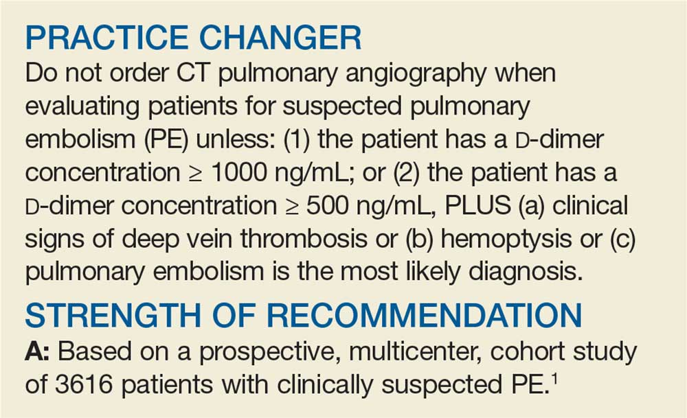
Penny E, a 48-year-old woman with a history of asthma, presents with wheezing and respiratory distress. There are no clinical signs of deep vein thrombosis or hemoptysis. PE is not your most likely diagnosis, but it is included in the differential, so you order a D
PE is the third most common type of cardiovascular disease after coronary artery disease and stroke, with an estimated incidence in the United States of 1-2/1000 individuals and a 30-day mortality rate between 10% and 30%.2 Improved adherence to a clinical decision support system has been shown to significantly decrease the number of diagnostic tests performed and the number of diagnostic failures.3
A diagnostic algorithm that includes the Wells criteria and a
Further, it is common for a
Three items of the original Wells criteria—clinical signs of deep vein thrombosis, hemoptysis, and whether PE is the most likely diagnosis—are the most predictive for PE.8 The development of a more efficient algorithm based on these 3 items that uses differential D
STUDY SUMMARY
Simplified algorithm diagnoses PE with fewer CTPAs
The YEARS study was a prospective cohort study conducted in 12 hospitals in the Netherlands that included 3616 patients with clinically suspected PE.1 A total of 151 patients met exclusion criteria (life expectancy < 3 months, ongoing anticoagulation treatment, pregnancy, and contraindication to CTPA). Investigators managed the remaining 3465 study patients according to the YEARS algorithm, which calls for obtaining a
PE was considered excluded if a patient had a
[polldaddy:10428150]
Continue to: Of the 1743 patients...
Of the 1743 patients who had none of the 3 YEARS items, 1320 had a D
Eighteen of the 2964 patients who had PE ruled out by the YEARS algorithm at baseline were found to have symptomatic VTE during the follow-up period (0.61%), with 6 patients (0.20%) sustaining a fatal PE. The 3-month incidence of VTE in patients who did not have CTPA was 0.43%, which is similar to the 0.34% reported in a previous meta-analysis of the Wells rule algorithm.13 Overall, fatal PE occurred in 0.3% of patients in the YEARS cohort vs 0.6% in a meta-analysis of studies using standard algorithms.14
Using an intention-to-diagnose analysis, 1611 (46%) patients did not have a CTPA indicated by the YEARS algorithm compared with 1174 (34%) using the Wells algorithm, for an absolute difference of 13% and estimated cost savings of $283,176 in this sample. The per-protocol analysis also had a decrease of CTPA examinations in favor of the YEARS algorithm, ruling out 1651 (48%) patients—a decrease of 14% and an estimated savings of $309,096.
WHAT’S NEW
High-level evidence says 14% fewer CTPAs
The YEARS study provides a high level of evidence that a new, simple diagnostic algorithm can reliably and efficiently exclude PE and decrease the need for CTPA by 14% (absolute difference) when compared with using the Wells rule and fixed
CAVEATS
No adjusting D -dimer for age
The YEARS criteria do not consider an age-adjusted
Continue to: CHALLENGES TO IMPLEMENTATION
CHALLENGES TO IMPLEMENTATION
None to speak of
We see no challenges to the implementation of this recommendation.
ACKNOWLEDGEMENT
The PURLs Surveillance System was supported in part by Grant Number UL1RR024999 from the National Center For Research Resources, a Clinical Translational Science Award to the University of Chicago. The content is solely the responsibility of the authors and does not necessarily represent the official views of the National Center For Research Resources or the National Institutes of Health.
Copyright © 2019. The Family Physicians Inquiries Network. All rights reserved.
Reprinted with permission from the Family Physicians Inquiries Network and The Journal of Family Practice (2019;68[5]:286-287,295).
1. van der Hulle T, Cheung WY, Kooij S, et al; YEARS study group. Simplified diagnostic management of suspected pulmonary embolism (the YEARS study): a prospective, multicentre, cohort study. Lancet. 2017;390:289-297.
2. Beckman MG, Hooper WC, Critchley SE, et al. Venous thromboembolism: a public health concern. Am J Prev Med. 2010;38(suppl 4):S495-S501.
3. Douma RA, Mos ICM, Erkens PMG, et al; Prometheus Study Group. Performance of 4 clinical decision rules in the diagnostic management of acute pulmonary embolism. Ann Intern Med. 2011;154:709-718.
4. van Es N, van der Hulle T, van Es J, et al. Wells Rule and d -dimer testing to rule out pulmonary embolism: a systematic review and individual-patient data meta-analysis. Ann Intern Med. 2016;165:253-261.
5. Roy P-M, Meyer G, Vielle B, et al; EMDEPU Study Group. Appropriateness of diagnostic management and outcomes of suspected pulmonary embolism. Ann Intern Med. 2006;144:157-164.
6. Newnham M, Stone H, Summerfield R, et al. Performance of algorithms and pre-test probability scores is often overlooked in the diagnosis of pulmonary embolism. BMJ. 2013;346:f1557.
7. Righini M, Van Es J, Den Exter PL, et al. Age-adjusted d -dimer cutoff levels to rule out pulmonary embolism. JAMA. 2014;311:1117-1124.
8. van Es J, Beenen LFM, Douma RA, et al. A simple decision rule including d -dimer to reduce the need for computed tomography scanning in patients with suspected pulmonary embolism. J Thromb Haemost. 2015;13:1428-1435.
9. Kooiman J, Klok FA, Mos ICM, et al. Incidence and predictors of contrast-induced nephropathy following CT-angiography for clinically suspected acute pulmonary embolism. J Thromb Haemost. 2010;8:409-411.
10. Sarma A, Heilbrun ME, Conner KE, et al. Radiation and chest CT scan examinations: what do we know? Chest. 2012;142:750-760.
11. Berrington de González A, Mahesh M, Kim KP, et al. Projected cancer risks from computed tomographic scans performed in the United States in 2007. Arch Intern Med. 2009;169:2071-2077.
12. Verma K, Legnani C, Palareti G. Cost-minimization analysis of venous thromboembolism diagnosis: comparison of standalone imaging with a strategy incorporating d -dimer for exclusion of venous thromboembolism. Res Pract Thromb Haemost. 2017;1:57-61.
13. Pasha SM, Klok FA, Snoep JD, et al. Safety of excluding acute pulmonary embolism based on an unlikely clinical probability by the Wells rule and normal d -dimer concentration: a meta-analysis. Thromb Res. 2010;125:e123-e127.
14. Mos ICM, Klok FA, Kroft LJM, et al. Safety of ruling out acute pulmonary embolism by normal computed tomography pulmonary angiography in patients with an indication for computed tomography: systematic review and meta-analysis. J Thromb Haemost. 2009;7:1491-1498.
Penny E, a 48-year-old woman with a history of asthma, presents with wheezing and respiratory distress. There are no clinical signs of deep vein thrombosis or hemoptysis. PE is not your most likely diagnosis, but it is included in the differential, so you order a D
PE is the third most common type of cardiovascular disease after coronary artery disease and stroke, with an estimated incidence in the United States of 1-2/1000 individuals and a 30-day mortality rate between 10% and 30%.2 Improved adherence to a clinical decision support system has been shown to significantly decrease the number of diagnostic tests performed and the number of diagnostic failures.3
A diagnostic algorithm that includes the Wells criteria and a
Further, it is common for a
Three items of the original Wells criteria—clinical signs of deep vein thrombosis, hemoptysis, and whether PE is the most likely diagnosis—are the most predictive for PE.8 The development of a more efficient algorithm based on these 3 items that uses differential D
STUDY SUMMARY
Simplified algorithm diagnoses PE with fewer CTPAs
The YEARS study was a prospective cohort study conducted in 12 hospitals in the Netherlands that included 3616 patients with clinically suspected PE.1 A total of 151 patients met exclusion criteria (life expectancy < 3 months, ongoing anticoagulation treatment, pregnancy, and contraindication to CTPA). Investigators managed the remaining 3465 study patients according to the YEARS algorithm, which calls for obtaining a
PE was considered excluded if a patient had a
[polldaddy:10428150]
Continue to: Of the 1743 patients...
Of the 1743 patients who had none of the 3 YEARS items, 1320 had a D
Eighteen of the 2964 patients who had PE ruled out by the YEARS algorithm at baseline were found to have symptomatic VTE during the follow-up period (0.61%), with 6 patients (0.20%) sustaining a fatal PE. The 3-month incidence of VTE in patients who did not have CTPA was 0.43%, which is similar to the 0.34% reported in a previous meta-analysis of the Wells rule algorithm.13 Overall, fatal PE occurred in 0.3% of patients in the YEARS cohort vs 0.6% in a meta-analysis of studies using standard algorithms.14
Using an intention-to-diagnose analysis, 1611 (46%) patients did not have a CTPA indicated by the YEARS algorithm compared with 1174 (34%) using the Wells algorithm, for an absolute difference of 13% and estimated cost savings of $283,176 in this sample. The per-protocol analysis also had a decrease of CTPA examinations in favor of the YEARS algorithm, ruling out 1651 (48%) patients—a decrease of 14% and an estimated savings of $309,096.
WHAT’S NEW
High-level evidence says 14% fewer CTPAs
The YEARS study provides a high level of evidence that a new, simple diagnostic algorithm can reliably and efficiently exclude PE and decrease the need for CTPA by 14% (absolute difference) when compared with using the Wells rule and fixed
CAVEATS
No adjusting D -dimer for age
The YEARS criteria do not consider an age-adjusted
Continue to: CHALLENGES TO IMPLEMENTATION
CHALLENGES TO IMPLEMENTATION
None to speak of
We see no challenges to the implementation of this recommendation.
ACKNOWLEDGEMENT
The PURLs Surveillance System was supported in part by Grant Number UL1RR024999 from the National Center For Research Resources, a Clinical Translational Science Award to the University of Chicago. The content is solely the responsibility of the authors and does not necessarily represent the official views of the National Center For Research Resources or the National Institutes of Health.
Copyright © 2019. The Family Physicians Inquiries Network. All rights reserved.
Reprinted with permission from the Family Physicians Inquiries Network and The Journal of Family Practice (2019;68[5]:286-287,295).
Penny E, a 48-year-old woman with a history of asthma, presents with wheezing and respiratory distress. There are no clinical signs of deep vein thrombosis or hemoptysis. PE is not your most likely diagnosis, but it is included in the differential, so you order a D
PE is the third most common type of cardiovascular disease after coronary artery disease and stroke, with an estimated incidence in the United States of 1-2/1000 individuals and a 30-day mortality rate between 10% and 30%.2 Improved adherence to a clinical decision support system has been shown to significantly decrease the number of diagnostic tests performed and the number of diagnostic failures.3
A diagnostic algorithm that includes the Wells criteria and a
Further, it is common for a
Three items of the original Wells criteria—clinical signs of deep vein thrombosis, hemoptysis, and whether PE is the most likely diagnosis—are the most predictive for PE.8 The development of a more efficient algorithm based on these 3 items that uses differential D
STUDY SUMMARY
Simplified algorithm diagnoses PE with fewer CTPAs
The YEARS study was a prospective cohort study conducted in 12 hospitals in the Netherlands that included 3616 patients with clinically suspected PE.1 A total of 151 patients met exclusion criteria (life expectancy < 3 months, ongoing anticoagulation treatment, pregnancy, and contraindication to CTPA). Investigators managed the remaining 3465 study patients according to the YEARS algorithm, which calls for obtaining a
PE was considered excluded if a patient had a
[polldaddy:10428150]
Continue to: Of the 1743 patients...
Of the 1743 patients who had none of the 3 YEARS items, 1320 had a D
Eighteen of the 2964 patients who had PE ruled out by the YEARS algorithm at baseline were found to have symptomatic VTE during the follow-up period (0.61%), with 6 patients (0.20%) sustaining a fatal PE. The 3-month incidence of VTE in patients who did not have CTPA was 0.43%, which is similar to the 0.34% reported in a previous meta-analysis of the Wells rule algorithm.13 Overall, fatal PE occurred in 0.3% of patients in the YEARS cohort vs 0.6% in a meta-analysis of studies using standard algorithms.14
Using an intention-to-diagnose analysis, 1611 (46%) patients did not have a CTPA indicated by the YEARS algorithm compared with 1174 (34%) using the Wells algorithm, for an absolute difference of 13% and estimated cost savings of $283,176 in this sample. The per-protocol analysis also had a decrease of CTPA examinations in favor of the YEARS algorithm, ruling out 1651 (48%) patients—a decrease of 14% and an estimated savings of $309,096.
WHAT’S NEW
High-level evidence says 14% fewer CTPAs
The YEARS study provides a high level of evidence that a new, simple diagnostic algorithm can reliably and efficiently exclude PE and decrease the need for CTPA by 14% (absolute difference) when compared with using the Wells rule and fixed
CAVEATS
No adjusting D -dimer for age
The YEARS criteria do not consider an age-adjusted
Continue to: CHALLENGES TO IMPLEMENTATION
CHALLENGES TO IMPLEMENTATION
None to speak of
We see no challenges to the implementation of this recommendation.
ACKNOWLEDGEMENT
The PURLs Surveillance System was supported in part by Grant Number UL1RR024999 from the National Center For Research Resources, a Clinical Translational Science Award to the University of Chicago. The content is solely the responsibility of the authors and does not necessarily represent the official views of the National Center For Research Resources or the National Institutes of Health.
Copyright © 2019. The Family Physicians Inquiries Network. All rights reserved.
Reprinted with permission from the Family Physicians Inquiries Network and The Journal of Family Practice (2019;68[5]:286-287,295).
1. van der Hulle T, Cheung WY, Kooij S, et al; YEARS study group. Simplified diagnostic management of suspected pulmonary embolism (the YEARS study): a prospective, multicentre, cohort study. Lancet. 2017;390:289-297.
2. Beckman MG, Hooper WC, Critchley SE, et al. Venous thromboembolism: a public health concern. Am J Prev Med. 2010;38(suppl 4):S495-S501.
3. Douma RA, Mos ICM, Erkens PMG, et al; Prometheus Study Group. Performance of 4 clinical decision rules in the diagnostic management of acute pulmonary embolism. Ann Intern Med. 2011;154:709-718.
4. van Es N, van der Hulle T, van Es J, et al. Wells Rule and d -dimer testing to rule out pulmonary embolism: a systematic review and individual-patient data meta-analysis. Ann Intern Med. 2016;165:253-261.
5. Roy P-M, Meyer G, Vielle B, et al; EMDEPU Study Group. Appropriateness of diagnostic management and outcomes of suspected pulmonary embolism. Ann Intern Med. 2006;144:157-164.
6. Newnham M, Stone H, Summerfield R, et al. Performance of algorithms and pre-test probability scores is often overlooked in the diagnosis of pulmonary embolism. BMJ. 2013;346:f1557.
7. Righini M, Van Es J, Den Exter PL, et al. Age-adjusted d -dimer cutoff levels to rule out pulmonary embolism. JAMA. 2014;311:1117-1124.
8. van Es J, Beenen LFM, Douma RA, et al. A simple decision rule including d -dimer to reduce the need for computed tomography scanning in patients with suspected pulmonary embolism. J Thromb Haemost. 2015;13:1428-1435.
9. Kooiman J, Klok FA, Mos ICM, et al. Incidence and predictors of contrast-induced nephropathy following CT-angiography for clinically suspected acute pulmonary embolism. J Thromb Haemost. 2010;8:409-411.
10. Sarma A, Heilbrun ME, Conner KE, et al. Radiation and chest CT scan examinations: what do we know? Chest. 2012;142:750-760.
11. Berrington de González A, Mahesh M, Kim KP, et al. Projected cancer risks from computed tomographic scans performed in the United States in 2007. Arch Intern Med. 2009;169:2071-2077.
12. Verma K, Legnani C, Palareti G. Cost-minimization analysis of venous thromboembolism diagnosis: comparison of standalone imaging with a strategy incorporating d -dimer for exclusion of venous thromboembolism. Res Pract Thromb Haemost. 2017;1:57-61.
13. Pasha SM, Klok FA, Snoep JD, et al. Safety of excluding acute pulmonary embolism based on an unlikely clinical probability by the Wells rule and normal d -dimer concentration: a meta-analysis. Thromb Res. 2010;125:e123-e127.
14. Mos ICM, Klok FA, Kroft LJM, et al. Safety of ruling out acute pulmonary embolism by normal computed tomography pulmonary angiography in patients with an indication for computed tomography: systematic review and meta-analysis. J Thromb Haemost. 2009;7:1491-1498.
1. van der Hulle T, Cheung WY, Kooij S, et al; YEARS study group. Simplified diagnostic management of suspected pulmonary embolism (the YEARS study): a prospective, multicentre, cohort study. Lancet. 2017;390:289-297.
2. Beckman MG, Hooper WC, Critchley SE, et al. Venous thromboembolism: a public health concern. Am J Prev Med. 2010;38(suppl 4):S495-S501.
3. Douma RA, Mos ICM, Erkens PMG, et al; Prometheus Study Group. Performance of 4 clinical decision rules in the diagnostic management of acute pulmonary embolism. Ann Intern Med. 2011;154:709-718.
4. van Es N, van der Hulle T, van Es J, et al. Wells Rule and d -dimer testing to rule out pulmonary embolism: a systematic review and individual-patient data meta-analysis. Ann Intern Med. 2016;165:253-261.
5. Roy P-M, Meyer G, Vielle B, et al; EMDEPU Study Group. Appropriateness of diagnostic management and outcomes of suspected pulmonary embolism. Ann Intern Med. 2006;144:157-164.
6. Newnham M, Stone H, Summerfield R, et al. Performance of algorithms and pre-test probability scores is often overlooked in the diagnosis of pulmonary embolism. BMJ. 2013;346:f1557.
7. Righini M, Van Es J, Den Exter PL, et al. Age-adjusted d -dimer cutoff levels to rule out pulmonary embolism. JAMA. 2014;311:1117-1124.
8. van Es J, Beenen LFM, Douma RA, et al. A simple decision rule including d -dimer to reduce the need for computed tomography scanning in patients with suspected pulmonary embolism. J Thromb Haemost. 2015;13:1428-1435.
9. Kooiman J, Klok FA, Mos ICM, et al. Incidence and predictors of contrast-induced nephropathy following CT-angiography for clinically suspected acute pulmonary embolism. J Thromb Haemost. 2010;8:409-411.
10. Sarma A, Heilbrun ME, Conner KE, et al. Radiation and chest CT scan examinations: what do we know? Chest. 2012;142:750-760.
11. Berrington de González A, Mahesh M, Kim KP, et al. Projected cancer risks from computed tomographic scans performed in the United States in 2007. Arch Intern Med. 2009;169:2071-2077.
12. Verma K, Legnani C, Palareti G. Cost-minimization analysis of venous thromboembolism diagnosis: comparison of standalone imaging with a strategy incorporating d -dimer for exclusion of venous thromboembolism. Res Pract Thromb Haemost. 2017;1:57-61.
13. Pasha SM, Klok FA, Snoep JD, et al. Safety of excluding acute pulmonary embolism based on an unlikely clinical probability by the Wells rule and normal d -dimer concentration: a meta-analysis. Thromb Res. 2010;125:e123-e127.
14. Mos ICM, Klok FA, Kroft LJM, et al. Safety of ruling out acute pulmonary embolism by normal computed tomography pulmonary angiography in patients with an indication for computed tomography: systematic review and meta-analysis. J Thromb Haemost. 2009;7:1491-1498.

PRIMA study: Niraparib maintenance improves PFS in advanced OC
BARCELONA – Niraparib significantly improves progression-free survival when given after first-line chemotherapy in patients with advanced ovarian cancer, according to “potentially practice-changing” results from the phase 3 PRIMA/ENGOT-OV26/GOG-3012 study.

Overall progression-free survival (PFS) in 484 patients randomized to receive the poly-ADP ribose polymerase inhibitor (PARPi) niraparib was 13.8 months, compared with 8.2 months in 244 patients who received placebo (hazard ratio, 0.62), Antonio González-Martin, MD, PhD, reported at the European Society for Medical Oncology Congress.
The findings were published simultaneously online in the New England Journal of Medicine (N Engl J Med. 2019 Sep 28. doi: 10.1056/NEJMoa1910962).
In patients at high risk for progression based on homologous recombination deficiency (HRd) – defined by certain tumor factors or the presence of BRCA mutation (BRCAm), PFS was 21.9 vs. 10.4 months in the treatment (n = 245) vs. placebo (n = 125) groups, respectively (HR, 0.43), said Dr. González-Martin of Grupo Español de Investigación en Cáncer de Ovario (GEICO), medical oncology department, Clínica Universidad de Navarra, Madrid.
“At 18 months, which means approximately 2 years after the initiation of chemotherapy, 42% of patients treated with niraparib remained alive and progression free,” he said, adding that 59% of the HRd patients remained alive and progression free at 18 months.
Exploratory analyses showed that the niraparib benefits occurred across all prespecified patient subgroups, including those aged 65 and older vs. those under age 65, those with stage III vs. stage IV disease at diagnosis, those receiving vs. not receiving neoadjuvant chemotherapy, those with complete response (CR) vs. partial response (PR) as their best response to platinum chemotherapy, and those with HRd who had BRCAm vs. BRCA wild type (BRCAwt) tumors, he said.
The hazard ratios for the HRd BRCAm vs. BRCAwt tumors were 0.40 and 0.50, respectively.
“So the benefit of niraparib in the HRd tumor is not driven only by the BRCA-mutated patients,” he said. “Importantly, we also saw benefit in the group of patients with tumors that were [homologous recombination] proficient (HRp), with a reduction in the risk of progression of 32%.”
For the key secondary endpoint of overall survival, a preplanned interim analysis showed that 84% vs. 77% in the niraparib and placebo groups, respectively, were alive at 2 years; in the HRd and HRp groups, those rates were 91% vs. 85% and 81% vs. 59%, respectively.
Participants in the double-blind trial had newly diagnosed, advanced high-grade serous or endometrioid ovarian, primary peritoneal, or fallopian tube cancer; their mean age was 62 years; and they had experienced a CR (69%) or PR (31%) to first-line platinum-based chemotherapy. Overall, 35% had stage IV disease and 67% received neoadjuvant chemotherapy. They were randomized 2:1 to once-daily niraparib at a starting dose of 300 mg or 200 mg depending on body weight and platelet count, with those weighing 77 kg or greater and with platelet count of 150,000/mcL or less starting at the higher dose, and those weighing less than 77 kg and/or with platelet count less than 150,000/mcL starting at the lower dose.
All subgroups showed a sustained and durable treatment effect, and although most patients experienced treatment-related adverse events (TRAEs), those were “manageable with dose interruption or dose reduction,” Dr. González-Martin said.
Discontinuations due to TRAEs occurred in 12% vs. 2.5% in the treatment vs. placebo groups, and this was consistent with prior niraparib experience, he said, adding that no niraparib-related deaths were reported and no new safety signals were identified.
The findings are notable, because the recurrence rate after standard first-line platinum-based chemotherapy in women with advanced ovarian cancer is estimated at up to 85%, and while certain subgroups of patients have options for maintenance therapy, there remains a high unmet need for others, he explained.
For example olaparib is an option, but only for tumors with BRCA mutation, and bevacizumab can be used, but “may be limited due to safety concerns in some patients and also due to limited data from randomized trials in the neoadjuvant setting,” he said.
As a result, surveillance after chemotherapy is the approach used for many patients, he added.
Niraparib is the first oral PARPi approved for maintenance in patients with recurrent ovarian cancer, regardless of BRCA mutation status; in the NOVA study, it demonstrated efficacy after platinum chemotherapy in all biomarker populations, and in the QUADRA study it showed benefit in patients who received at least three prior therapies.
The current study was designed to test the efficacy and safety of niraparib therapy after response to platinum-based chemotherapy in patients with newly diagnosed advanced ovarian cancer, including those at high risk of relapse.
“Niraparib is the first PARP inhibitor that has demonstrated benefit after front-line platinum-based chemotherapy across all the biomarker subgroups, regardless of BRCA status, consistent with data from the recurrent setting,” Dr. González-Martin said, adding that patients with ovarian cancer at the highest risk of early disease progression obtained significant benefit. “What does this mean for our patients and our practice? Based on these results, niraparib after first-line platinum chemotherapy should be considered a new standard of care.”
Invited discussant Ana Oaknin, MD, PhD, head of the gynecologic cancer program at Vall d’Hebron Institute of Oncology, Vall d’Hebron University Hospital, Barcelona, called the findings “striking” and noted that they, along with those from the PAOLA-1/ENGOT-Ov25 trial demonstrating a PFS benefit with the addition of olaparib to bevacizumab maintenance therapy after first-line platinum-based chemotherapy in advanced ovarian cancer, represent important advances.
“We are witnessing a paradigm shift in the first-line treatment of advanced ovarian cancer patients,” she said.
Are the findings of these trials clinically meaningful enough to justify the addition of PARPi maintenance therapy after first-line chemotherapy therapy as a new standard of care?
“Yes, but while the benefit is clinically meaningful in the overall population, we should consider PFS outcomes according to the biomarker status in the selection of optimal therapy; companion diagnostic tests will be needed,” she said.
The PRIMA/ENGOT-OV26/GOG-3012 study was sponsored by TESARO. Dr. González-Martin reported relationships with numerous pharmaceutical companies.
SOURCE: González-Martin A et al. ESMO 2019: Abstract LBA1.
BARCELONA – Niraparib significantly improves progression-free survival when given after first-line chemotherapy in patients with advanced ovarian cancer, according to “potentially practice-changing” results from the phase 3 PRIMA/ENGOT-OV26/GOG-3012 study.
Overall progression-free survival (PFS) in 484 patients randomized to receive the poly-ADP ribose polymerase inhibitor (PARPi) niraparib was 13.8 months, compared with 8.2 months in 244 patients who received placebo (hazard ratio, 0.62), Antonio González-Martin, MD, PhD, reported at the European Society for Medical Oncology Congress.
The findings were published simultaneously online in the New England Journal of Medicine (N Engl J Med. 2019 Sep 28. doi: 10.1056/NEJMoa1910962).
In patients at high risk for progression based on homologous recombination deficiency (HRd) – defined by certain tumor factors or the presence of BRCA mutation (BRCAm), PFS was 21.9 vs. 10.4 months in the treatment (n = 245) vs. placebo (n = 125) groups, respectively (HR, 0.43), said Dr. González-Martin of Grupo Español de Investigación en Cáncer de Ovario (GEICO), medical oncology department, Clínica Universidad de Navarra, Madrid.
“At 18 months, which means approximately 2 years after the initiation of chemotherapy, 42% of patients treated with niraparib remained alive and progression free,” he said, adding that 59% of the HRd patients remained alive and progression free at 18 months.
Exploratory analyses showed that the niraparib benefits occurred across all prespecified patient subgroups, including those aged 65 and older vs. those under age 65, those with stage III vs. stage IV disease at diagnosis, those receiving vs. not receiving neoadjuvant chemotherapy, those with complete response (CR) vs. partial response (PR) as their best response to platinum chemotherapy, and those with HRd who had BRCAm vs. BRCA wild type (BRCAwt) tumors, he said.
The hazard ratios for the HRd BRCAm vs. BRCAwt tumors were 0.40 and 0.50, respectively.
“So the benefit of niraparib in the HRd tumor is not driven only by the BRCA-mutated patients,” he said. “Importantly, we also saw benefit in the group of patients with tumors that were [homologous recombination] proficient (HRp), with a reduction in the risk of progression of 32%.”
For the key secondary endpoint of overall survival, a preplanned interim analysis showed that 84% vs. 77% in the niraparib and placebo groups, respectively, were alive at 2 years; in the HRd and HRp groups, those rates were 91% vs. 85% and 81% vs. 59%, respectively.
Participants in the double-blind trial had newly diagnosed, advanced high-grade serous or endometrioid ovarian, primary peritoneal, or fallopian tube cancer; their mean age was 62 years; and they had experienced a CR (69%) or PR (31%) to first-line platinum-based chemotherapy. Overall, 35% had stage IV disease and 67% received neoadjuvant chemotherapy. They were randomized 2:1 to once-daily niraparib at a starting dose of 300 mg or 200 mg depending on body weight and platelet count, with those weighing 77 kg or greater and with platelet count of 150,000/mcL or less starting at the higher dose, and those weighing less than 77 kg and/or with platelet count less than 150,000/mcL starting at the lower dose.
All subgroups showed a sustained and durable treatment effect, and although most patients experienced treatment-related adverse events (TRAEs), those were “manageable with dose interruption or dose reduction,” Dr. González-Martin said.
Discontinuations due to TRAEs occurred in 12% vs. 2.5% in the treatment vs. placebo groups, and this was consistent with prior niraparib experience, he said, adding that no niraparib-related deaths were reported and no new safety signals were identified.
The findings are notable, because the recurrence rate after standard first-line platinum-based chemotherapy in women with advanced ovarian cancer is estimated at up to 85%, and while certain subgroups of patients have options for maintenance therapy, there remains a high unmet need for others, he explained.
For example olaparib is an option, but only for tumors with BRCA mutation, and bevacizumab can be used, but “may be limited due to safety concerns in some patients and also due to limited data from randomized trials in the neoadjuvant setting,” he said.
As a result, surveillance after chemotherapy is the approach used for many patients, he added.
Niraparib is the first oral PARPi approved for maintenance in patients with recurrent ovarian cancer, regardless of BRCA mutation status; in the NOVA study, it demonstrated efficacy after platinum chemotherapy in all biomarker populations, and in the QUADRA study it showed benefit in patients who received at least three prior therapies.
The current study was designed to test the efficacy and safety of niraparib therapy after response to platinum-based chemotherapy in patients with newly diagnosed advanced ovarian cancer, including those at high risk of relapse.
“Niraparib is the first PARP inhibitor that has demonstrated benefit after front-line platinum-based chemotherapy across all the biomarker subgroups, regardless of BRCA status, consistent with data from the recurrent setting,” Dr. González-Martin said, adding that patients with ovarian cancer at the highest risk of early disease progression obtained significant benefit. “What does this mean for our patients and our practice? Based on these results, niraparib after first-line platinum chemotherapy should be considered a new standard of care.”
Invited discussant Ana Oaknin, MD, PhD, head of the gynecologic cancer program at Vall d’Hebron Institute of Oncology, Vall d’Hebron University Hospital, Barcelona, called the findings “striking” and noted that they, along with those from the PAOLA-1/ENGOT-Ov25 trial demonstrating a PFS benefit with the addition of olaparib to bevacizumab maintenance therapy after first-line platinum-based chemotherapy in advanced ovarian cancer, represent important advances.
“We are witnessing a paradigm shift in the first-line treatment of advanced ovarian cancer patients,” she said.
Are the findings of these trials clinically meaningful enough to justify the addition of PARPi maintenance therapy after first-line chemotherapy therapy as a new standard of care?
“Yes, but while the benefit is clinically meaningful in the overall population, we should consider PFS outcomes according to the biomarker status in the selection of optimal therapy; companion diagnostic tests will be needed,” she said.
The PRIMA/ENGOT-OV26/GOG-3012 study was sponsored by TESARO. Dr. González-Martin reported relationships with numerous pharmaceutical companies.
SOURCE: González-Martin A et al. ESMO 2019: Abstract LBA1.
BARCELONA – Niraparib significantly improves progression-free survival when given after first-line chemotherapy in patients with advanced ovarian cancer, according to “potentially practice-changing” results from the phase 3 PRIMA/ENGOT-OV26/GOG-3012 study.
Overall progression-free survival (PFS) in 484 patients randomized to receive the poly-ADP ribose polymerase inhibitor (PARPi) niraparib was 13.8 months, compared with 8.2 months in 244 patients who received placebo (hazard ratio, 0.62), Antonio González-Martin, MD, PhD, reported at the European Society for Medical Oncology Congress.
The findings were published simultaneously online in the New England Journal of Medicine (N Engl J Med. 2019 Sep 28. doi: 10.1056/NEJMoa1910962).
In patients at high risk for progression based on homologous recombination deficiency (HRd) – defined by certain tumor factors or the presence of BRCA mutation (BRCAm), PFS was 21.9 vs. 10.4 months in the treatment (n = 245) vs. placebo (n = 125) groups, respectively (HR, 0.43), said Dr. González-Martin of Grupo Español de Investigación en Cáncer de Ovario (GEICO), medical oncology department, Clínica Universidad de Navarra, Madrid.
“At 18 months, which means approximately 2 years after the initiation of chemotherapy, 42% of patients treated with niraparib remained alive and progression free,” he said, adding that 59% of the HRd patients remained alive and progression free at 18 months.
Exploratory analyses showed that the niraparib benefits occurred across all prespecified patient subgroups, including those aged 65 and older vs. those under age 65, those with stage III vs. stage IV disease at diagnosis, those receiving vs. not receiving neoadjuvant chemotherapy, those with complete response (CR) vs. partial response (PR) as their best response to platinum chemotherapy, and those with HRd who had BRCAm vs. BRCA wild type (BRCAwt) tumors, he said.
The hazard ratios for the HRd BRCAm vs. BRCAwt tumors were 0.40 and 0.50, respectively.
“So the benefit of niraparib in the HRd tumor is not driven only by the BRCA-mutated patients,” he said. “Importantly, we also saw benefit in the group of patients with tumors that were [homologous recombination] proficient (HRp), with a reduction in the risk of progression of 32%.”
For the key secondary endpoint of overall survival, a preplanned interim analysis showed that 84% vs. 77% in the niraparib and placebo groups, respectively, were alive at 2 years; in the HRd and HRp groups, those rates were 91% vs. 85% and 81% vs. 59%, respectively.
Participants in the double-blind trial had newly diagnosed, advanced high-grade serous or endometrioid ovarian, primary peritoneal, or fallopian tube cancer; their mean age was 62 years; and they had experienced a CR (69%) or PR (31%) to first-line platinum-based chemotherapy. Overall, 35% had stage IV disease and 67% received neoadjuvant chemotherapy. They were randomized 2:1 to once-daily niraparib at a starting dose of 300 mg or 200 mg depending on body weight and platelet count, with those weighing 77 kg or greater and with platelet count of 150,000/mcL or less starting at the higher dose, and those weighing less than 77 kg and/or with platelet count less than 150,000/mcL starting at the lower dose.
All subgroups showed a sustained and durable treatment effect, and although most patients experienced treatment-related adverse events (TRAEs), those were “manageable with dose interruption or dose reduction,” Dr. González-Martin said.
Discontinuations due to TRAEs occurred in 12% vs. 2.5% in the treatment vs. placebo groups, and this was consistent with prior niraparib experience, he said, adding that no niraparib-related deaths were reported and no new safety signals were identified.
The findings are notable, because the recurrence rate after standard first-line platinum-based chemotherapy in women with advanced ovarian cancer is estimated at up to 85%, and while certain subgroups of patients have options for maintenance therapy, there remains a high unmet need for others, he explained.
For example olaparib is an option, but only for tumors with BRCA mutation, and bevacizumab can be used, but “may be limited due to safety concerns in some patients and also due to limited data from randomized trials in the neoadjuvant setting,” he said.
As a result, surveillance after chemotherapy is the approach used for many patients, he added.
Niraparib is the first oral PARPi approved for maintenance in patients with recurrent ovarian cancer, regardless of BRCA mutation status; in the NOVA study, it demonstrated efficacy after platinum chemotherapy in all biomarker populations, and in the QUADRA study it showed benefit in patients who received at least three prior therapies.
The current study was designed to test the efficacy and safety of niraparib therapy after response to platinum-based chemotherapy in patients with newly diagnosed advanced ovarian cancer, including those at high risk of relapse.
“Niraparib is the first PARP inhibitor that has demonstrated benefit after front-line platinum-based chemotherapy across all the biomarker subgroups, regardless of BRCA status, consistent with data from the recurrent setting,” Dr. González-Martin said, adding that patients with ovarian cancer at the highest risk of early disease progression obtained significant benefit. “What does this mean for our patients and our practice? Based on these results, niraparib after first-line platinum chemotherapy should be considered a new standard of care.”
Invited discussant Ana Oaknin, MD, PhD, head of the gynecologic cancer program at Vall d’Hebron Institute of Oncology, Vall d’Hebron University Hospital, Barcelona, called the findings “striking” and noted that they, along with those from the PAOLA-1/ENGOT-Ov25 trial demonstrating a PFS benefit with the addition of olaparib to bevacizumab maintenance therapy after first-line platinum-based chemotherapy in advanced ovarian cancer, represent important advances.
“We are witnessing a paradigm shift in the first-line treatment of advanced ovarian cancer patients,” she said.
Are the findings of these trials clinically meaningful enough to justify the addition of PARPi maintenance therapy after first-line chemotherapy therapy as a new standard of care?
“Yes, but while the benefit is clinically meaningful in the overall population, we should consider PFS outcomes according to the biomarker status in the selection of optimal therapy; companion diagnostic tests will be needed,” she said.
The PRIMA/ENGOT-OV26/GOG-3012 study was sponsored by TESARO. Dr. González-Martin reported relationships with numerous pharmaceutical companies.
SOURCE: González-Martin A et al. ESMO 2019: Abstract LBA1.
REPORTING FROM ESMO 2019

Larval Tick Infestation Causing an Eruption of Pruritic Papules and Pustules
Case Reports
Patient 1
A 65-year-old woman presented to the dermatology clinic in July with a pruritic rash of 2 days’ duration that started on the back and spread diffusely. The patient gardened regularly. Physical examination showed inflammatory papules and pustules on the back (Figure 1), as well as the groin, breasts, and ears. There was a punctate black dot in the center of some papules, and dermoscopy revealed ticks (Figure 2). Removal and microscopic examination confirmed larval-stage lone star ticks (Figure 3). The patient was prescribed topical steroids for pruritus as well as oral doxycycline for prophylaxis against tick-borne illnesses.



Patient 2
A 54-year-old man presented to the same clinic in July with pruritic lesions on the back, legs, ankles, and scrotum of 3 days’ duration that first appeared 24 hours after performing yardwork. Physical examination revealed diffusely distributed papules, pustules, and vesicles on the back (Figure 4). Some papules featured a punctate black dot in the center (similar to patient 1), and dermoscopy again revealed ticks. Removal and microscopic examination confirmed larval-stage ticks. The patient was treated with topical steroids and oral antihistamines for pruritus as well as prophylactic oral doxycycline.

Comment
Ticks are well-known human parasites, representing the second most common vector of human infectious disease.1 Ticks have 3 motile stages: larva (or “seed”), nymph, and adult. They can bite humans during all stages. Larval ticks, distinguished by having 6 legs rather than 8 legs in nymphs and adults, can attack in droves and cause an infestation that presents as diffuse, pruritic, erythematous papules and pustules.2-4 The first report of larval tick infestation in humans may have been in 1728 by William Byrd who described finding ticks on the skin that were too small to see without a microscope.5
Identification
The ticks in both of our cases were lone star ticks (Amblyomma americanum). The larval stage of A americanum is a proven cause of cutaneous reaction.6,7 A PubMed search of articles indexed for MEDLINE as well as a Google Scholar search using the terms tick, seed tick, or tick bite in combination with rash, eruption, infestation, papule, pustule, or pruritic revealed 6 reported cases of larval tick infestation in the literature (including our case); 5 were caused by A americanum and 1 by Ixodes dammini (now known as Ixodes scapularis); all occurred in July or August.3,7-10 This time frame is consistent with the general tick life cycle across species: Adults feed from April to June, then lay eggs that hatch into larval ticks within 4 to 6 weeks. After hatching, larval ticks climb grass and weeds awaiting a passing host.4
Diagnosis
Larval tick infestation remains a frequently misdiagnosed etiology of diffuse pruritic papules and pustules, especially in urban settings where physicians are less likely to be familiar with this type of manifestation.3,9-11 Larval ticks are submillimeter in size and difficult to appreciate with the naked eye, contributing to misdiagnosis. A punctate black dot may sometimes be seen in papules; however, dermoscopy is critical for accurate diagnosis, as hemorrhagic crust is a frequent misdiagnosis.
Management
In addition to symptomatic therapy, both of our patients received doxycycline as antibiotic prophylaxis for tick-borne illnesses given that a high number of ticks had been attached for more than 2 days.12,13 Antibiotic prophylaxis for tick-borne illness is controversial. The exception is Lyme disease transmitted by nymphal or adult I scapularis when specific conditions are met: the bite must have occurred in an endemic area, doxycycline cannot be contraindicated, estimated duration of attachment is at least 36 hours, and prophylaxis must be started within 72 hours of tick removal.13 There are no official recommendations for the A americanum species or for larval-stage ticks of any species. Larval-stage ticks acting as vectors for disease transmission is not well documented in recent literature, and there currently is limited evidence supporting prophylactic antibiotics for larval tick bites. The presence of spotted fever rickettsioses has been reported (with the exception of Rickettsia rickettsii and Ehrlichia chaffeensis) in larval A americanum ticks, suggesting a theoretical possibility that they could act as disease vectors.3,8,11,14-17 At a minimum, both prompt tick removal and close patient follow-up is warranted.
Conclusion
Human infestation with larval ticks is a common occurrence but can present a diagnostic challenge to an unfamiliar physician. We encourage consideration of larval tick infestation as the etiology of multiple or diffuse pruritic papules with a history of outdoor exposure.
- Sonenshine DE. Biology of Ticks. New York, NY: Oxford University; 1991.
- Alexander JOD. The effects of tick bites. In: Alexander JOD. Arthropods and Human Skin. London, England: Springer London; 1984:363-382.
- Duckworth PF Jr, Hayden GF, Reed CN. Human infestation by Amblyomma americanum larvae (“seed ticks”). South Med J. 1985;78:751-753.
- Parola P, Raoult D. Ticks and tickborne bacterial diseases in humans: an emerging infectious threat. Clin Infect Dis. 2001;32:897-928.
- Cropley TG. William Byrd on ticks, 1728. Arch Dermatol. 2009;145:187.
- Goddard J. A ten-year study of tick biting in Mississippi: implications for human disease transmission. J Agromedicine. 2002;8:25-32.
- Goddard J, Portugal JS. Cutaneous lesions due to bites by larval Amblyomma americanum ticks. JAMA Dermatol. 2015;151:1373-1375.
- Fibeger EA, Erickson QL, Weintraub BD, et al. Larval tick infestation: a case report and review of tick-borne disease. Cutis. 2008;82:38-46.
- Jones BE. Human ‘seed tick’ infestation: Amblyomma americanum larvae. Arch Dermatol. 1981;117:812-814.
- Fisher EJ, Mo J, Lucky AW. Multiple pruritic papules from lone star tick larvae bites. Arch Dermatol. 2006;142:491-494.
- Culp JS. Seed ticks. Am Fam Physician. 1987;36:121-123.
- Perea AE, Hinckley AF, Mead PS. Tick bite prophylaxis: results from a 2012 survey of healthcare providers. Zoonoses Public Health. 2015;62:388-392.
- Tick bites/prevention. Centers for Disease Control and Prevention website. https://www.cdc.gov/ticks/tickbornediseases/tick-bites-prevention.html. Revised January 10, 2019. Accessed September 17, 2019.
- Moncayo AC, Cohen SB, Fritzen CM, et al. Absence of Rickettsia rickettsii and occurrence of other spotted fever group rickettsiae in ticks from Tennessee. Am J Trop Med Hyg. 2010;83:653-657.
- Castellaw AH, Showers J, Goddard J, et al. Detection of vector-borne agents in lone star ticks, Amblyomma americanum (Acari: Ixodidae), from Mississippi. J Med Entomol. 2010;47:473-476.
- Stromdahl EY, Vince MA, Billingsley PM, et al. Rickettsia amblyommii infecting Amblyomma americanum larvae. Vector Borne Zoonotic Dis. 2008;8:15-24.
- Long SW, Zhang X, Zhang J, et al. Evaluation of transovarial transmission and transmissibility of Ehrlichia chaffeensis (Rickettsiales: Anaplasmataceae) in Amblyomma americanum (Acari: Ixodidae). J Med Entomol. 2003;40:1000-1004.
Case Reports
Patient 1
A 65-year-old woman presented to the dermatology clinic in July with a pruritic rash of 2 days’ duration that started on the back and spread diffusely. The patient gardened regularly. Physical examination showed inflammatory papules and pustules on the back (Figure 1), as well as the groin, breasts, and ears. There was a punctate black dot in the center of some papules, and dermoscopy revealed ticks (Figure 2). Removal and microscopic examination confirmed larval-stage lone star ticks (Figure 3). The patient was prescribed topical steroids for pruritus as well as oral doxycycline for prophylaxis against tick-borne illnesses.
Patient 2
A 54-year-old man presented to the same clinic in July with pruritic lesions on the back, legs, ankles, and scrotum of 3 days’ duration that first appeared 24 hours after performing yardwork. Physical examination revealed diffusely distributed papules, pustules, and vesicles on the back (Figure 4). Some papules featured a punctate black dot in the center (similar to patient 1), and dermoscopy again revealed ticks. Removal and microscopic examination confirmed larval-stage ticks. The patient was treated with topical steroids and oral antihistamines for pruritus as well as prophylactic oral doxycycline.
Comment
Ticks are well-known human parasites, representing the second most common vector of human infectious disease.1 Ticks have 3 motile stages: larva (or “seed”), nymph, and adult. They can bite humans during all stages. Larval ticks, distinguished by having 6 legs rather than 8 legs in nymphs and adults, can attack in droves and cause an infestation that presents as diffuse, pruritic, erythematous papules and pustules.2-4 The first report of larval tick infestation in humans may have been in 1728 by William Byrd who described finding ticks on the skin that were too small to see without a microscope.5
Identification
The ticks in both of our cases were lone star ticks (Amblyomma americanum). The larval stage of A americanum is a proven cause of cutaneous reaction.6,7 A PubMed search of articles indexed for MEDLINE as well as a Google Scholar search using the terms tick, seed tick, or tick bite in combination with rash, eruption, infestation, papule, pustule, or pruritic revealed 6 reported cases of larval tick infestation in the literature (including our case); 5 were caused by A americanum and 1 by Ixodes dammini (now known as Ixodes scapularis); all occurred in July or August.3,7-10 This time frame is consistent with the general tick life cycle across species: Adults feed from April to June, then lay eggs that hatch into larval ticks within 4 to 6 weeks. After hatching, larval ticks climb grass and weeds awaiting a passing host.4
Diagnosis
Larval tick infestation remains a frequently misdiagnosed etiology of diffuse pruritic papules and pustules, especially in urban settings where physicians are less likely to be familiar with this type of manifestation.3,9-11 Larval ticks are submillimeter in size and difficult to appreciate with the naked eye, contributing to misdiagnosis. A punctate black dot may sometimes be seen in papules; however, dermoscopy is critical for accurate diagnosis, as hemorrhagic crust is a frequent misdiagnosis.
Management
In addition to symptomatic therapy, both of our patients received doxycycline as antibiotic prophylaxis for tick-borne illnesses given that a high number of ticks had been attached for more than 2 days.12,13 Antibiotic prophylaxis for tick-borne illness is controversial. The exception is Lyme disease transmitted by nymphal or adult I scapularis when specific conditions are met: the bite must have occurred in an endemic area, doxycycline cannot be contraindicated, estimated duration of attachment is at least 36 hours, and prophylaxis must be started within 72 hours of tick removal.13 There are no official recommendations for the A americanum species or for larval-stage ticks of any species. Larval-stage ticks acting as vectors for disease transmission is not well documented in recent literature, and there currently is limited evidence supporting prophylactic antibiotics for larval tick bites. The presence of spotted fever rickettsioses has been reported (with the exception of Rickettsia rickettsii and Ehrlichia chaffeensis) in larval A americanum ticks, suggesting a theoretical possibility that they could act as disease vectors.3,8,11,14-17 At a minimum, both prompt tick removal and close patient follow-up is warranted.
Conclusion
Human infestation with larval ticks is a common occurrence but can present a diagnostic challenge to an unfamiliar physician. We encourage consideration of larval tick infestation as the etiology of multiple or diffuse pruritic papules with a history of outdoor exposure.
Case Reports
Patient 1
A 65-year-old woman presented to the dermatology clinic in July with a pruritic rash of 2 days’ duration that started on the back and spread diffusely. The patient gardened regularly. Physical examination showed inflammatory papules and pustules on the back (Figure 1), as well as the groin, breasts, and ears. There was a punctate black dot in the center of some papules, and dermoscopy revealed ticks (Figure 2). Removal and microscopic examination confirmed larval-stage lone star ticks (Figure 3). The patient was prescribed topical steroids for pruritus as well as oral doxycycline for prophylaxis against tick-borne illnesses.
Patient 2
A 54-year-old man presented to the same clinic in July with pruritic lesions on the back, legs, ankles, and scrotum of 3 days’ duration that first appeared 24 hours after performing yardwork. Physical examination revealed diffusely distributed papules, pustules, and vesicles on the back (Figure 4). Some papules featured a punctate black dot in the center (similar to patient 1), and dermoscopy again revealed ticks. Removal and microscopic examination confirmed larval-stage ticks. The patient was treated with topical steroids and oral antihistamines for pruritus as well as prophylactic oral doxycycline.
Comment
Ticks are well-known human parasites, representing the second most common vector of human infectious disease.1 Ticks have 3 motile stages: larva (or “seed”), nymph, and adult. They can bite humans during all stages. Larval ticks, distinguished by having 6 legs rather than 8 legs in nymphs and adults, can attack in droves and cause an infestation that presents as diffuse, pruritic, erythematous papules and pustules.2-4 The first report of larval tick infestation in humans may have been in 1728 by William Byrd who described finding ticks on the skin that were too small to see without a microscope.5
Identification
The ticks in both of our cases were lone star ticks (Amblyomma americanum). The larval stage of A americanum is a proven cause of cutaneous reaction.6,7 A PubMed search of articles indexed for MEDLINE as well as a Google Scholar search using the terms tick, seed tick, or tick bite in combination with rash, eruption, infestation, papule, pustule, or pruritic revealed 6 reported cases of larval tick infestation in the literature (including our case); 5 were caused by A americanum and 1 by Ixodes dammini (now known as Ixodes scapularis); all occurred in July or August.3,7-10 This time frame is consistent with the general tick life cycle across species: Adults feed from April to June, then lay eggs that hatch into larval ticks within 4 to 6 weeks. After hatching, larval ticks climb grass and weeds awaiting a passing host.4
Diagnosis
Larval tick infestation remains a frequently misdiagnosed etiology of diffuse pruritic papules and pustules, especially in urban settings where physicians are less likely to be familiar with this type of manifestation.3,9-11 Larval ticks are submillimeter in size and difficult to appreciate with the naked eye, contributing to misdiagnosis. A punctate black dot may sometimes be seen in papules; however, dermoscopy is critical for accurate diagnosis, as hemorrhagic crust is a frequent misdiagnosis.
Management
In addition to symptomatic therapy, both of our patients received doxycycline as antibiotic prophylaxis for tick-borne illnesses given that a high number of ticks had been attached for more than 2 days.12,13 Antibiotic prophylaxis for tick-borne illness is controversial. The exception is Lyme disease transmitted by nymphal or adult I scapularis when specific conditions are met: the bite must have occurred in an endemic area, doxycycline cannot be contraindicated, estimated duration of attachment is at least 36 hours, and prophylaxis must be started within 72 hours of tick removal.13 There are no official recommendations for the A americanum species or for larval-stage ticks of any species. Larval-stage ticks acting as vectors for disease transmission is not well documented in recent literature, and there currently is limited evidence supporting prophylactic antibiotics for larval tick bites. The presence of spotted fever rickettsioses has been reported (with the exception of Rickettsia rickettsii and Ehrlichia chaffeensis) in larval A americanum ticks, suggesting a theoretical possibility that they could act as disease vectors.3,8,11,14-17 At a minimum, both prompt tick removal and close patient follow-up is warranted.
Conclusion
Human infestation with larval ticks is a common occurrence but can present a diagnostic challenge to an unfamiliar physician. We encourage consideration of larval tick infestation as the etiology of multiple or diffuse pruritic papules with a history of outdoor exposure.
- Sonenshine DE. Biology of Ticks. New York, NY: Oxford University; 1991.
- Alexander JOD. The effects of tick bites. In: Alexander JOD. Arthropods and Human Skin. London, England: Springer London; 1984:363-382.
- Duckworth PF Jr, Hayden GF, Reed CN. Human infestation by Amblyomma americanum larvae (“seed ticks”). South Med J. 1985;78:751-753.
- Parola P, Raoult D. Ticks and tickborne bacterial diseases in humans: an emerging infectious threat. Clin Infect Dis. 2001;32:897-928.
- Cropley TG. William Byrd on ticks, 1728. Arch Dermatol. 2009;145:187.
- Goddard J. A ten-year study of tick biting in Mississippi: implications for human disease transmission. J Agromedicine. 2002;8:25-32.
- Goddard J, Portugal JS. Cutaneous lesions due to bites by larval Amblyomma americanum ticks. JAMA Dermatol. 2015;151:1373-1375.
- Fibeger EA, Erickson QL, Weintraub BD, et al. Larval tick infestation: a case report and review of tick-borne disease. Cutis. 2008;82:38-46.
- Jones BE. Human ‘seed tick’ infestation: Amblyomma americanum larvae. Arch Dermatol. 1981;117:812-814.
- Fisher EJ, Mo J, Lucky AW. Multiple pruritic papules from lone star tick larvae bites. Arch Dermatol. 2006;142:491-494.
- Culp JS. Seed ticks. Am Fam Physician. 1987;36:121-123.
- Perea AE, Hinckley AF, Mead PS. Tick bite prophylaxis: results from a 2012 survey of healthcare providers. Zoonoses Public Health. 2015;62:388-392.
- Tick bites/prevention. Centers for Disease Control and Prevention website. https://www.cdc.gov/ticks/tickbornediseases/tick-bites-prevention.html. Revised January 10, 2019. Accessed September 17, 2019.
- Moncayo AC, Cohen SB, Fritzen CM, et al. Absence of Rickettsia rickettsii and occurrence of other spotted fever group rickettsiae in ticks from Tennessee. Am J Trop Med Hyg. 2010;83:653-657.
- Castellaw AH, Showers J, Goddard J, et al. Detection of vector-borne agents in lone star ticks, Amblyomma americanum (Acari: Ixodidae), from Mississippi. J Med Entomol. 2010;47:473-476.
- Stromdahl EY, Vince MA, Billingsley PM, et al. Rickettsia amblyommii infecting Amblyomma americanum larvae. Vector Borne Zoonotic Dis. 2008;8:15-24.
- Long SW, Zhang X, Zhang J, et al. Evaluation of transovarial transmission and transmissibility of Ehrlichia chaffeensis (Rickettsiales: Anaplasmataceae) in Amblyomma americanum (Acari: Ixodidae). J Med Entomol. 2003;40:1000-1004.
- Sonenshine DE. Biology of Ticks. New York, NY: Oxford University; 1991.
- Alexander JOD. The effects of tick bites. In: Alexander JOD. Arthropods and Human Skin. London, England: Springer London; 1984:363-382.
- Duckworth PF Jr, Hayden GF, Reed CN. Human infestation by Amblyomma americanum larvae (“seed ticks”). South Med J. 1985;78:751-753.
- Parola P, Raoult D. Ticks and tickborne bacterial diseases in humans: an emerging infectious threat. Clin Infect Dis. 2001;32:897-928.
- Cropley TG. William Byrd on ticks, 1728. Arch Dermatol. 2009;145:187.
- Goddard J. A ten-year study of tick biting in Mississippi: implications for human disease transmission. J Agromedicine. 2002;8:25-32.
- Goddard J, Portugal JS. Cutaneous lesions due to bites by larval Amblyomma americanum ticks. JAMA Dermatol. 2015;151:1373-1375.
- Fibeger EA, Erickson QL, Weintraub BD, et al. Larval tick infestation: a case report and review of tick-borne disease. Cutis. 2008;82:38-46.
- Jones BE. Human ‘seed tick’ infestation: Amblyomma americanum larvae. Arch Dermatol. 1981;117:812-814.
- Fisher EJ, Mo J, Lucky AW. Multiple pruritic papules from lone star tick larvae bites. Arch Dermatol. 2006;142:491-494.
- Culp JS. Seed ticks. Am Fam Physician. 1987;36:121-123.
- Perea AE, Hinckley AF, Mead PS. Tick bite prophylaxis: results from a 2012 survey of healthcare providers. Zoonoses Public Health. 2015;62:388-392.
- Tick bites/prevention. Centers for Disease Control and Prevention website. https://www.cdc.gov/ticks/tickbornediseases/tick-bites-prevention.html. Revised January 10, 2019. Accessed September 17, 2019.
- Moncayo AC, Cohen SB, Fritzen CM, et al. Absence of Rickettsia rickettsii and occurrence of other spotted fever group rickettsiae in ticks from Tennessee. Am J Trop Med Hyg. 2010;83:653-657.
- Castellaw AH, Showers J, Goddard J, et al. Detection of vector-borne agents in lone star ticks, Amblyomma americanum (Acari: Ixodidae), from Mississippi. J Med Entomol. 2010;47:473-476.
- Stromdahl EY, Vince MA, Billingsley PM, et al. Rickettsia amblyommii infecting Amblyomma americanum larvae. Vector Borne Zoonotic Dis. 2008;8:15-24.
- Long SW, Zhang X, Zhang J, et al. Evaluation of transovarial transmission and transmissibility of Ehrlichia chaffeensis (Rickettsiales: Anaplasmataceae) in Amblyomma americanum (Acari: Ixodidae). J Med Entomol. 2003;40:1000-1004.
Practice Points
- Larval (“seed”) ticks can attack in droves, causing a widespread rash consisting of pruritic erythematous papules and pustules.
- Tiny black dots can be seen in some papules, which are the seed ticks themselves. Careful dermoscopic examination is critical to avoid easy misdiagnosis as hemorrhagic crust.
- We encourage providers to include larval tick infestation in the differential for eruptive pruritic papules and pustules with a history of outdoor exposure, especially during the summer months.

Infographic: Monitoring Acne Patients on Oral Therapy Survey Results and Practice Recommendations
Corticosteroid use in pregnancy linked to preterm birth
Pregnant women taking oral corticosteroids for rheumatoid arthritis may be at increased risk of preterm birth, according to research published online Sept. 30 in Rheumatology.

A study of 528 pregnant women with rheumatoid arthritis enrolled in the MotherToBaby Pregnancy Studies found that those taking a daily dose of 10 mg or more of prednisone equivalent – representing a mean cumulative dose of 2,208.6 mg over the first 139 days of pregnancy – had 4.77-fold higher odds of preterm birth, compared with those not taking oral corticosteroids. Women on medium doses – with a mean cumulative dose of 883 mg – had 81% higher odds of preterm birth, while those on low cumulative doses of 264.9 mg showed a nonsignificant 38% increase in preterm birth risk.
Women who did not use oral corticosteroids before day 140 of pregnancy had a 2.2% risk of early preterm birth. Among women with low use of oral corticosteroids, the risk was 3.4%, among those with medium use the risk was 3.3%, but among those with high use the risk was 26.7%.
After day 140 of gestation, there was a nonsignificant 64% increase in the risk for preterm birth with any use of oral corticosteroids, compared with no use. But among women taking 10 mg or more of prednisone equivalent per day, the risk was 2.45-fold higher, whereas those taking under 10 mg showed no significant increase in risk.
“Systemic corticosteroid use has been associated with serious infection in pregnant women and serious and nonserious infection in individuals with autoimmune diseases, independent of other immunosuppressive medications, especially for doses of 10 mg of prednisone equivalent per day and greater,” wrote Kristin Palmsten, ScD, a research investigator with HealthPartners Institute in Minneapolis, Minn., and coauthors.
Given that intrauterine infection is believed to contribute to preterm birth, some have suggested that the immunosuppressive effects of oral corticosteroids could be associated with an increased risk of preterm birth because of subclinical intra-amniotic infection, they wrote.
However, they noted that there was a lack of information on the effect of dose and timing of oral corticosteroids during pregnancy on the risk of preterm birth.
The authors acknowledged that dosage of oral corticosteroids during pregnancy was linked to disease activity, which was itself associated with preterm birth risk. They adjusted for self-assessed rheumatoid arthritis severity at enrollment, which was generally during the first trimester, and found that this did attenuate the association with preterm birth.
“Ideally, we would have measures of disease severity at the time of every medication start, stop, or dose change to account for time-varying confounding later in pregnancy,” they wrote.
The study did not find any effect of biologic or nonbiologic disease-modifying antirheumatic drugs, either before or after the first 140 days of gestation.
The authors also looked at pregnancy outcomes among women with inflammatory bowel disease and asthma who were taking corticosteroids for those conditions.
While noting that these estimates were “imprecise,” they did see the suggestion of an increase in preterm birth among women taking oral corticosteroids for asthma, especially when used in the first half of pregnancy. There was also a suggestion of increased preterm birth risk associated with high oral corticosteroid use for inflammatory bowel disease, but these estimates were unadjusted, they noted.
“Overall, IBD and asthma exploratory analyses align with the direction of the associations in the RA analysis despite limitations of precision and inability to adjust for IBD severity,” they wrote.
The conclusions to be drawn from the study are limited by its small size, the investigators noted, as well as a lack of information on the type of rheumatoid arthritis and longitudinal disease severity. They added that while the hypothesized mechanism of action linking oral corticosteroid use to preterm birth was subclinical intrauterine infection, they did not have access to placental pathology to confirm this.
The study was supported by the National Institutes of Health, and the MotherToBaby Pregnancy Studies are supported by research grants from a number of pharmaceutical companies. No other conflicts of interest were declared.
SOURCE: Palmsten K et al. Rheumatology 2019 Sep 30. doi: 10.1093/rheumatology/kez405.
Pregnant women taking oral corticosteroids for rheumatoid arthritis may be at increased risk of preterm birth, according to research published online Sept. 30 in Rheumatology.
A study of 528 pregnant women with rheumatoid arthritis enrolled in the MotherToBaby Pregnancy Studies found that those taking a daily dose of 10 mg or more of prednisone equivalent – representing a mean cumulative dose of 2,208.6 mg over the first 139 days of pregnancy – had 4.77-fold higher odds of preterm birth, compared with those not taking oral corticosteroids. Women on medium doses – with a mean cumulative dose of 883 mg – had 81% higher odds of preterm birth, while those on low cumulative doses of 264.9 mg showed a nonsignificant 38% increase in preterm birth risk.
Women who did not use oral corticosteroids before day 140 of pregnancy had a 2.2% risk of early preterm birth. Among women with low use of oral corticosteroids, the risk was 3.4%, among those with medium use the risk was 3.3%, but among those with high use the risk was 26.7%.
After day 140 of gestation, there was a nonsignificant 64% increase in the risk for preterm birth with any use of oral corticosteroids, compared with no use. But among women taking 10 mg or more of prednisone equivalent per day, the risk was 2.45-fold higher, whereas those taking under 10 mg showed no significant increase in risk.
“Systemic corticosteroid use has been associated with serious infection in pregnant women and serious and nonserious infection in individuals with autoimmune diseases, independent of other immunosuppressive medications, especially for doses of 10 mg of prednisone equivalent per day and greater,” wrote Kristin Palmsten, ScD, a research investigator with HealthPartners Institute in Minneapolis, Minn., and coauthors.
Given that intrauterine infection is believed to contribute to preterm birth, some have suggested that the immunosuppressive effects of oral corticosteroids could be associated with an increased risk of preterm birth because of subclinical intra-amniotic infection, they wrote.
However, they noted that there was a lack of information on the effect of dose and timing of oral corticosteroids during pregnancy on the risk of preterm birth.
The authors acknowledged that dosage of oral corticosteroids during pregnancy was linked to disease activity, which was itself associated with preterm birth risk. They adjusted for self-assessed rheumatoid arthritis severity at enrollment, which was generally during the first trimester, and found that this did attenuate the association with preterm birth.
“Ideally, we would have measures of disease severity at the time of every medication start, stop, or dose change to account for time-varying confounding later in pregnancy,” they wrote.
The study did not find any effect of biologic or nonbiologic disease-modifying antirheumatic drugs, either before or after the first 140 days of gestation.
The authors also looked at pregnancy outcomes among women with inflammatory bowel disease and asthma who were taking corticosteroids for those conditions.
While noting that these estimates were “imprecise,” they did see the suggestion of an increase in preterm birth among women taking oral corticosteroids for asthma, especially when used in the first half of pregnancy. There was also a suggestion of increased preterm birth risk associated with high oral corticosteroid use for inflammatory bowel disease, but these estimates were unadjusted, they noted.
“Overall, IBD and asthma exploratory analyses align with the direction of the associations in the RA analysis despite limitations of precision and inability to adjust for IBD severity,” they wrote.
The conclusions to be drawn from the study are limited by its small size, the investigators noted, as well as a lack of information on the type of rheumatoid arthritis and longitudinal disease severity. They added that while the hypothesized mechanism of action linking oral corticosteroid use to preterm birth was subclinical intrauterine infection, they did not have access to placental pathology to confirm this.
The study was supported by the National Institutes of Health, and the MotherToBaby Pregnancy Studies are supported by research grants from a number of pharmaceutical companies. No other conflicts of interest were declared.
SOURCE: Palmsten K et al. Rheumatology 2019 Sep 30. doi: 10.1093/rheumatology/kez405.
Pregnant women taking oral corticosteroids for rheumatoid arthritis may be at increased risk of preterm birth, according to research published online Sept. 30 in Rheumatology.
A study of 528 pregnant women with rheumatoid arthritis enrolled in the MotherToBaby Pregnancy Studies found that those taking a daily dose of 10 mg or more of prednisone equivalent – representing a mean cumulative dose of 2,208.6 mg over the first 139 days of pregnancy – had 4.77-fold higher odds of preterm birth, compared with those not taking oral corticosteroids. Women on medium doses – with a mean cumulative dose of 883 mg – had 81% higher odds of preterm birth, while those on low cumulative doses of 264.9 mg showed a nonsignificant 38% increase in preterm birth risk.
Women who did not use oral corticosteroids before day 140 of pregnancy had a 2.2% risk of early preterm birth. Among women with low use of oral corticosteroids, the risk was 3.4%, among those with medium use the risk was 3.3%, but among those with high use the risk was 26.7%.
After day 140 of gestation, there was a nonsignificant 64% increase in the risk for preterm birth with any use of oral corticosteroids, compared with no use. But among women taking 10 mg or more of prednisone equivalent per day, the risk was 2.45-fold higher, whereas those taking under 10 mg showed no significant increase in risk.
“Systemic corticosteroid use has been associated with serious infection in pregnant women and serious and nonserious infection in individuals with autoimmune diseases, independent of other immunosuppressive medications, especially for doses of 10 mg of prednisone equivalent per day and greater,” wrote Kristin Palmsten, ScD, a research investigator with HealthPartners Institute in Minneapolis, Minn., and coauthors.
Given that intrauterine infection is believed to contribute to preterm birth, some have suggested that the immunosuppressive effects of oral corticosteroids could be associated with an increased risk of preterm birth because of subclinical intra-amniotic infection, they wrote.
However, they noted that there was a lack of information on the effect of dose and timing of oral corticosteroids during pregnancy on the risk of preterm birth.
The authors acknowledged that dosage of oral corticosteroids during pregnancy was linked to disease activity, which was itself associated with preterm birth risk. They adjusted for self-assessed rheumatoid arthritis severity at enrollment, which was generally during the first trimester, and found that this did attenuate the association with preterm birth.
“Ideally, we would have measures of disease severity at the time of every medication start, stop, or dose change to account for time-varying confounding later in pregnancy,” they wrote.
The study did not find any effect of biologic or nonbiologic disease-modifying antirheumatic drugs, either before or after the first 140 days of gestation.
The authors also looked at pregnancy outcomes among women with inflammatory bowel disease and asthma who were taking corticosteroids for those conditions.
While noting that these estimates were “imprecise,” they did see the suggestion of an increase in preterm birth among women taking oral corticosteroids for asthma, especially when used in the first half of pregnancy. There was also a suggestion of increased preterm birth risk associated with high oral corticosteroid use for inflammatory bowel disease, but these estimates were unadjusted, they noted.
“Overall, IBD and asthma exploratory analyses align with the direction of the associations in the RA analysis despite limitations of precision and inability to adjust for IBD severity,” they wrote.
The conclusions to be drawn from the study are limited by its small size, the investigators noted, as well as a lack of information on the type of rheumatoid arthritis and longitudinal disease severity. They added that while the hypothesized mechanism of action linking oral corticosteroid use to preterm birth was subclinical intrauterine infection, they did not have access to placental pathology to confirm this.
The study was supported by the National Institutes of Health, and the MotherToBaby Pregnancy Studies are supported by research grants from a number of pharmaceutical companies. No other conflicts of interest were declared.
SOURCE: Palmsten K et al. Rheumatology 2019 Sep 30. doi: 10.1093/rheumatology/kez405.
FROM RHEUMATOLOGY

Baseline neurofilament light levels track with brain volume loss in MS
STOCKHOLM – according to a recent study. The correlation between CSF NfL at baseline and the percent of total brain volume change was statistically significant and persisted over time, said Alok Bhan, MD, in an interview at the annual congress of the European Committee for Treatment and Research in Multiple Sclerosis.
“We found a significant correlation between loss of total brain volume with baseline CSF NfL at the 5- and 10-year follow-up,” reported Dr. Bhan. At the 5-year mark, the correlation coefficient (r) was –0.430 (P = .003); at 10 years, the correlation coefficient was –0.395 (P = .042).
Higher CSF levels of the neuropeptide “thereby predicted steeper loss of brain volume over a 10-year disease course, and baseline CSF NfL could thus be a prognostic factor for later MRI-confirmed progression,” reported Dr. Bhan, of the department of neurology at Stavanger (Norway) University Hospital, and his coinvestigators.
Magnetic resonance imaging, the current standard in tracking MS lesions, has limitations, noted the investigators. “MRI pathology is only revealed after the injury has manifested itself, and as it is commonly being conducted no more frequent[ly] than annually, lesions may have developed up to a year” before they are evident on MRI scans, they said.
Concerns about long-term effects of the gadolinium deposition that results from frequent scans are rising as well, said Dr. Bhan. For these reasons, he said, MRI is an imperfect biomarker for MS progression.
NfL is a polypeptide in neurons that has been proposed as prognostic in MS, but “longitudinal data regarding its prognostic value” are still scarce, said Dr. Bhan.
To help fill that knowledge gap, the study aimed to assess how NfL obtained from CSF at the time of MS diagnosis correlated with MRI-determined loss of brain volume over a period of 10 years.
Participants’ brain atrophy was assessed using MRI performed at baseline and at 5 and 10 years using the same protocol on a 1.5 Tesla scanner. The protocol included dual spin echo T2-weighted imaging, 3-D T1-weighted imaging, and dual spin echo T1-weighted imaging. In addition to obtaining both global and regional changes in brain volume, the investigators also calculated regional volumes for subcortical deep gray matter.
Cerebrospinal fluid obtained from the lumbar puncture performed at the time of baseline assessment was the source for the single NfL value, obtained by use of a commercially available enzyme-linked immunosorbent assay.
Looking at volume loss in specific brain regions, significant correlations were seen between volume loss in the deep gray matter at both 5 and 10 years (r = –0.430 and –0.395; P = .006 and .042, respectively). At 10 years, pallidal nuclei and hippocampal volume losses also correlated significantly with baseline CSF NfL (r = –0.445 and –0.472; P = .020 and .013, respectively).
Attrition was substantial in the cohort, with the initial 44 patients dropping to 39 at 5 years and 27 by 10 years. Of those 44 initial enrollees, 30 were female, and the mean age was about 42 years at baseline. Duration of MS was a median 60 months at the time of enrollment, and the mean Extended Disability Status Scale (EDSS) score was 3.5 at baseline. Most patients (n = 35) had relapsing-remitting MS.
Just 16% of enrollees were on disease-modifying therapy at baseline, but this figure climbed to 52% by the end of the study period, said Dr. Bhan, a trend that in part reflected changes in practice patterns over the study period.
“Our study is yet another report that supports the use of CSF NfL in the routine work-up of newly diagnosed MS,” noted Dr. Bhan and his coauthors. Having a baseline CSF NfL level “would give patients an indication of future disease burden 10 years in advance,” thereby adding to the prognostic toolkit, they said.
Dr. Bhan reported receiving research funding from Novartis Norway, and two other coauthors reported several financial relationships with pharmaceutical companies.
SOURCE: Bhan A et al. ECTRIMS 2019, Abstract P592.
STOCKHOLM – according to a recent study. The correlation between CSF NfL at baseline and the percent of total brain volume change was statistically significant and persisted over time, said Alok Bhan, MD, in an interview at the annual congress of the European Committee for Treatment and Research in Multiple Sclerosis.
“We found a significant correlation between loss of total brain volume with baseline CSF NfL at the 5- and 10-year follow-up,” reported Dr. Bhan. At the 5-year mark, the correlation coefficient (r) was –0.430 (P = .003); at 10 years, the correlation coefficient was –0.395 (P = .042).
Higher CSF levels of the neuropeptide “thereby predicted steeper loss of brain volume over a 10-year disease course, and baseline CSF NfL could thus be a prognostic factor for later MRI-confirmed progression,” reported Dr. Bhan, of the department of neurology at Stavanger (Norway) University Hospital, and his coinvestigators.
Magnetic resonance imaging, the current standard in tracking MS lesions, has limitations, noted the investigators. “MRI pathology is only revealed after the injury has manifested itself, and as it is commonly being conducted no more frequent[ly] than annually, lesions may have developed up to a year” before they are evident on MRI scans, they said.
Concerns about long-term effects of the gadolinium deposition that results from frequent scans are rising as well, said Dr. Bhan. For these reasons, he said, MRI is an imperfect biomarker for MS progression.
NfL is a polypeptide in neurons that has been proposed as prognostic in MS, but “longitudinal data regarding its prognostic value” are still scarce, said Dr. Bhan.
To help fill that knowledge gap, the study aimed to assess how NfL obtained from CSF at the time of MS diagnosis correlated with MRI-determined loss of brain volume over a period of 10 years.
Participants’ brain atrophy was assessed using MRI performed at baseline and at 5 and 10 years using the same protocol on a 1.5 Tesla scanner. The protocol included dual spin echo T2-weighted imaging, 3-D T1-weighted imaging, and dual spin echo T1-weighted imaging. In addition to obtaining both global and regional changes in brain volume, the investigators also calculated regional volumes for subcortical deep gray matter.
Cerebrospinal fluid obtained from the lumbar puncture performed at the time of baseline assessment was the source for the single NfL value, obtained by use of a commercially available enzyme-linked immunosorbent assay.
Looking at volume loss in specific brain regions, significant correlations were seen between volume loss in the deep gray matter at both 5 and 10 years (r = –0.430 and –0.395; P = .006 and .042, respectively). At 10 years, pallidal nuclei and hippocampal volume losses also correlated significantly with baseline CSF NfL (r = –0.445 and –0.472; P = .020 and .013, respectively).
Attrition was substantial in the cohort, with the initial 44 patients dropping to 39 at 5 years and 27 by 10 years. Of those 44 initial enrollees, 30 were female, and the mean age was about 42 years at baseline. Duration of MS was a median 60 months at the time of enrollment, and the mean Extended Disability Status Scale (EDSS) score was 3.5 at baseline. Most patients (n = 35) had relapsing-remitting MS.
Just 16% of enrollees were on disease-modifying therapy at baseline, but this figure climbed to 52% by the end of the study period, said Dr. Bhan, a trend that in part reflected changes in practice patterns over the study period.
“Our study is yet another report that supports the use of CSF NfL in the routine work-up of newly diagnosed MS,” noted Dr. Bhan and his coauthors. Having a baseline CSF NfL level “would give patients an indication of future disease burden 10 years in advance,” thereby adding to the prognostic toolkit, they said.
Dr. Bhan reported receiving research funding from Novartis Norway, and two other coauthors reported several financial relationships with pharmaceutical companies.
SOURCE: Bhan A et al. ECTRIMS 2019, Abstract P592.
STOCKHOLM – according to a recent study. The correlation between CSF NfL at baseline and the percent of total brain volume change was statistically significant and persisted over time, said Alok Bhan, MD, in an interview at the annual congress of the European Committee for Treatment and Research in Multiple Sclerosis.
“We found a significant correlation between loss of total brain volume with baseline CSF NfL at the 5- and 10-year follow-up,” reported Dr. Bhan. At the 5-year mark, the correlation coefficient (r) was –0.430 (P = .003); at 10 years, the correlation coefficient was –0.395 (P = .042).
Higher CSF levels of the neuropeptide “thereby predicted steeper loss of brain volume over a 10-year disease course, and baseline CSF NfL could thus be a prognostic factor for later MRI-confirmed progression,” reported Dr. Bhan, of the department of neurology at Stavanger (Norway) University Hospital, and his coinvestigators.
Magnetic resonance imaging, the current standard in tracking MS lesions, has limitations, noted the investigators. “MRI pathology is only revealed after the injury has manifested itself, and as it is commonly being conducted no more frequent[ly] than annually, lesions may have developed up to a year” before they are evident on MRI scans, they said.
Concerns about long-term effects of the gadolinium deposition that results from frequent scans are rising as well, said Dr. Bhan. For these reasons, he said, MRI is an imperfect biomarker for MS progression.
NfL is a polypeptide in neurons that has been proposed as prognostic in MS, but “longitudinal data regarding its prognostic value” are still scarce, said Dr. Bhan.
To help fill that knowledge gap, the study aimed to assess how NfL obtained from CSF at the time of MS diagnosis correlated with MRI-determined loss of brain volume over a period of 10 years.
Participants’ brain atrophy was assessed using MRI performed at baseline and at 5 and 10 years using the same protocol on a 1.5 Tesla scanner. The protocol included dual spin echo T2-weighted imaging, 3-D T1-weighted imaging, and dual spin echo T1-weighted imaging. In addition to obtaining both global and regional changes in brain volume, the investigators also calculated regional volumes for subcortical deep gray matter.
Cerebrospinal fluid obtained from the lumbar puncture performed at the time of baseline assessment was the source for the single NfL value, obtained by use of a commercially available enzyme-linked immunosorbent assay.
Looking at volume loss in specific brain regions, significant correlations were seen between volume loss in the deep gray matter at both 5 and 10 years (r = –0.430 and –0.395; P = .006 and .042, respectively). At 10 years, pallidal nuclei and hippocampal volume losses also correlated significantly with baseline CSF NfL (r = –0.445 and –0.472; P = .020 and .013, respectively).
Attrition was substantial in the cohort, with the initial 44 patients dropping to 39 at 5 years and 27 by 10 years. Of those 44 initial enrollees, 30 were female, and the mean age was about 42 years at baseline. Duration of MS was a median 60 months at the time of enrollment, and the mean Extended Disability Status Scale (EDSS) score was 3.5 at baseline. Most patients (n = 35) had relapsing-remitting MS.
Just 16% of enrollees were on disease-modifying therapy at baseline, but this figure climbed to 52% by the end of the study period, said Dr. Bhan, a trend that in part reflected changes in practice patterns over the study period.
“Our study is yet another report that supports the use of CSF NfL in the routine work-up of newly diagnosed MS,” noted Dr. Bhan and his coauthors. Having a baseline CSF NfL level “would give patients an indication of future disease burden 10 years in advance,” thereby adding to the prognostic toolkit, they said.
Dr. Bhan reported receiving research funding from Novartis Norway, and two other coauthors reported several financial relationships with pharmaceutical companies.
SOURCE: Bhan A et al. ECTRIMS 2019, Abstract P592.
REPORTING FROM ECTRIMS 2019
Smoking Out the Truth About Pot and Cancer
MINNEAPOLIS -- Medical professionals within the US Department of Veterans Affairs (VA) can’t prescribe cannabis or certify patients to be able to get it. VA pharmacists can’t dispense it. Still, “we’re asked about it plenty,” a hospice and palliative care specialist told colleagues, at the annual meeting of the Association of VA Hematology/Oncology (AVAHO).
That brings up a big question, said Michael Stellini, MD, MS, FACP, FAAHPM, of Wayne State University, Karmanos Cancer Center, and the John D. Dingell VA Medical Center, in Detroit Michigan: “Should sick people be smoking pot?”
Even the question itself isn’t a simple one to answer since smoking isn’t the only way to consume cannabis for medical purposes. And figuring out the best advice is difficult. As Dr. Stellini said, there’s plenty of uncertainty about crucial cannabis topics like safety and benefits.
Dr. Stellini offered a number of facts and tips about cannabis in medicine.
Understand ‘qualifying conditions’ in your state
In states with legal medical marijuana, he said, physicians do not prescribe marijuana. However, they may certify that patients are eligible to get the drug for medical purposes if they meet certain qualifications.
A typical list of qualifying conditions includes diseases such as cancer, glaucoma, HIV/AIDS and Crohn’s disease. Qualifying conditions also tend to include treatments for severe diseases that produce wasting syndrome, severe and chronic pain, severe nausea, seizures and severe and persistent muscle spasm.
In Michigan, where Dr. Stellini practices, a panel in 2018 approved a long list of added qualifying conditions such as chronic pain, obsessive compulsive disorder and arthritis. But the panel rejected other conditions such as anxiety, asthma, panic attacks and schizophrenia.
Vaporizers are an alternative to joints, but...
Vaporizers are commonly used as an alternative to smoking marijuana joints, Dr. Stellini said, and they don’t significantly release tars or much if any carbon monoxide. While research is limited, he said, use of vaporizers hasn’t been linked to more lung cancer or chronic obstructive pulmonary disease.
“Vaping” is another option, but it’s been linked to dozens of deaths and hundreds of cases of illness in recent weeks. Many patients have reported using products that contain THC, a component of marijuana.
Other delivery methods exist
Marijuana can be ingested in liquid and solid food. “But edibles can have a slow onset of action compared to vaporizing or smoking,” Dr. Stellini said. “You might over-indulge. When users get to their steady state, they might have some adverse effects [AEs].”
Marijuana still has risks
Cannabis use has a long list of well-known AEs linked to the THC component. The most common are drowsiness, fatigue, dizziness, dry mouth, anxiety, cognitive effects, cough, and nausea, Dr. Stellini said. More serious AEs such as psychosis have been reported.
And, of course, users of cannabis with THC get high if they use enough.
A 2017 National Academies of Sciences, Engineering and Medicine report linked cannabis use to a higher risk of motor vehicle accidents. Still, Dr. Stellini said, “it’s relatively safe with respect to mortality, especially compared to opioids.”1
Risk of use in cancer may be low
Research suggest that patients with cancer use cannabis as much as other people and perhaps even more, Dr. Stellini said. But are they facing any extra risks? In general, he said, it doesn’t appear that way.
Cannabis seems to be safe when used with chemotherapy, he said, and drug-drug interactions in cancer appear to be rare. Some studies have suggested that cannabinoids—a component of marijuana—may be an effective treatment for chemotherapy-induced peripheral neuropathy.
However, he said, 1 study has raised a red flag about a possible interaction with cancer immunotherapy. Researchers found evidence that patients who used cannabis had lower tumor response rates to nivolomab for advanced melanoma, non-small cell lung cancer, and renal clear cell carcinoma. However, survival wasn’t affected.2
Meanwhile, he said, there’s no strong evidence that cannabis is a useful treatment for cancer, he said, although it’s worth investigating.
Cannabidiol is the hot new product
Cannabidiol, also known as CBD, has become hugely popular, Dr. Stellini said. It is derived from hemp and doesn’t cause a “buzz” like cannabis.
Due to lack of regulation, he said, buyers should beware. And, he said, CBD has multiple EAs. Standard doses can cause drowsiness, fatigue, dizziness, dry mouth, hypotension and lightheadedness.
Dr. Stellini reports no relevant disclosures.
1. National Academies of Sciences, Engineering, and Medicine. The Health Effects of Cannabis and Cannabinoids: The Current State of Evidence and Recommendations for Research. Washington, DC: The National Academies Press; 2017.
2. Taha T, Meiri D, Talhamy S, Wollner M, Peer A, Bar-Sela G. Cannabis impacts tumor response rate to nivolumab in patients with advanced malignancies. Oncologist. 2019;24(4):549-554.
MINNEAPOLIS -- Medical professionals within the US Department of Veterans Affairs (VA) can’t prescribe cannabis or certify patients to be able to get it. VA pharmacists can’t dispense it. Still, “we’re asked about it plenty,” a hospice and palliative care specialist told colleagues, at the annual meeting of the Association of VA Hematology/Oncology (AVAHO).
That brings up a big question, said Michael Stellini, MD, MS, FACP, FAAHPM, of Wayne State University, Karmanos Cancer Center, and the John D. Dingell VA Medical Center, in Detroit Michigan: “Should sick people be smoking pot?”
Even the question itself isn’t a simple one to answer since smoking isn’t the only way to consume cannabis for medical purposes. And figuring out the best advice is difficult. As Dr. Stellini said, there’s plenty of uncertainty about crucial cannabis topics like safety and benefits.
Dr. Stellini offered a number of facts and tips about cannabis in medicine.
Understand ‘qualifying conditions’ in your state
In states with legal medical marijuana, he said, physicians do not prescribe marijuana. However, they may certify that patients are eligible to get the drug for medical purposes if they meet certain qualifications.
A typical list of qualifying conditions includes diseases such as cancer, glaucoma, HIV/AIDS and Crohn’s disease. Qualifying conditions also tend to include treatments for severe diseases that produce wasting syndrome, severe and chronic pain, severe nausea, seizures and severe and persistent muscle spasm.
In Michigan, where Dr. Stellini practices, a panel in 2018 approved a long list of added qualifying conditions such as chronic pain, obsessive compulsive disorder and arthritis. But the panel rejected other conditions such as anxiety, asthma, panic attacks and schizophrenia.
Vaporizers are an alternative to joints, but...
Vaporizers are commonly used as an alternative to smoking marijuana joints, Dr. Stellini said, and they don’t significantly release tars or much if any carbon monoxide. While research is limited, he said, use of vaporizers hasn’t been linked to more lung cancer or chronic obstructive pulmonary disease.
“Vaping” is another option, but it’s been linked to dozens of deaths and hundreds of cases of illness in recent weeks. Many patients have reported using products that contain THC, a component of marijuana.
Other delivery methods exist
Marijuana can be ingested in liquid and solid food. “But edibles can have a slow onset of action compared to vaporizing or smoking,” Dr. Stellini said. “You might over-indulge. When users get to their steady state, they might have some adverse effects [AEs].”
Marijuana still has risks
Cannabis use has a long list of well-known AEs linked to the THC component. The most common are drowsiness, fatigue, dizziness, dry mouth, anxiety, cognitive effects, cough, and nausea, Dr. Stellini said. More serious AEs such as psychosis have been reported.
And, of course, users of cannabis with THC get high if they use enough.
A 2017 National Academies of Sciences, Engineering and Medicine report linked cannabis use to a higher risk of motor vehicle accidents. Still, Dr. Stellini said, “it’s relatively safe with respect to mortality, especially compared to opioids.”1
Risk of use in cancer may be low
Research suggest that patients with cancer use cannabis as much as other people and perhaps even more, Dr. Stellini said. But are they facing any extra risks? In general, he said, it doesn’t appear that way.
Cannabis seems to be safe when used with chemotherapy, he said, and drug-drug interactions in cancer appear to be rare. Some studies have suggested that cannabinoids—a component of marijuana—may be an effective treatment for chemotherapy-induced peripheral neuropathy.
However, he said, 1 study has raised a red flag about a possible interaction with cancer immunotherapy. Researchers found evidence that patients who used cannabis had lower tumor response rates to nivolomab for advanced melanoma, non-small cell lung cancer, and renal clear cell carcinoma. However, survival wasn’t affected.2
Meanwhile, he said, there’s no strong evidence that cannabis is a useful treatment for cancer, he said, although it’s worth investigating.
Cannabidiol is the hot new product
Cannabidiol, also known as CBD, has become hugely popular, Dr. Stellini said. It is derived from hemp and doesn’t cause a “buzz” like cannabis.
Due to lack of regulation, he said, buyers should beware. And, he said, CBD has multiple EAs. Standard doses can cause drowsiness, fatigue, dizziness, dry mouth, hypotension and lightheadedness.
Dr. Stellini reports no relevant disclosures.
MINNEAPOLIS -- Medical professionals within the US Department of Veterans Affairs (VA) can’t prescribe cannabis or certify patients to be able to get it. VA pharmacists can’t dispense it. Still, “we’re asked about it plenty,” a hospice and palliative care specialist told colleagues, at the annual meeting of the Association of VA Hematology/Oncology (AVAHO).
That brings up a big question, said Michael Stellini, MD, MS, FACP, FAAHPM, of Wayne State University, Karmanos Cancer Center, and the John D. Dingell VA Medical Center, in Detroit Michigan: “Should sick people be smoking pot?”
Even the question itself isn’t a simple one to answer since smoking isn’t the only way to consume cannabis for medical purposes. And figuring out the best advice is difficult. As Dr. Stellini said, there’s plenty of uncertainty about crucial cannabis topics like safety and benefits.
Dr. Stellini offered a number of facts and tips about cannabis in medicine.
Understand ‘qualifying conditions’ in your state
In states with legal medical marijuana, he said, physicians do not prescribe marijuana. However, they may certify that patients are eligible to get the drug for medical purposes if they meet certain qualifications.
A typical list of qualifying conditions includes diseases such as cancer, glaucoma, HIV/AIDS and Crohn’s disease. Qualifying conditions also tend to include treatments for severe diseases that produce wasting syndrome, severe and chronic pain, severe nausea, seizures and severe and persistent muscle spasm.
In Michigan, where Dr. Stellini practices, a panel in 2018 approved a long list of added qualifying conditions such as chronic pain, obsessive compulsive disorder and arthritis. But the panel rejected other conditions such as anxiety, asthma, panic attacks and schizophrenia.
Vaporizers are an alternative to joints, but...
Vaporizers are commonly used as an alternative to smoking marijuana joints, Dr. Stellini said, and they don’t significantly release tars or much if any carbon monoxide. While research is limited, he said, use of vaporizers hasn’t been linked to more lung cancer or chronic obstructive pulmonary disease.
“Vaping” is another option, but it’s been linked to dozens of deaths and hundreds of cases of illness in recent weeks. Many patients have reported using products that contain THC, a component of marijuana.
Other delivery methods exist
Marijuana can be ingested in liquid and solid food. “But edibles can have a slow onset of action compared to vaporizing or smoking,” Dr. Stellini said. “You might over-indulge. When users get to their steady state, they might have some adverse effects [AEs].”
Marijuana still has risks
Cannabis use has a long list of well-known AEs linked to the THC component. The most common are drowsiness, fatigue, dizziness, dry mouth, anxiety, cognitive effects, cough, and nausea, Dr. Stellini said. More serious AEs such as psychosis have been reported.
And, of course, users of cannabis with THC get high if they use enough.
A 2017 National Academies of Sciences, Engineering and Medicine report linked cannabis use to a higher risk of motor vehicle accidents. Still, Dr. Stellini said, “it’s relatively safe with respect to mortality, especially compared to opioids.”1
Risk of use in cancer may be low
Research suggest that patients with cancer use cannabis as much as other people and perhaps even more, Dr. Stellini said. But are they facing any extra risks? In general, he said, it doesn’t appear that way.
Cannabis seems to be safe when used with chemotherapy, he said, and drug-drug interactions in cancer appear to be rare. Some studies have suggested that cannabinoids—a component of marijuana—may be an effective treatment for chemotherapy-induced peripheral neuropathy.
However, he said, 1 study has raised a red flag about a possible interaction with cancer immunotherapy. Researchers found evidence that patients who used cannabis had lower tumor response rates to nivolomab for advanced melanoma, non-small cell lung cancer, and renal clear cell carcinoma. However, survival wasn’t affected.2
Meanwhile, he said, there’s no strong evidence that cannabis is a useful treatment for cancer, he said, although it’s worth investigating.
Cannabidiol is the hot new product
Cannabidiol, also known as CBD, has become hugely popular, Dr. Stellini said. It is derived from hemp and doesn’t cause a “buzz” like cannabis.
Due to lack of regulation, he said, buyers should beware. And, he said, CBD has multiple EAs. Standard doses can cause drowsiness, fatigue, dizziness, dry mouth, hypotension and lightheadedness.
Dr. Stellini reports no relevant disclosures.
1. National Academies of Sciences, Engineering, and Medicine. The Health Effects of Cannabis and Cannabinoids: The Current State of Evidence and Recommendations for Research. Washington, DC: The National Academies Press; 2017.
2. Taha T, Meiri D, Talhamy S, Wollner M, Peer A, Bar-Sela G. Cannabis impacts tumor response rate to nivolumab in patients with advanced malignancies. Oncologist. 2019;24(4):549-554.
1. National Academies of Sciences, Engineering, and Medicine. The Health Effects of Cannabis and Cannabinoids: The Current State of Evidence and Recommendations for Research. Washington, DC: The National Academies Press; 2017.
2. Taha T, Meiri D, Talhamy S, Wollner M, Peer A, Bar-Sela G. Cannabis impacts tumor response rate to nivolumab in patients with advanced malignancies. Oncologist. 2019;24(4):549-554.