User login
AbbVie, Samsung Bioepis settle suits with delayed U.S. entry for adalimumab biosimilar
A new adalimumab biosimilar will become available in the European Union later this year, but a court settlement will keep Samsung Bioepis’ competitor off U.S. shelves until 2023.
Under the settlement, AbbVie, which manufactures adalimumab (Humira), will grant Bioepis and its partner, Biogen, a nonexclusive license to the intellectual property relating to the antibody. Bioepis’ version, dubbed SB5 (Imraldi), will enter global markets in a staggered fashion, according to an AbbVie press statement. In most countries in the European Union, the license period will begin on Oct. 16, 2018. In the United States, Samsung Bioepis’ license period will begin on June 30, 2023, according to the Abbvie statement.
Biogen and Bioepis hailed the settlement as a victory, but Imraldi won’t be the first Humira biosimilar to break into the U.S. market. Last September, AbbVie settled a similar suit with Amgen, granting patent licenses for the global use and sale of its anti–tumor necrosis factor–alpha antibody, Amgevita/Amjevita. Amgen expects to launch Amgevita in Europe on Oct. 16, 2018, and Amjevita in the United States on Jan. 31, 2023. Samsung Bioepis’ U.S. license date will not be accelerated upon Amgen’s entry.
Ian Henshaw, Biogen’s global head of biosimilars, said the deal further strengthens the company’s European biosimilars reach.
“Biogen is a leader in the emerging field of biosimilars through Samsung Bioepis, our joint venture with Samsung BioLogics,” Mr. Henshaw said in a press statement. “Biogen already markets two biosimilars in Europe and the planned introduction of Imraldi on Oct. 16 could potentially expand patient choice by offering physicians more options to meet the needs of patients while delivering significant savings to healthcare systems.”
AbbVie focused on the settlement as a global recognition of its leadership role in developing the anti-TNF-alpha antibody.
“The Samsung Bioepis settlement reflects the strength and breadth of AbbVie’s intellectual property,” Laura Schumacher, the company’s general counsel, said in the Abbvie statement. “We continue to believe biosimilars will play an important role in our healthcare system, but we also believe it is important to protect our investment in innovation. This agreement accomplishes both objectives.”
Samsung Bioepis will pay royalties to AbbVie for licensing its adalimumab patents once its biosimilar product is launched. As is the case with the prior Amgen resolution, AbbVie will not make any payments to Samsung Bioepis. “All litigation pending between the parties, as well as all litigation with Samsung Bioepis’ European partner, Biogen, will be dismissed. The precise terms of the agreements are confidential,” the Abbvie statement said.
The settlement brings to a closing a flurry of lawsuits Samsung Bioepis filed against AbbVie in 2017.
A new adalimumab biosimilar will become available in the European Union later this year, but a court settlement will keep Samsung Bioepis’ competitor off U.S. shelves until 2023.
Under the settlement, AbbVie, which manufactures adalimumab (Humira), will grant Bioepis and its partner, Biogen, a nonexclusive license to the intellectual property relating to the antibody. Bioepis’ version, dubbed SB5 (Imraldi), will enter global markets in a staggered fashion, according to an AbbVie press statement. In most countries in the European Union, the license period will begin on Oct. 16, 2018. In the United States, Samsung Bioepis’ license period will begin on June 30, 2023, according to the Abbvie statement.
Biogen and Bioepis hailed the settlement as a victory, but Imraldi won’t be the first Humira biosimilar to break into the U.S. market. Last September, AbbVie settled a similar suit with Amgen, granting patent licenses for the global use and sale of its anti–tumor necrosis factor–alpha antibody, Amgevita/Amjevita. Amgen expects to launch Amgevita in Europe on Oct. 16, 2018, and Amjevita in the United States on Jan. 31, 2023. Samsung Bioepis’ U.S. license date will not be accelerated upon Amgen’s entry.
Ian Henshaw, Biogen’s global head of biosimilars, said the deal further strengthens the company’s European biosimilars reach.
“Biogen is a leader in the emerging field of biosimilars through Samsung Bioepis, our joint venture with Samsung BioLogics,” Mr. Henshaw said in a press statement. “Biogen already markets two biosimilars in Europe and the planned introduction of Imraldi on Oct. 16 could potentially expand patient choice by offering physicians more options to meet the needs of patients while delivering significant savings to healthcare systems.”
AbbVie focused on the settlement as a global recognition of its leadership role in developing the anti-TNF-alpha antibody.
“The Samsung Bioepis settlement reflects the strength and breadth of AbbVie’s intellectual property,” Laura Schumacher, the company’s general counsel, said in the Abbvie statement. “We continue to believe biosimilars will play an important role in our healthcare system, but we also believe it is important to protect our investment in innovation. This agreement accomplishes both objectives.”
Samsung Bioepis will pay royalties to AbbVie for licensing its adalimumab patents once its biosimilar product is launched. As is the case with the prior Amgen resolution, AbbVie will not make any payments to Samsung Bioepis. “All litigation pending between the parties, as well as all litigation with Samsung Bioepis’ European partner, Biogen, will be dismissed. The precise terms of the agreements are confidential,” the Abbvie statement said.
The settlement brings to a closing a flurry of lawsuits Samsung Bioepis filed against AbbVie in 2017.
A new adalimumab biosimilar will become available in the European Union later this year, but a court settlement will keep Samsung Bioepis’ competitor off U.S. shelves until 2023.
Under the settlement, AbbVie, which manufactures adalimumab (Humira), will grant Bioepis and its partner, Biogen, a nonexclusive license to the intellectual property relating to the antibody. Bioepis’ version, dubbed SB5 (Imraldi), will enter global markets in a staggered fashion, according to an AbbVie press statement. In most countries in the European Union, the license period will begin on Oct. 16, 2018. In the United States, Samsung Bioepis’ license period will begin on June 30, 2023, according to the Abbvie statement.
Biogen and Bioepis hailed the settlement as a victory, but Imraldi won’t be the first Humira biosimilar to break into the U.S. market. Last September, AbbVie settled a similar suit with Amgen, granting patent licenses for the global use and sale of its anti–tumor necrosis factor–alpha antibody, Amgevita/Amjevita. Amgen expects to launch Amgevita in Europe on Oct. 16, 2018, and Amjevita in the United States on Jan. 31, 2023. Samsung Bioepis’ U.S. license date will not be accelerated upon Amgen’s entry.
Ian Henshaw, Biogen’s global head of biosimilars, said the deal further strengthens the company’s European biosimilars reach.
“Biogen is a leader in the emerging field of biosimilars through Samsung Bioepis, our joint venture with Samsung BioLogics,” Mr. Henshaw said in a press statement. “Biogen already markets two biosimilars in Europe and the planned introduction of Imraldi on Oct. 16 could potentially expand patient choice by offering physicians more options to meet the needs of patients while delivering significant savings to healthcare systems.”
AbbVie focused on the settlement as a global recognition of its leadership role in developing the anti-TNF-alpha antibody.
“The Samsung Bioepis settlement reflects the strength and breadth of AbbVie’s intellectual property,” Laura Schumacher, the company’s general counsel, said in the Abbvie statement. “We continue to believe biosimilars will play an important role in our healthcare system, but we also believe it is important to protect our investment in innovation. This agreement accomplishes both objectives.”
Samsung Bioepis will pay royalties to AbbVie for licensing its adalimumab patents once its biosimilar product is launched. As is the case with the prior Amgen resolution, AbbVie will not make any payments to Samsung Bioepis. “All litigation pending between the parties, as well as all litigation with Samsung Bioepis’ European partner, Biogen, will be dismissed. The precise terms of the agreements are confidential,” the Abbvie statement said.
The settlement brings to a closing a flurry of lawsuits Samsung Bioepis filed against AbbVie in 2017.
Consider hydroxychloroquine in treating pediatric alopecia areata
according to Duri Yun, MD, of the University of Chicago Medicine, and associates.
In a retrospective review published in Pediatric Dermatology, nine children aged 6-16 years with AA and diverse ethnicities were treated with hydroxychloroquine between July 1, 2013, and July 1, 2015; all had failed multiple previous treatment modalities. In patient 1, hydroxychloroquine therapy was initiated, fine hair regrowth occurred after 5 months of therapy and was maintained, with dosage tapered to 200 mg once daily after 1 year. After 2 years of therapy, hair had nearly completely regrown. Similar results occurred in patient 2, who had nearly complete hair loss within 2 weeks of initiating hydroxychloroquine. Steady regrowth continued to near-complete regrowth after 1 year of treatment, when dosage was tapered to 200 mg once daily.
Four patients (44%) had no evidence of regrowth after 4-6 months of hydroxychloroquine therapy so they discontinued therapy. The most common adverse events while taking hydroxychloroquine were abdominal pain in two patients (22%) and headache in two patients (22%).
“In the context of children with severe AA failing multiple first-line therapies, our findings suggest that there may be a subgroup that benefits from therapy with hydroxychloroquine,” the researchers concluded. “Determining which factors might predict response to various therapies will come from combined efforts to conduct well-controlled clinical trials of treatments for AA.”
SOURCE: Yun D et al., Pediatr Dermatol. 2018 Mar 25. doi: 10.1111/pde.13451.
according to Duri Yun, MD, of the University of Chicago Medicine, and associates.
In a retrospective review published in Pediatric Dermatology, nine children aged 6-16 years with AA and diverse ethnicities were treated with hydroxychloroquine between July 1, 2013, and July 1, 2015; all had failed multiple previous treatment modalities. In patient 1, hydroxychloroquine therapy was initiated, fine hair regrowth occurred after 5 months of therapy and was maintained, with dosage tapered to 200 mg once daily after 1 year. After 2 years of therapy, hair had nearly completely regrown. Similar results occurred in patient 2, who had nearly complete hair loss within 2 weeks of initiating hydroxychloroquine. Steady regrowth continued to near-complete regrowth after 1 year of treatment, when dosage was tapered to 200 mg once daily.
Four patients (44%) had no evidence of regrowth after 4-6 months of hydroxychloroquine therapy so they discontinued therapy. The most common adverse events while taking hydroxychloroquine were abdominal pain in two patients (22%) and headache in two patients (22%).
“In the context of children with severe AA failing multiple first-line therapies, our findings suggest that there may be a subgroup that benefits from therapy with hydroxychloroquine,” the researchers concluded. “Determining which factors might predict response to various therapies will come from combined efforts to conduct well-controlled clinical trials of treatments for AA.”
SOURCE: Yun D et al., Pediatr Dermatol. 2018 Mar 25. doi: 10.1111/pde.13451.
according to Duri Yun, MD, of the University of Chicago Medicine, and associates.
In a retrospective review published in Pediatric Dermatology, nine children aged 6-16 years with AA and diverse ethnicities were treated with hydroxychloroquine between July 1, 2013, and July 1, 2015; all had failed multiple previous treatment modalities. In patient 1, hydroxychloroquine therapy was initiated, fine hair regrowth occurred after 5 months of therapy and was maintained, with dosage tapered to 200 mg once daily after 1 year. After 2 years of therapy, hair had nearly completely regrown. Similar results occurred in patient 2, who had nearly complete hair loss within 2 weeks of initiating hydroxychloroquine. Steady regrowth continued to near-complete regrowth after 1 year of treatment, when dosage was tapered to 200 mg once daily.
Four patients (44%) had no evidence of regrowth after 4-6 months of hydroxychloroquine therapy so they discontinued therapy. The most common adverse events while taking hydroxychloroquine were abdominal pain in two patients (22%) and headache in two patients (22%).
“In the context of children with severe AA failing multiple first-line therapies, our findings suggest that there may be a subgroup that benefits from therapy with hydroxychloroquine,” the researchers concluded. “Determining which factors might predict response to various therapies will come from combined efforts to conduct well-controlled clinical trials of treatments for AA.”
SOURCE: Yun D et al., Pediatr Dermatol. 2018 Mar 25. doi: 10.1111/pde.13451.
FROM PEDIATRIC DERMATOLOGY

Prioritize topical treatments in pregnant women
SAN DIEGO – “Some doctors don’t treat them at all, or they undertreat because they’re afraid of prescribing anything,” said dermatologist Kelly H. Tyler, MD.
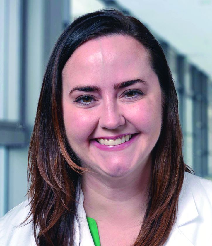
But the Food and Drug Administration’s new system of prescription drug labeling for pregnant women offers improved insight for dermatologists, and a variety of alternatives are available to commonly used dermatologic drugs that pose risks, said Dr. Tyler, who has a unique perspective on this issue: She used to practice as an ob.gyn.
In her presentation, she provided the following tips when caring for pregnant patients.
Check the prescription drug labeling for pregnant and lactating women
Prior to 2015, the FDA used a letter ratings to denote the risk of medications during pregnancy. The ratings – A, B, C, D, and X – go from “A” showing no fetal risk in controlled studies to “X” in which a drug is contraindicated because “there is no reason to risk use of drug in pregnancy.”
This rating system is problematic, Dr. Tyler said, since it’s imprecise. She noted that about two-thirds of medications have a risk rating of C, which means “Risk cannot be ruled out; human studies may or may not show risk; potential benefits may justify potential risk.” But, she asked, “How are you supposed to know what to do with that kind of information?”
By June 30, 2015, FDA had retired the letter classifications and replaced this system with the Pregnancy and Lactation Labeling final rule, which requires that labels include a pregnancy section with information on pregnancy exposure registries, in addition to risk summaries, clinical considerations, and available data. There are also sections on lactation, including nursing mothers, and females and males of reproductive potential. (This information appears in the prescription drug labeling sections 8.1-8.3.) Labels for medications approved prior to June 30, 2001 did not need to be updated. Labels for newer medications are required to comply within 3-5 years of the 2015 policy change.
Ask female patients about sexual activity
It’s important to ask all premenopausal female patients whether they’re sexually active and whether they are using birth control, trying to conceive, or are currently pregnant. If they’re pregnant, what trimester are they in?
Keep in mind, she said, that women can be pregnant for quite some time without knowing it. Home pregnancy tests may not show positive results for up to 5 weeks after conception, she said. But the embryonic/organogenesis stage – from 2 to 8 weeks – is the most important period for a pregnant mother avoid drugs that could damage the unborn child and is “when you want to avoid most medications,” she said.
Risks don’t end after a few months, however. Dr. Tyler urged dermatologists to keep in mind that the brain, teeth, and bones remain susceptible to damage from teratogenic medications after the eighth week.
Keep topical medications in mind
When it comes to pregnant women, “for most dermatologic therapies, topical medications will be your No. 1 choice” because of less absorption, Dr. Tyler said.
But there are exceptions, she noted. In acne and rosacea, for example, there’s controversy about the safety of topical retinoids. “I would just urge you to not use those medications in pregnancy because you don’t really need to use them,” Dr. Tyler said. “You have other options that are safer.”
Adapalene and tretinoin are listed as category C (risk cannot be ruled out in pregnancy) and topical tazarotene is category X (contraindicated in pregnancy) because of retinoid-like anomalies in animal studies, she said.
Avoid 3 major contraindicated systemic drugs
Isotretinoin, acitretin, and methotrexate are “absolutely contraindicated” for anyone who is pregnant or could become pregnant, Dr. Tyler said.
Use some systemic drugs with caution
Tetracyclines are category D (positive evidence of risk to human fetus, but benefits may outweigh risks of drug) with the highest risk in the second and third trimesters, Dr. Tyler said. “If for some reason you have a patient who’s on tetracycline, stop before she’s in her second trimester.”
She recommended avoiding the antibiotic erythromycin because of studies hinting at risks when used early in pregnancy. Spironolactone is theoretically risky after the week 8 of pregnancy, she said. “Because of the animal studies, we typically do not use this during pregnancy.”
Psoriasis often improves during pregnancy
Psoriasis improves during pregnancy in about half of women with psoriasis and worsens in about 20%, Dr. Tyler said. During pregnancy, topical treatments are the first-line treatment, said Dr. Tyler, who recommends that treatment should begin with topical steroids, then calcipotriene or tacrolimus if needed (J Am Acad Dermatol. 2013 Apr;68[4]:663-71). Cyclosporine is an option, as is phototherapy, she said. In an interview, she noted that phototherapy (narrowband UVB) would be considered next-line therapy in patients who are past the first 28 days of gestation and have failed topical therapy, given they are taking adequate folic acid supplementation, which is present in prenatal vitamins.
“As for biologics, you have to go with older ones that have more data. When we look at newer medications, we don’t know a lot about them,” she said during the presentation.
It’s okay to prescribe oral steroids and antihistamines
Oral steroids are safe during pregnancy, Dr. Tyler said, but “just be judicious” with moderate doses and short durations.
Antihistamines are also appropriate, she said, but be aware of the potential for neonatal sedation during lactation.
As for antibiotics for bacterial infections, azithromycin, penicillins, and cephalosporins are all category B (no risk to human fetus despite possible animal risk; or no risk in animal studies and human studies not done), as are all topical antibiotics except dapsone, which is category C because of a theoretical risk of neonatal hyperbilirubinemia if used near the time of delivery.
Hydroxychloroquine may be appropriate for connective tissue disease, she said, although steroids may be a better option in some cases.
And topical antifungals are considered safer for fungal diseases than systemic medications. She said she prefers clotrimazole and oxiconazole, both category B.
Finally, Dr. Tyler recommended permethrin (category B) for parasitic infections since it has been used extensively in pregnancy without a sign of risk and is the preferred treatment for scabies. It’s a better option than ivermectin, she said.
Postpone surgery until at least the second trimester
If it’s not possible to delay nonemergent dermatologic surgery until after pregnancy, she recommended performing procedures during the second trimester. Destruction of local lesions, however, is safe without anesthesia.
“In summary,” when treating pregnant patients, Dr. Tyler said, “a conservative approach is always best, topical medications are always first-line for any condition, and certain oral medications are safe.”
Dr. Tyler reported no relevant disclosures.
SAN DIEGO – “Some doctors don’t treat them at all, or they undertreat because they’re afraid of prescribing anything,” said dermatologist Kelly H. Tyler, MD.
But the Food and Drug Administration’s new system of prescription drug labeling for pregnant women offers improved insight for dermatologists, and a variety of alternatives are available to commonly used dermatologic drugs that pose risks, said Dr. Tyler, who has a unique perspective on this issue: She used to practice as an ob.gyn.
In her presentation, she provided the following tips when caring for pregnant patients.
Check the prescription drug labeling for pregnant and lactating women
Prior to 2015, the FDA used a letter ratings to denote the risk of medications during pregnancy. The ratings – A, B, C, D, and X – go from “A” showing no fetal risk in controlled studies to “X” in which a drug is contraindicated because “there is no reason to risk use of drug in pregnancy.”
This rating system is problematic, Dr. Tyler said, since it’s imprecise. She noted that about two-thirds of medications have a risk rating of C, which means “Risk cannot be ruled out; human studies may or may not show risk; potential benefits may justify potential risk.” But, she asked, “How are you supposed to know what to do with that kind of information?”
By June 30, 2015, FDA had retired the letter classifications and replaced this system with the Pregnancy and Lactation Labeling final rule, which requires that labels include a pregnancy section with information on pregnancy exposure registries, in addition to risk summaries, clinical considerations, and available data. There are also sections on lactation, including nursing mothers, and females and males of reproductive potential. (This information appears in the prescription drug labeling sections 8.1-8.3.) Labels for medications approved prior to June 30, 2001 did not need to be updated. Labels for newer medications are required to comply within 3-5 years of the 2015 policy change.
Ask female patients about sexual activity
It’s important to ask all premenopausal female patients whether they’re sexually active and whether they are using birth control, trying to conceive, or are currently pregnant. If they’re pregnant, what trimester are they in?
Keep in mind, she said, that women can be pregnant for quite some time without knowing it. Home pregnancy tests may not show positive results for up to 5 weeks after conception, she said. But the embryonic/organogenesis stage – from 2 to 8 weeks – is the most important period for a pregnant mother avoid drugs that could damage the unborn child and is “when you want to avoid most medications,” she said.
Risks don’t end after a few months, however. Dr. Tyler urged dermatologists to keep in mind that the brain, teeth, and bones remain susceptible to damage from teratogenic medications after the eighth week.
Keep topical medications in mind
When it comes to pregnant women, “for most dermatologic therapies, topical medications will be your No. 1 choice” because of less absorption, Dr. Tyler said.
But there are exceptions, she noted. In acne and rosacea, for example, there’s controversy about the safety of topical retinoids. “I would just urge you to not use those medications in pregnancy because you don’t really need to use them,” Dr. Tyler said. “You have other options that are safer.”
Adapalene and tretinoin are listed as category C (risk cannot be ruled out in pregnancy) and topical tazarotene is category X (contraindicated in pregnancy) because of retinoid-like anomalies in animal studies, she said.
Avoid 3 major contraindicated systemic drugs
Isotretinoin, acitretin, and methotrexate are “absolutely contraindicated” for anyone who is pregnant or could become pregnant, Dr. Tyler said.
Use some systemic drugs with caution
Tetracyclines are category D (positive evidence of risk to human fetus, but benefits may outweigh risks of drug) with the highest risk in the second and third trimesters, Dr. Tyler said. “If for some reason you have a patient who’s on tetracycline, stop before she’s in her second trimester.”
She recommended avoiding the antibiotic erythromycin because of studies hinting at risks when used early in pregnancy. Spironolactone is theoretically risky after the week 8 of pregnancy, she said. “Because of the animal studies, we typically do not use this during pregnancy.”
Psoriasis often improves during pregnancy
Psoriasis improves during pregnancy in about half of women with psoriasis and worsens in about 20%, Dr. Tyler said. During pregnancy, topical treatments are the first-line treatment, said Dr. Tyler, who recommends that treatment should begin with topical steroids, then calcipotriene or tacrolimus if needed (J Am Acad Dermatol. 2013 Apr;68[4]:663-71). Cyclosporine is an option, as is phototherapy, she said. In an interview, she noted that phototherapy (narrowband UVB) would be considered next-line therapy in patients who are past the first 28 days of gestation and have failed topical therapy, given they are taking adequate folic acid supplementation, which is present in prenatal vitamins.
“As for biologics, you have to go with older ones that have more data. When we look at newer medications, we don’t know a lot about them,” she said during the presentation.
It’s okay to prescribe oral steroids and antihistamines
Oral steroids are safe during pregnancy, Dr. Tyler said, but “just be judicious” with moderate doses and short durations.
Antihistamines are also appropriate, she said, but be aware of the potential for neonatal sedation during lactation.
As for antibiotics for bacterial infections, azithromycin, penicillins, and cephalosporins are all category B (no risk to human fetus despite possible animal risk; or no risk in animal studies and human studies not done), as are all topical antibiotics except dapsone, which is category C because of a theoretical risk of neonatal hyperbilirubinemia if used near the time of delivery.
Hydroxychloroquine may be appropriate for connective tissue disease, she said, although steroids may be a better option in some cases.
And topical antifungals are considered safer for fungal diseases than systemic medications. She said she prefers clotrimazole and oxiconazole, both category B.
Finally, Dr. Tyler recommended permethrin (category B) for parasitic infections since it has been used extensively in pregnancy without a sign of risk and is the preferred treatment for scabies. It’s a better option than ivermectin, she said.
Postpone surgery until at least the second trimester
If it’s not possible to delay nonemergent dermatologic surgery until after pregnancy, she recommended performing procedures during the second trimester. Destruction of local lesions, however, is safe without anesthesia.
“In summary,” when treating pregnant patients, Dr. Tyler said, “a conservative approach is always best, topical medications are always first-line for any condition, and certain oral medications are safe.”
Dr. Tyler reported no relevant disclosures.
SAN DIEGO – “Some doctors don’t treat them at all, or they undertreat because they’re afraid of prescribing anything,” said dermatologist Kelly H. Tyler, MD.
But the Food and Drug Administration’s new system of prescription drug labeling for pregnant women offers improved insight for dermatologists, and a variety of alternatives are available to commonly used dermatologic drugs that pose risks, said Dr. Tyler, who has a unique perspective on this issue: She used to practice as an ob.gyn.
In her presentation, she provided the following tips when caring for pregnant patients.
Check the prescription drug labeling for pregnant and lactating women
Prior to 2015, the FDA used a letter ratings to denote the risk of medications during pregnancy. The ratings – A, B, C, D, and X – go from “A” showing no fetal risk in controlled studies to “X” in which a drug is contraindicated because “there is no reason to risk use of drug in pregnancy.”
This rating system is problematic, Dr. Tyler said, since it’s imprecise. She noted that about two-thirds of medications have a risk rating of C, which means “Risk cannot be ruled out; human studies may or may not show risk; potential benefits may justify potential risk.” But, she asked, “How are you supposed to know what to do with that kind of information?”
By June 30, 2015, FDA had retired the letter classifications and replaced this system with the Pregnancy and Lactation Labeling final rule, which requires that labels include a pregnancy section with information on pregnancy exposure registries, in addition to risk summaries, clinical considerations, and available data. There are also sections on lactation, including nursing mothers, and females and males of reproductive potential. (This information appears in the prescription drug labeling sections 8.1-8.3.) Labels for medications approved prior to June 30, 2001 did not need to be updated. Labels for newer medications are required to comply within 3-5 years of the 2015 policy change.
Ask female patients about sexual activity
It’s important to ask all premenopausal female patients whether they’re sexually active and whether they are using birth control, trying to conceive, or are currently pregnant. If they’re pregnant, what trimester are they in?
Keep in mind, she said, that women can be pregnant for quite some time without knowing it. Home pregnancy tests may not show positive results for up to 5 weeks after conception, she said. But the embryonic/organogenesis stage – from 2 to 8 weeks – is the most important period for a pregnant mother avoid drugs that could damage the unborn child and is “when you want to avoid most medications,” she said.
Risks don’t end after a few months, however. Dr. Tyler urged dermatologists to keep in mind that the brain, teeth, and bones remain susceptible to damage from teratogenic medications after the eighth week.
Keep topical medications in mind
When it comes to pregnant women, “for most dermatologic therapies, topical medications will be your No. 1 choice” because of less absorption, Dr. Tyler said.
But there are exceptions, she noted. In acne and rosacea, for example, there’s controversy about the safety of topical retinoids. “I would just urge you to not use those medications in pregnancy because you don’t really need to use them,” Dr. Tyler said. “You have other options that are safer.”
Adapalene and tretinoin are listed as category C (risk cannot be ruled out in pregnancy) and topical tazarotene is category X (contraindicated in pregnancy) because of retinoid-like anomalies in animal studies, she said.
Avoid 3 major contraindicated systemic drugs
Isotretinoin, acitretin, and methotrexate are “absolutely contraindicated” for anyone who is pregnant or could become pregnant, Dr. Tyler said.
Use some systemic drugs with caution
Tetracyclines are category D (positive evidence of risk to human fetus, but benefits may outweigh risks of drug) with the highest risk in the second and third trimesters, Dr. Tyler said. “If for some reason you have a patient who’s on tetracycline, stop before she’s in her second trimester.”
She recommended avoiding the antibiotic erythromycin because of studies hinting at risks when used early in pregnancy. Spironolactone is theoretically risky after the week 8 of pregnancy, she said. “Because of the animal studies, we typically do not use this during pregnancy.”
Psoriasis often improves during pregnancy
Psoriasis improves during pregnancy in about half of women with psoriasis and worsens in about 20%, Dr. Tyler said. During pregnancy, topical treatments are the first-line treatment, said Dr. Tyler, who recommends that treatment should begin with topical steroids, then calcipotriene or tacrolimus if needed (J Am Acad Dermatol. 2013 Apr;68[4]:663-71). Cyclosporine is an option, as is phototherapy, she said. In an interview, she noted that phototherapy (narrowband UVB) would be considered next-line therapy in patients who are past the first 28 days of gestation and have failed topical therapy, given they are taking adequate folic acid supplementation, which is present in prenatal vitamins.
“As for biologics, you have to go with older ones that have more data. When we look at newer medications, we don’t know a lot about them,” she said during the presentation.
It’s okay to prescribe oral steroids and antihistamines
Oral steroids are safe during pregnancy, Dr. Tyler said, but “just be judicious” with moderate doses and short durations.
Antihistamines are also appropriate, she said, but be aware of the potential for neonatal sedation during lactation.
As for antibiotics for bacterial infections, azithromycin, penicillins, and cephalosporins are all category B (no risk to human fetus despite possible animal risk; or no risk in animal studies and human studies not done), as are all topical antibiotics except dapsone, which is category C because of a theoretical risk of neonatal hyperbilirubinemia if used near the time of delivery.
Hydroxychloroquine may be appropriate for connective tissue disease, she said, although steroids may be a better option in some cases.
And topical antifungals are considered safer for fungal diseases than systemic medications. She said she prefers clotrimazole and oxiconazole, both category B.
Finally, Dr. Tyler recommended permethrin (category B) for parasitic infections since it has been used extensively in pregnancy without a sign of risk and is the preferred treatment for scabies. It’s a better option than ivermectin, she said.
Postpone surgery until at least the second trimester
If it’s not possible to delay nonemergent dermatologic surgery until after pregnancy, she recommended performing procedures during the second trimester. Destruction of local lesions, however, is safe without anesthesia.
“In summary,” when treating pregnant patients, Dr. Tyler said, “a conservative approach is always best, topical medications are always first-line for any condition, and certain oral medications are safe.”
Dr. Tyler reported no relevant disclosures.
EXPERT ANALYSIS FROM AAD 18
VIDEO: Cervical cancer laparotomy outperforms minimally invasive surgery
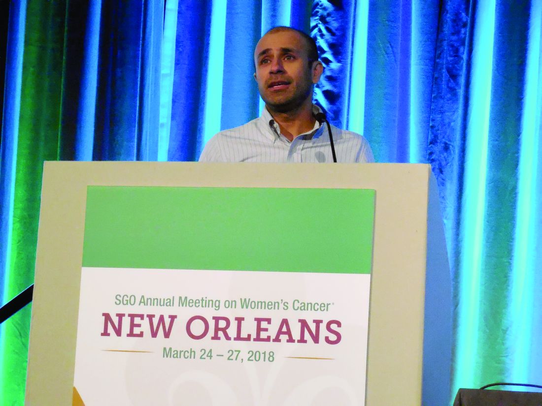
NEW ORLEANS – Use of minimally invasive radical hysterectomy to treat early-stage cervical cancer has grown over the past decade, and in current U.S. practice, roughly half of these cases are done with a minimally-invasive approach, with the rest done by conventional laparotomy. But the first data ever reported from a large, prospective trial that compared the efficacy of both methods for cervical cancer had the unexpected finding that disease-free survival following minimally invasive procedures significantly lagged behind radical hysterectomies done by open laparotomy, Pedro T. Ramirez, MD, said at the annual meeting of the Society of Gynecologic Oncology.
Just after this report came results from a second study that used propensity score–adjusted observational data from the National Cancer Database and found significantly worse overall survival following minimally invasive radical hysterectomy for early-stage cervical cancer, compared with laparotomy, said J. Alejandro Rauh-Hain, MD, a gynecologic oncologist at the University of Texas MD Anderson Cancer Center in Houston.
Both findings were “very surprising,” said Dr. Rauh-Hain in a video interview. “I was pretty sure we’d see no difference” in outcomes between minimally invasive radical hysterectomies and the same surgery either done by laparoscope or robotically assisted.
Prior prospective comparisons of minimally invasive and open surgical methods for other cancer types, including endometrial, gastric, and ovarian, showed no differences in cancer recurrences and survival, which led to widening use of minimally invasive surgery (MIS) for cervical cancer despite no direct evidence supporting equivalence, Dr. Rauh-Hain noted. “We adopted it with no data. It made sense that cervical cancer would be the same as endometrial cancer,” he explained.
The Laparoscopic Approach to Cervical Cancer (LACC) trial ran at 33 centers in 12 countries, including six U.S. centers. The study randomized women during 2008-2017 who had stage 1A1, 1A2, or 1B1 cervical cancer to either MIS or open surgery for a radical hysterectomy. Each participating center had to submit to a trial review committee full case records for 10 patients and unedited surgical videos of two patients who had previously undergone a minimally invasive radical hysterectomy at the center to document local prowess with MIS.
Dr. Ramirez and his colleagues designed LACC to prove the noninferiority of MIS and calculated an expected enrollment of 740 patients based on statistical expectations, but the study stopped early after enrolling 631 patients because of the adverse outcomes identified in the MIS patients, with a median follow-up of 2.5 years instead of the planned follow-up of 4.5 years. The study reached the 4.5-year follow-up in about 39% of patients. Of the 312 patients randomized to undergo laparotomy, 88% actually underwent the surgery; of the 319 patients randomized to MIS, 91% received this surgery, with 16% of the MIS procedures done using robotic assistance.
The study’s primary endpoint was disease-free survival at 4.5 years, which occurred in 86% of the MIS patients and in 96.5% of the laparotomy patients, a difference that failed to meet the study’s prespecified definition of noninferiority for MIS, reported Dr. Ramirez, a professor of gynecologic oncology and director of Minimally Invasive Surgery Research and Education at the MD Anderson Cancer Center. In addition, several secondary analyses of the data all showed starkly superior outcomes in the laparotomy subgroup.
Disease-free survival among all patients regardless of follow-up duration occurred in 98% of laparotomy patients and 92% of MIS patients, which translated into a 3.74 hazard ratio (P = .002) for disease recurrence or death among the MIS patients when compared with laparotomy patients. The all-cause mortality rates were 1% in the laparotomy patients and 6% among the MIS patients, a hazard ratio of 6.00 (P = .004). The risk of local or regional recurrences was more than fourfold higher in the MIS patients. A blinded, central panel adjudicated all recurrences identified during the study.
The LACC results “should be discussed with patients scheduled to undergo radical hysterectomy” for cervical cancer, Dr. Ramirez concluded.
The observational data from the National Cancer Database used in the analysis led by Dr. Rauh-Hain came from 2,221 patients hospitalized and treated with radical hysterectomy and pelvic lymph node dissection at a U.S. center during 2010-2012 for either stage 1A2 or 1B1 cervical cancer. Among these patients, 47.5% underwent MIS, with 79% of those procedures done with robotic assistance, while the other 52.5% underwent open laparotomy, Dr. Rauh-Hain reported. Additional analysis of data from this database by the researchers showed that, although the first report of MIS for radical hysterectomy appeared in 1992, the approach remained largely unused in U.S. practice until 2007, when use of MIS began to sharply rise. By 2010, about a third of radical hysterectomies for cervical cancer involved MIS, and usage increased still further during 2011 and 2012 to produce a nearly 48% rate during the 3-year study period.
The primary endpoint of Dr. Rauh-Hain’s analysis was overall survival following propensity-score matching of the MIS and laparotomy patients using 13 demographic and clinical criteria. The analysis showed 4-year mortality rates of 5.8% among the laparotomy patients and 8.4% among the MIS patients, which calculated to a relatively increased mortality hazard from MIS of 48% (P = .02).
Dr. Rauh-Hain also reported results from an interrupted time series analysis using data from the Surveillance, Epidemiology, and End Results database of the National Cancer Institute. This analysis compared annual 4-year relative survival rates among women undergoing radical hysterectomy for cervical cancer and found that, after survival rates showed a gradual, steady rise during the years culminating in 2006, once MIS began being more widely used in 2007 survival rates began to drop, with a statistically significant annualized decline of 1% through 2010.
Based on the results from both studies, “at MD Anderson we discuss the results with patients,” with the consequence that the percentage of patients treated with laparotomy is now increasing, Dr. Rauh-Hain said. The results from both studies “are concerning,” he explained.
SOURCE: Ramirez PT and Rauh-Hain JA. SGO 2018, Late-Breaking Abstracts 1 and 2.
The findings from these studies appear valid and should be discussed with patients.
The findings raise a major question: Why has minimally invasive surgery (MIS) led to worse survival rates than laparotomy? Several possible explanations can be hypothesized: The uterine manipulator used in MIS led to local spread of cancer cells; MIS involves a learning curve and initial attempts at MIS did not remove enough of the tumor; and MIS led to increased exposure of the peritoneal cavity to the cancer. The findings also raise another question: Why has MIS for cervical cancer performed less well than MIS for cancers from other organs, such as endometrial and prostate?
The overall survival results from the LACC trial calculate out to 4.75 added deaths per year for every 1,000 patients treated with MIS, compared with laparoscopy. But the National Inpatient Sample data suggest that MIS cuts mortality by about two deaths per year per 1,000 patients, compared with laparotomy, and mortality data from a different analysis (Gynecol Oncol. 2012 Oct;127[1]:11-7) suggest that MIS might prevent six deaths annually for every 1,000 patients, compared with laparotomy. Overall, these three sets of findings suggest roughly comparable mortality outcomes from MIS and laparotomy, but with MIS having the bonus of fewer complications and less need for transfusions.
The cautions and concerns raised by the LACC trial and Dr. Rauh-Hain’s analysis of observational data cannot be easily dismissed. We need to figure out why the results from both studies show worse survival and recurrence rates with MIS, and we need to identify whether subgroups of patients exist who might clearly benefit from either the MIS or open-surgery approach.
Shitanshu Uppal, MD , is a gynecologic oncologist at the University of Michigan in Ann Arbor. He made these comments as designated discussant for the two studies. He had no disclosures.
The findings from these studies appear valid and should be discussed with patients.
The findings raise a major question: Why has minimally invasive surgery (MIS) led to worse survival rates than laparotomy? Several possible explanations can be hypothesized: The uterine manipulator used in MIS led to local spread of cancer cells; MIS involves a learning curve and initial attempts at MIS did not remove enough of the tumor; and MIS led to increased exposure of the peritoneal cavity to the cancer. The findings also raise another question: Why has MIS for cervical cancer performed less well than MIS for cancers from other organs, such as endometrial and prostate?
The overall survival results from the LACC trial calculate out to 4.75 added deaths per year for every 1,000 patients treated with MIS, compared with laparoscopy. But the National Inpatient Sample data suggest that MIS cuts mortality by about two deaths per year per 1,000 patients, compared with laparotomy, and mortality data from a different analysis (Gynecol Oncol. 2012 Oct;127[1]:11-7) suggest that MIS might prevent six deaths annually for every 1,000 patients, compared with laparotomy. Overall, these three sets of findings suggest roughly comparable mortality outcomes from MIS and laparotomy, but with MIS having the bonus of fewer complications and less need for transfusions.
The cautions and concerns raised by the LACC trial and Dr. Rauh-Hain’s analysis of observational data cannot be easily dismissed. We need to figure out why the results from both studies show worse survival and recurrence rates with MIS, and we need to identify whether subgroups of patients exist who might clearly benefit from either the MIS or open-surgery approach.
Shitanshu Uppal, MD , is a gynecologic oncologist at the University of Michigan in Ann Arbor. He made these comments as designated discussant for the two studies. He had no disclosures.
The findings from these studies appear valid and should be discussed with patients.
The findings raise a major question: Why has minimally invasive surgery (MIS) led to worse survival rates than laparotomy? Several possible explanations can be hypothesized: The uterine manipulator used in MIS led to local spread of cancer cells; MIS involves a learning curve and initial attempts at MIS did not remove enough of the tumor; and MIS led to increased exposure of the peritoneal cavity to the cancer. The findings also raise another question: Why has MIS for cervical cancer performed less well than MIS for cancers from other organs, such as endometrial and prostate?
The overall survival results from the LACC trial calculate out to 4.75 added deaths per year for every 1,000 patients treated with MIS, compared with laparoscopy. But the National Inpatient Sample data suggest that MIS cuts mortality by about two deaths per year per 1,000 patients, compared with laparotomy, and mortality data from a different analysis (Gynecol Oncol. 2012 Oct;127[1]:11-7) suggest that MIS might prevent six deaths annually for every 1,000 patients, compared with laparotomy. Overall, these three sets of findings suggest roughly comparable mortality outcomes from MIS and laparotomy, but with MIS having the bonus of fewer complications and less need for transfusions.
The cautions and concerns raised by the LACC trial and Dr. Rauh-Hain’s analysis of observational data cannot be easily dismissed. We need to figure out why the results from both studies show worse survival and recurrence rates with MIS, and we need to identify whether subgroups of patients exist who might clearly benefit from either the MIS or open-surgery approach.
Shitanshu Uppal, MD , is a gynecologic oncologist at the University of Michigan in Ann Arbor. He made these comments as designated discussant for the two studies. He had no disclosures.
NEW ORLEANS – Use of minimally invasive radical hysterectomy to treat early-stage cervical cancer has grown over the past decade, and in current U.S. practice, roughly half of these cases are done with a minimally-invasive approach, with the rest done by conventional laparotomy. But the first data ever reported from a large, prospective trial that compared the efficacy of both methods for cervical cancer had the unexpected finding that disease-free survival following minimally invasive procedures significantly lagged behind radical hysterectomies done by open laparotomy, Pedro T. Ramirez, MD, said at the annual meeting of the Society of Gynecologic Oncology.
Just after this report came results from a second study that used propensity score–adjusted observational data from the National Cancer Database and found significantly worse overall survival following minimally invasive radical hysterectomy for early-stage cervical cancer, compared with laparotomy, said J. Alejandro Rauh-Hain, MD, a gynecologic oncologist at the University of Texas MD Anderson Cancer Center in Houston.
Both findings were “very surprising,” said Dr. Rauh-Hain in a video interview. “I was pretty sure we’d see no difference” in outcomes between minimally invasive radical hysterectomies and the same surgery either done by laparoscope or robotically assisted.
Prior prospective comparisons of minimally invasive and open surgical methods for other cancer types, including endometrial, gastric, and ovarian, showed no differences in cancer recurrences and survival, which led to widening use of minimally invasive surgery (MIS) for cervical cancer despite no direct evidence supporting equivalence, Dr. Rauh-Hain noted. “We adopted it with no data. It made sense that cervical cancer would be the same as endometrial cancer,” he explained.
The Laparoscopic Approach to Cervical Cancer (LACC) trial ran at 33 centers in 12 countries, including six U.S. centers. The study randomized women during 2008-2017 who had stage 1A1, 1A2, or 1B1 cervical cancer to either MIS or open surgery for a radical hysterectomy. Each participating center had to submit to a trial review committee full case records for 10 patients and unedited surgical videos of two patients who had previously undergone a minimally invasive radical hysterectomy at the center to document local prowess with MIS.
Dr. Ramirez and his colleagues designed LACC to prove the noninferiority of MIS and calculated an expected enrollment of 740 patients based on statistical expectations, but the study stopped early after enrolling 631 patients because of the adverse outcomes identified in the MIS patients, with a median follow-up of 2.5 years instead of the planned follow-up of 4.5 years. The study reached the 4.5-year follow-up in about 39% of patients. Of the 312 patients randomized to undergo laparotomy, 88% actually underwent the surgery; of the 319 patients randomized to MIS, 91% received this surgery, with 16% of the MIS procedures done using robotic assistance.
The study’s primary endpoint was disease-free survival at 4.5 years, which occurred in 86% of the MIS patients and in 96.5% of the laparotomy patients, a difference that failed to meet the study’s prespecified definition of noninferiority for MIS, reported Dr. Ramirez, a professor of gynecologic oncology and director of Minimally Invasive Surgery Research and Education at the MD Anderson Cancer Center. In addition, several secondary analyses of the data all showed starkly superior outcomes in the laparotomy subgroup.
Disease-free survival among all patients regardless of follow-up duration occurred in 98% of laparotomy patients and 92% of MIS patients, which translated into a 3.74 hazard ratio (P = .002) for disease recurrence or death among the MIS patients when compared with laparotomy patients. The all-cause mortality rates were 1% in the laparotomy patients and 6% among the MIS patients, a hazard ratio of 6.00 (P = .004). The risk of local or regional recurrences was more than fourfold higher in the MIS patients. A blinded, central panel adjudicated all recurrences identified during the study.
The LACC results “should be discussed with patients scheduled to undergo radical hysterectomy” for cervical cancer, Dr. Ramirez concluded.
The observational data from the National Cancer Database used in the analysis led by Dr. Rauh-Hain came from 2,221 patients hospitalized and treated with radical hysterectomy and pelvic lymph node dissection at a U.S. center during 2010-2012 for either stage 1A2 or 1B1 cervical cancer. Among these patients, 47.5% underwent MIS, with 79% of those procedures done with robotic assistance, while the other 52.5% underwent open laparotomy, Dr. Rauh-Hain reported. Additional analysis of data from this database by the researchers showed that, although the first report of MIS for radical hysterectomy appeared in 1992, the approach remained largely unused in U.S. practice until 2007, when use of MIS began to sharply rise. By 2010, about a third of radical hysterectomies for cervical cancer involved MIS, and usage increased still further during 2011 and 2012 to produce a nearly 48% rate during the 3-year study period.
The primary endpoint of Dr. Rauh-Hain’s analysis was overall survival following propensity-score matching of the MIS and laparotomy patients using 13 demographic and clinical criteria. The analysis showed 4-year mortality rates of 5.8% among the laparotomy patients and 8.4% among the MIS patients, which calculated to a relatively increased mortality hazard from MIS of 48% (P = .02).
Dr. Rauh-Hain also reported results from an interrupted time series analysis using data from the Surveillance, Epidemiology, and End Results database of the National Cancer Institute. This analysis compared annual 4-year relative survival rates among women undergoing radical hysterectomy for cervical cancer and found that, after survival rates showed a gradual, steady rise during the years culminating in 2006, once MIS began being more widely used in 2007 survival rates began to drop, with a statistically significant annualized decline of 1% through 2010.
Based on the results from both studies, “at MD Anderson we discuss the results with patients,” with the consequence that the percentage of patients treated with laparotomy is now increasing, Dr. Rauh-Hain said. The results from both studies “are concerning,” he explained.
SOURCE: Ramirez PT and Rauh-Hain JA. SGO 2018, Late-Breaking Abstracts 1 and 2.
NEW ORLEANS – Use of minimally invasive radical hysterectomy to treat early-stage cervical cancer has grown over the past decade, and in current U.S. practice, roughly half of these cases are done with a minimally-invasive approach, with the rest done by conventional laparotomy. But the first data ever reported from a large, prospective trial that compared the efficacy of both methods for cervical cancer had the unexpected finding that disease-free survival following minimally invasive procedures significantly lagged behind radical hysterectomies done by open laparotomy, Pedro T. Ramirez, MD, said at the annual meeting of the Society of Gynecologic Oncology.
Just after this report came results from a second study that used propensity score–adjusted observational data from the National Cancer Database and found significantly worse overall survival following minimally invasive radical hysterectomy for early-stage cervical cancer, compared with laparotomy, said J. Alejandro Rauh-Hain, MD, a gynecologic oncologist at the University of Texas MD Anderson Cancer Center in Houston.
Both findings were “very surprising,” said Dr. Rauh-Hain in a video interview. “I was pretty sure we’d see no difference” in outcomes between minimally invasive radical hysterectomies and the same surgery either done by laparoscope or robotically assisted.
Prior prospective comparisons of minimally invasive and open surgical methods for other cancer types, including endometrial, gastric, and ovarian, showed no differences in cancer recurrences and survival, which led to widening use of minimally invasive surgery (MIS) for cervical cancer despite no direct evidence supporting equivalence, Dr. Rauh-Hain noted. “We adopted it with no data. It made sense that cervical cancer would be the same as endometrial cancer,” he explained.
The Laparoscopic Approach to Cervical Cancer (LACC) trial ran at 33 centers in 12 countries, including six U.S. centers. The study randomized women during 2008-2017 who had stage 1A1, 1A2, or 1B1 cervical cancer to either MIS or open surgery for a radical hysterectomy. Each participating center had to submit to a trial review committee full case records for 10 patients and unedited surgical videos of two patients who had previously undergone a minimally invasive radical hysterectomy at the center to document local prowess with MIS.
Dr. Ramirez and his colleagues designed LACC to prove the noninferiority of MIS and calculated an expected enrollment of 740 patients based on statistical expectations, but the study stopped early after enrolling 631 patients because of the adverse outcomes identified in the MIS patients, with a median follow-up of 2.5 years instead of the planned follow-up of 4.5 years. The study reached the 4.5-year follow-up in about 39% of patients. Of the 312 patients randomized to undergo laparotomy, 88% actually underwent the surgery; of the 319 patients randomized to MIS, 91% received this surgery, with 16% of the MIS procedures done using robotic assistance.
The study’s primary endpoint was disease-free survival at 4.5 years, which occurred in 86% of the MIS patients and in 96.5% of the laparotomy patients, a difference that failed to meet the study’s prespecified definition of noninferiority for MIS, reported Dr. Ramirez, a professor of gynecologic oncology and director of Minimally Invasive Surgery Research and Education at the MD Anderson Cancer Center. In addition, several secondary analyses of the data all showed starkly superior outcomes in the laparotomy subgroup.
Disease-free survival among all patients regardless of follow-up duration occurred in 98% of laparotomy patients and 92% of MIS patients, which translated into a 3.74 hazard ratio (P = .002) for disease recurrence or death among the MIS patients when compared with laparotomy patients. The all-cause mortality rates were 1% in the laparotomy patients and 6% among the MIS patients, a hazard ratio of 6.00 (P = .004). The risk of local or regional recurrences was more than fourfold higher in the MIS patients. A blinded, central panel adjudicated all recurrences identified during the study.
The LACC results “should be discussed with patients scheduled to undergo radical hysterectomy” for cervical cancer, Dr. Ramirez concluded.
The observational data from the National Cancer Database used in the analysis led by Dr. Rauh-Hain came from 2,221 patients hospitalized and treated with radical hysterectomy and pelvic lymph node dissection at a U.S. center during 2010-2012 for either stage 1A2 or 1B1 cervical cancer. Among these patients, 47.5% underwent MIS, with 79% of those procedures done with robotic assistance, while the other 52.5% underwent open laparotomy, Dr. Rauh-Hain reported. Additional analysis of data from this database by the researchers showed that, although the first report of MIS for radical hysterectomy appeared in 1992, the approach remained largely unused in U.S. practice until 2007, when use of MIS began to sharply rise. By 2010, about a third of radical hysterectomies for cervical cancer involved MIS, and usage increased still further during 2011 and 2012 to produce a nearly 48% rate during the 3-year study period.
The primary endpoint of Dr. Rauh-Hain’s analysis was overall survival following propensity-score matching of the MIS and laparotomy patients using 13 demographic and clinical criteria. The analysis showed 4-year mortality rates of 5.8% among the laparotomy patients and 8.4% among the MIS patients, which calculated to a relatively increased mortality hazard from MIS of 48% (P = .02).
Dr. Rauh-Hain also reported results from an interrupted time series analysis using data from the Surveillance, Epidemiology, and End Results database of the National Cancer Institute. This analysis compared annual 4-year relative survival rates among women undergoing radical hysterectomy for cervical cancer and found that, after survival rates showed a gradual, steady rise during the years culminating in 2006, once MIS began being more widely used in 2007 survival rates began to drop, with a statistically significant annualized decline of 1% through 2010.
Based on the results from both studies, “at MD Anderson we discuss the results with patients,” with the consequence that the percentage of patients treated with laparotomy is now increasing, Dr. Rauh-Hain said. The results from both studies “are concerning,” he explained.
SOURCE: Ramirez PT and Rauh-Hain JA. SGO 2018, Late-Breaking Abstracts 1 and 2.
REPORTING FROM SGO 2018
Key clinical point: Laparotomy produced better survival than did minimally invasive surgery for cervical cancer.
Major finding: Disease-free survival after 4.5 years was 96.5% with laparotomy and 86.0% with minimally invasive surgery.
Study details: LACC was a multicenter, randomized trial with 631 patients. The observational study included 2,221 patients from the National Cancer Database during 2010-2012.
Disclosures: Dr. Ramirez and Dr. Rauh-Hain had no disclosures.
Source: Ramirez PT and Rauh-Hain JA. SGO 2018, Late-Breaking Abstracts 1 and 2.

Homework
How do you feel about homework? Do you think your school-age patients are given too much homework? Would they be better off spending their after-school time at home in free play or exploring nonacademic interests? Or, do you feel the school day is too short to adequately cover what a well-educated child needs to know? Doesn’t homework foster good independent work habits and discipline?
Do you have fond memories of doing homework? Are you glad those days of bringing home an hour or 3 of extra work are behind you? Maybe they aren’t behind you. Are you still spending an hour or more getting stuff done at home you didn’t get done in the office?
Primary care pediatrics has never promised its practitioners that they will arrive at home at the end of the workday free of unfinished business. If you have after-hours call responsibilities, there always have been phone calls, decisions to make, and trips to EDs and delivery rooms. Even if you are fortunate enough to not have after-hours call responsibilities, there are certainly evenings when you are nagged by second thoughts and worries about troublesome patients you have seen during the day. Did you make the correct diagnosis or forget to order a critical lab test?
This kind of homework is expected. It’s what you signed up for. But with experience, you learn how to provide better anticipatory guidance that can decrease the number of after-hours calls. You can minimize, but never eliminate, second-guessing by learning to make wiser diagnostic and therapeutic decisions.
However, arriving home with a laptop or notebook loaded with unfinished electronic health records and work-related emails is not what you thought primary care pediatrics was about ... and it didn’t used to be. For the first 35 years of practice, when I saw my last patient, my office work was over. If I wasn’t on call, I could enjoy the entire evening with my family uninterrupted.
But change happens. Coincident with the launch of a new computer system, my workday became an hour longer so that I could complete my electronic office notes before I went home. For some of my colleagues, this unwelcome addition ran more than an hour and a half or 2 hours, and many of them leapt at the practice administrator’s offer to link their home computers with our new office EHR. Buried in what sounded like a good deal to them, I could hear the creaky opening of a Pandora’s box.

Dr. Wilkoff practiced primary care pediatrics in Brunswick, Maine, for nearly 40 years. He has authored several books on behavioral pediatrics, including “How to Say No to Your Toddler.” Email him at pdnews@mdedge.com.
How do you feel about homework? Do you think your school-age patients are given too much homework? Would they be better off spending their after-school time at home in free play or exploring nonacademic interests? Or, do you feel the school day is too short to adequately cover what a well-educated child needs to know? Doesn’t homework foster good independent work habits and discipline?
Do you have fond memories of doing homework? Are you glad those days of bringing home an hour or 3 of extra work are behind you? Maybe they aren’t behind you. Are you still spending an hour or more getting stuff done at home you didn’t get done in the office?
Primary care pediatrics has never promised its practitioners that they will arrive at home at the end of the workday free of unfinished business. If you have after-hours call responsibilities, there always have been phone calls, decisions to make, and trips to EDs and delivery rooms. Even if you are fortunate enough to not have after-hours call responsibilities, there are certainly evenings when you are nagged by second thoughts and worries about troublesome patients you have seen during the day. Did you make the correct diagnosis or forget to order a critical lab test?
This kind of homework is expected. It’s what you signed up for. But with experience, you learn how to provide better anticipatory guidance that can decrease the number of after-hours calls. You can minimize, but never eliminate, second-guessing by learning to make wiser diagnostic and therapeutic decisions.
However, arriving home with a laptop or notebook loaded with unfinished electronic health records and work-related emails is not what you thought primary care pediatrics was about ... and it didn’t used to be. For the first 35 years of practice, when I saw my last patient, my office work was over. If I wasn’t on call, I could enjoy the entire evening with my family uninterrupted.
But change happens. Coincident with the launch of a new computer system, my workday became an hour longer so that I could complete my electronic office notes before I went home. For some of my colleagues, this unwelcome addition ran more than an hour and a half or 2 hours, and many of them leapt at the practice administrator’s offer to link their home computers with our new office EHR. Buried in what sounded like a good deal to them, I could hear the creaky opening of a Pandora’s box.
Dr. Wilkoff practiced primary care pediatrics in Brunswick, Maine, for nearly 40 years. He has authored several books on behavioral pediatrics, including “How to Say No to Your Toddler.” Email him at pdnews@mdedge.com.
How do you feel about homework? Do you think your school-age patients are given too much homework? Would they be better off spending their after-school time at home in free play or exploring nonacademic interests? Or, do you feel the school day is too short to adequately cover what a well-educated child needs to know? Doesn’t homework foster good independent work habits and discipline?
Do you have fond memories of doing homework? Are you glad those days of bringing home an hour or 3 of extra work are behind you? Maybe they aren’t behind you. Are you still spending an hour or more getting stuff done at home you didn’t get done in the office?
Primary care pediatrics has never promised its practitioners that they will arrive at home at the end of the workday free of unfinished business. If you have after-hours call responsibilities, there always have been phone calls, decisions to make, and trips to EDs and delivery rooms. Even if you are fortunate enough to not have after-hours call responsibilities, there are certainly evenings when you are nagged by second thoughts and worries about troublesome patients you have seen during the day. Did you make the correct diagnosis or forget to order a critical lab test?
This kind of homework is expected. It’s what you signed up for. But with experience, you learn how to provide better anticipatory guidance that can decrease the number of after-hours calls. You can minimize, but never eliminate, second-guessing by learning to make wiser diagnostic and therapeutic decisions.
However, arriving home with a laptop or notebook loaded with unfinished electronic health records and work-related emails is not what you thought primary care pediatrics was about ... and it didn’t used to be. For the first 35 years of practice, when I saw my last patient, my office work was over. If I wasn’t on call, I could enjoy the entire evening with my family uninterrupted.
But change happens. Coincident with the launch of a new computer system, my workday became an hour longer so that I could complete my electronic office notes before I went home. For some of my colleagues, this unwelcome addition ran more than an hour and a half or 2 hours, and many of them leapt at the practice administrator’s offer to link their home computers with our new office EHR. Buried in what sounded like a good deal to them, I could hear the creaky opening of a Pandora’s box.
Dr. Wilkoff practiced primary care pediatrics in Brunswick, Maine, for nearly 40 years. He has authored several books on behavioral pediatrics, including “How to Say No to Your Toddler.” Email him at pdnews@mdedge.com.

AHRQ Practice Toolbox: Practice facilitation
This is the eighth in a series of articles from the National Center for Excellence in Primary Care Research (NCEPCR) in the Agency for Healthcare Research and Quality (AHRQ). This series introduces sets of tools and resources designed to help your practice.
Last month’s article discussed AHRQ’s tool and resources for practice transformation, or how to help make your practice ready for advances such as shared decision making, team-based care, and integrating behavioral health and primary care. Use of practice facilitation, an evidence-based approach to quality improvement, can be a key element to helping primary care practices make the necessary changes for transformation. Practice facilitators (also known as practice coaches, quality improvement coaches, or practice enhancement assistants) are specially trained individuals who assist primary care clinicians in research and quality improvement projects. They are distinguished from consultants through specialized training, broad scope of practice, and long-term relationships with an organization, its staff and clinicians, and its patients.
If you are wondering how a practice facilitator can help you, AHRQ offers How a Practice Facilitator Can Support Your Practice. This tip sheet provides ideas and techniques for primary care practices interested in getting started with quality improvement activities and describes the benefits of working with a practice facilitator. The tip sheet is distilled from a more extensive white paper on Engaging Primary Care Practices in Quality Improvement: Strategies for Practice Facilitators, which presents a framework for engaging primary care practices in quality improvement and provides strategies for sustained involvement in the undertaking.
For those with greater interest in implementation of practice facilitation, AHRQ offers the Practice Facilitation Handbook: Training Modules for New Facilitators and Their Trainers. The Handbook is designed to assist in the training of new practice facilitators as they begin to develop the knowledge and skills needed to support meaningful improvement in primary care practices. AHRQ built upon the modules in the Handbook with the Primary Care Practice Facilitation Curriculum. The Curriculum updates the modules in the Handbook and adds an additional twelve modules for a total of 32 modules. More importantly, some of the modules can be used by clinicians and staff as a sort of Quality Improvement 101. Modules on appreciative inquiry, approaches to quality improvement, redesigning work flow, root cause analysis, and measuring clinical performance can be useful to anyone interested in increasing their knowledge and skills for quality improvement.

Mr. McNellis is senior adviser for primary care at AHRQ and Dr. Ganiats is director of the National Center for Excellence in Primary Care Research at AHRQ.
Links from the AHRQ Web site:
How a Practice Facilitator Can Support Your Practice
Engaging Primary Care Practices in Quality Improvement: Strategies for Practice Facilitators
Primary Care Practice Facilitation Curriculum
Practice Facilitation Handbook
A How-To Guide on Developing and Running a Practice Facilitation Program
These and other tools can be found at the NCEPCR Web site.
This is the eighth in a series of articles from the National Center for Excellence in Primary Care Research (NCEPCR) in the Agency for Healthcare Research and Quality (AHRQ). This series introduces sets of tools and resources designed to help your practice.
Last month’s article discussed AHRQ’s tool and resources for practice transformation, or how to help make your practice ready for advances such as shared decision making, team-based care, and integrating behavioral health and primary care. Use of practice facilitation, an evidence-based approach to quality improvement, can be a key element to helping primary care practices make the necessary changes for transformation. Practice facilitators (also known as practice coaches, quality improvement coaches, or practice enhancement assistants) are specially trained individuals who assist primary care clinicians in research and quality improvement projects. They are distinguished from consultants through specialized training, broad scope of practice, and long-term relationships with an organization, its staff and clinicians, and its patients.
If you are wondering how a practice facilitator can help you, AHRQ offers How a Practice Facilitator Can Support Your Practice. This tip sheet provides ideas and techniques for primary care practices interested in getting started with quality improvement activities and describes the benefits of working with a practice facilitator. The tip sheet is distilled from a more extensive white paper on Engaging Primary Care Practices in Quality Improvement: Strategies for Practice Facilitators, which presents a framework for engaging primary care practices in quality improvement and provides strategies for sustained involvement in the undertaking.
For those with greater interest in implementation of practice facilitation, AHRQ offers the Practice Facilitation Handbook: Training Modules for New Facilitators and Their Trainers. The Handbook is designed to assist in the training of new practice facilitators as they begin to develop the knowledge and skills needed to support meaningful improvement in primary care practices. AHRQ built upon the modules in the Handbook with the Primary Care Practice Facilitation Curriculum. The Curriculum updates the modules in the Handbook and adds an additional twelve modules for a total of 32 modules. More importantly, some of the modules can be used by clinicians and staff as a sort of Quality Improvement 101. Modules on appreciative inquiry, approaches to quality improvement, redesigning work flow, root cause analysis, and measuring clinical performance can be useful to anyone interested in increasing their knowledge and skills for quality improvement.
Mr. McNellis is senior adviser for primary care at AHRQ and Dr. Ganiats is director of the National Center for Excellence in Primary Care Research at AHRQ.
Links from the AHRQ Web site:
How a Practice Facilitator Can Support Your Practice
Engaging Primary Care Practices in Quality Improvement: Strategies for Practice Facilitators
Primary Care Practice Facilitation Curriculum
Practice Facilitation Handbook
A How-To Guide on Developing and Running a Practice Facilitation Program
These and other tools can be found at the NCEPCR Web site.
This is the eighth in a series of articles from the National Center for Excellence in Primary Care Research (NCEPCR) in the Agency for Healthcare Research and Quality (AHRQ). This series introduces sets of tools and resources designed to help your practice.
Last month’s article discussed AHRQ’s tool and resources for practice transformation, or how to help make your practice ready for advances such as shared decision making, team-based care, and integrating behavioral health and primary care. Use of practice facilitation, an evidence-based approach to quality improvement, can be a key element to helping primary care practices make the necessary changes for transformation. Practice facilitators (also known as practice coaches, quality improvement coaches, or practice enhancement assistants) are specially trained individuals who assist primary care clinicians in research and quality improvement projects. They are distinguished from consultants through specialized training, broad scope of practice, and long-term relationships with an organization, its staff and clinicians, and its patients.
If you are wondering how a practice facilitator can help you, AHRQ offers How a Practice Facilitator Can Support Your Practice. This tip sheet provides ideas and techniques for primary care practices interested in getting started with quality improvement activities and describes the benefits of working with a practice facilitator. The tip sheet is distilled from a more extensive white paper on Engaging Primary Care Practices in Quality Improvement: Strategies for Practice Facilitators, which presents a framework for engaging primary care practices in quality improvement and provides strategies for sustained involvement in the undertaking.
For those with greater interest in implementation of practice facilitation, AHRQ offers the Practice Facilitation Handbook: Training Modules for New Facilitators and Their Trainers. The Handbook is designed to assist in the training of new practice facilitators as they begin to develop the knowledge and skills needed to support meaningful improvement in primary care practices. AHRQ built upon the modules in the Handbook with the Primary Care Practice Facilitation Curriculum. The Curriculum updates the modules in the Handbook and adds an additional twelve modules for a total of 32 modules. More importantly, some of the modules can be used by clinicians and staff as a sort of Quality Improvement 101. Modules on appreciative inquiry, approaches to quality improvement, redesigning work flow, root cause analysis, and measuring clinical performance can be useful to anyone interested in increasing their knowledge and skills for quality improvement.
Mr. McNellis is senior adviser for primary care at AHRQ and Dr. Ganiats is director of the National Center for Excellence in Primary Care Research at AHRQ.
Links from the AHRQ Web site:
How a Practice Facilitator Can Support Your Practice
Engaging Primary Care Practices in Quality Improvement: Strategies for Practice Facilitators
Primary Care Practice Facilitation Curriculum
Practice Facilitation Handbook
A How-To Guide on Developing and Running a Practice Facilitation Program
These and other tools can be found at the NCEPCR Web site.

Dramatic improvements reported after surgery for hidradenitis suppurativa
A retrospective German study found that the majority of , with many saying they no longer suffered from everyday impairment from the disease.
“We were able to show that surgical therapy resulted in convincing improvement of life quality and long-term results for HS that are at least as effective as biologicals,” the researchers wrote. The study was published online in the Journal of the European Academy of Dermatology and Venereology.
Lukas Kofler, MD, and associates from the department of dermatology at Eberhard Karls University’s University Medical Center, Tübingen, Germany, surveyed 910 of the facility’s patients who had undergone wide local excision for HS from 2006 to 2015. Surgery was “designed to reach into clinically disease-free subcutaneous fatty tissue,” followed by second intention healing, they wrote.
A total of 255 patients answered the survey, a response rate of 28%. There were 103 men and 152 women with a median age of 38 years (range, 14-66 years); 75% reported prior “nicotine abuse.” Almost half had been treated previously, most often with systemic antibiotics in 68%. The mean follow-up time was 57 months (range, 19-127 months);
All cases were Hurley grade III. Just over three-quarters of the patients described disease-related limitations in private life prior to surgery as “very strong” or “strong,” and 95% reported that their day-to-day life was impaired. Sixty percent said the disease impaired their work life (another 8% were not employed).
After surgery, 27% experienced postoperative complications, including minor bleeding, infection, and limited mobility; 65% experienced pain but 38% of the patients required analgesics postoperatively.
After surgery, 80% were satisfied or very satisfied with the results, and more than two-thirds were satisfied with the cosmetic results. Just over half said their private life was not impaired by the disease at all, compared with 3% who said so before surgery. After surgery, 20% reported being strongly or very strongly impaired, compared with 77% before surgery.

Nearly 70% reported that HS recurred after surgery, but 62% of those with recurrences said the disease was not as severe as before surgery.
“Surgery represents an important treatment option by itself but should also be part of combined therapeutic strategies, especially in severe disease stages. However, consistent approaches combining systemic and surgical treatments have not been established yet,” the authors noted.
The study had no funding source. The authors had no conflicts to disclose.
SOURCE: Kofler L et al. J Eur Acad Dermatol Venereol. 2018 Mar 23. doi: 10.1111/jdv.14892.
A retrospective German study found that the majority of , with many saying they no longer suffered from everyday impairment from the disease.
“We were able to show that surgical therapy resulted in convincing improvement of life quality and long-term results for HS that are at least as effective as biologicals,” the researchers wrote. The study was published online in the Journal of the European Academy of Dermatology and Venereology.
Lukas Kofler, MD, and associates from the department of dermatology at Eberhard Karls University’s University Medical Center, Tübingen, Germany, surveyed 910 of the facility’s patients who had undergone wide local excision for HS from 2006 to 2015. Surgery was “designed to reach into clinically disease-free subcutaneous fatty tissue,” followed by second intention healing, they wrote.
A total of 255 patients answered the survey, a response rate of 28%. There were 103 men and 152 women with a median age of 38 years (range, 14-66 years); 75% reported prior “nicotine abuse.” Almost half had been treated previously, most often with systemic antibiotics in 68%. The mean follow-up time was 57 months (range, 19-127 months);
All cases were Hurley grade III. Just over three-quarters of the patients described disease-related limitations in private life prior to surgery as “very strong” or “strong,” and 95% reported that their day-to-day life was impaired. Sixty percent said the disease impaired their work life (another 8% were not employed).
After surgery, 27% experienced postoperative complications, including minor bleeding, infection, and limited mobility; 65% experienced pain but 38% of the patients required analgesics postoperatively.
After surgery, 80% were satisfied or very satisfied with the results, and more than two-thirds were satisfied with the cosmetic results. Just over half said their private life was not impaired by the disease at all, compared with 3% who said so before surgery. After surgery, 20% reported being strongly or very strongly impaired, compared with 77% before surgery.
Nearly 70% reported that HS recurred after surgery, but 62% of those with recurrences said the disease was not as severe as before surgery.
“Surgery represents an important treatment option by itself but should also be part of combined therapeutic strategies, especially in severe disease stages. However, consistent approaches combining systemic and surgical treatments have not been established yet,” the authors noted.
The study had no funding source. The authors had no conflicts to disclose.
SOURCE: Kofler L et al. J Eur Acad Dermatol Venereol. 2018 Mar 23. doi: 10.1111/jdv.14892.
A retrospective German study found that the majority of , with many saying they no longer suffered from everyday impairment from the disease.
“We were able to show that surgical therapy resulted in convincing improvement of life quality and long-term results for HS that are at least as effective as biologicals,” the researchers wrote. The study was published online in the Journal of the European Academy of Dermatology and Venereology.
Lukas Kofler, MD, and associates from the department of dermatology at Eberhard Karls University’s University Medical Center, Tübingen, Germany, surveyed 910 of the facility’s patients who had undergone wide local excision for HS from 2006 to 2015. Surgery was “designed to reach into clinically disease-free subcutaneous fatty tissue,” followed by second intention healing, they wrote.
A total of 255 patients answered the survey, a response rate of 28%. There were 103 men and 152 women with a median age of 38 years (range, 14-66 years); 75% reported prior “nicotine abuse.” Almost half had been treated previously, most often with systemic antibiotics in 68%. The mean follow-up time was 57 months (range, 19-127 months);
All cases were Hurley grade III. Just over three-quarters of the patients described disease-related limitations in private life prior to surgery as “very strong” or “strong,” and 95% reported that their day-to-day life was impaired. Sixty percent said the disease impaired their work life (another 8% were not employed).
After surgery, 27% experienced postoperative complications, including minor bleeding, infection, and limited mobility; 65% experienced pain but 38% of the patients required analgesics postoperatively.
After surgery, 80% were satisfied or very satisfied with the results, and more than two-thirds were satisfied with the cosmetic results. Just over half said their private life was not impaired by the disease at all, compared with 3% who said so before surgery. After surgery, 20% reported being strongly or very strongly impaired, compared with 77% before surgery.
Nearly 70% reported that HS recurred after surgery, but 62% of those with recurrences said the disease was not as severe as before surgery.
“Surgery represents an important treatment option by itself but should also be part of combined therapeutic strategies, especially in severe disease stages. However, consistent approaches combining systemic and surgical treatments have not been established yet,” the authors noted.
The study had no funding source. The authors had no conflicts to disclose.
SOURCE: Kofler L et al. J Eur Acad Dermatol Venereol. 2018 Mar 23. doi: 10.1111/jdv.14892.
FROM THE JOURNAL OF THE EUROPEAN ACADEMY OF DERMATOLOGY AND VENEREOLOGY.
Key clinical point: Patients with hidradenitis suppurativa (HS) report dramatic improvement after radical surgery.
Major finding: The percentage reporting strong or very strong impairment of private life fell from 77% before surgery to 20% afterward.
Study details: A retrospective survey of 255 patients who had undergone surgery for HS (Hurley stage III).
Disclosures: The study had no funding source. The authors had no conflicts to disclose.
Source: Kofler L et al. J Eur Acad Dermatol Venereol. 2018 Mar 23. doi: 10.1111/jdv.14892.

Permanent His-bundle pacing superior to RV pacing
ORLANDO – Pacing at the bundle of His was associated with significantly reduced morbidity and mortality, compared with right ventricular pacing, over time in a large observational registry of patients needing a permanent pacemaker for bradycardia, Mohamed Abdelrahman, MD, reported at the annual meeting of the American College of Cardiology.
The superiority of His-bundle pacing (HBP) was concentrated in patients who required ventricular pacing more than 20% of the time. This finding is consistent with previous reports that even a modest utilization of ventricular pacing is sufficient to boost the risk of left ventricular dysfunction secondary to electrical and mechanical dyssynchrony, added Dr. Abdelrahman of the Geisinger Heart Institute in Danville, Pa.

The primary study endpoint was a composite of all-cause mortality, heart-failure hospitalization, and biventricular pacing upgrade. During a mean 2 years of follow-up, this endpoint was reached in 25% of the HBP group, compared with 32% of the RVP group, for a significant 29% relative risk reduction. In patients with a ventricular pacing burden greater than 20%, the primary endpoint occurred in 25% of the HBP group and 36% of patients with RVP, for a 35% relative risk reduction. However, in patients who required ventricular pacing less than 20% of the time, there was no significant difference in the primary outcome between the two groups.
Heart failure hospitalization occurred in 12.4% of the HBP group and 17.6% of the RVP patients, for a 37% relative risk reduction. In patients with ventricular pacing more than 20% of the time, the rates were 12.4% and 20.1%, for a 46% relative risk reduction in favor of HBP.
Among patients with a ventricular pacing burden of more than 20%, all-cause mortality occurred in 18% of the HBP group, compared with 23.7% of RVP-treated patients.
One patient in the HBP group required an upgrade to biventricular pacing, as did six patients in the RVP group. Lead revision was necessary in 14 patients in the HBP group, versus 2 in the RVP group. Pericardial effusion within the first month of pacemaker implantation occurred in three patients in the RVP group and did not occur in the HBP group.
Discussant Kristen K. Patton, MD, called the Geisinger work “a really wonderful study,” adding, “It’s incredibly difficult to overstate how excited we are in electrophysiology about His-bundle pacing and what a wonderfully elegant solution this is to the problem of pacing-induced dyssynchrony.
“Is there anything that gives you pause, any patients in whom the increased risk of revisions makes you think, ‘I shouldn’t do this in everyone?’ Because I can tell you, it’s hard not to want to do this in everyone,” said Dr. Patton, professor of medicine at the University of Washington, Seattle.
Dr. Abdelrahman replied that Geisinger electrophysiologists now utilize HBP in all patients who require a permanent pacemaker for bradycardia.
Session chair Martin B. Leon, MD, of Columbia University, New York, had a question: “This is such an important area. Why didn’t you do a randomized trial from the start?”
Dr. Abdelrahman’s senior coinvestigator, Pugazhendhi Vijayaraman, MD, explained: “His-bundle pacing has been around for the last 20 years. It’s had its ups and downs. In the last few years there’s been a groundswell of implanters doing His-bundle pacing. The number of implanters here and around the world is rapidly expanding. So we are ready for a randomized trial, and we’ve applied for funding from the National Institutes of Health. Industry support for this has not been forthcoming because His-bundle pacing does not seem to add to the value of a company’s portfolio, but more to better patient outcomes.”
He emphasized that, of the 14 patients in the HBP group who underwent lead revision, only 2 had absolute lead failure and loss of capture, underscoring the safety of this pacing strategy.
Dr. Abdelrahman reported having no financial conflicts of interest regarding the study.
SOURCE: Abdelrahman M. ACC 18.
ORLANDO – Pacing at the bundle of His was associated with significantly reduced morbidity and mortality, compared with right ventricular pacing, over time in a large observational registry of patients needing a permanent pacemaker for bradycardia, Mohamed Abdelrahman, MD, reported at the annual meeting of the American College of Cardiology.
The superiority of His-bundle pacing (HBP) was concentrated in patients who required ventricular pacing more than 20% of the time. This finding is consistent with previous reports that even a modest utilization of ventricular pacing is sufficient to boost the risk of left ventricular dysfunction secondary to electrical and mechanical dyssynchrony, added Dr. Abdelrahman of the Geisinger Heart Institute in Danville, Pa.
The primary study endpoint was a composite of all-cause mortality, heart-failure hospitalization, and biventricular pacing upgrade. During a mean 2 years of follow-up, this endpoint was reached in 25% of the HBP group, compared with 32% of the RVP group, for a significant 29% relative risk reduction. In patients with a ventricular pacing burden greater than 20%, the primary endpoint occurred in 25% of the HBP group and 36% of patients with RVP, for a 35% relative risk reduction. However, in patients who required ventricular pacing less than 20% of the time, there was no significant difference in the primary outcome between the two groups.
Heart failure hospitalization occurred in 12.4% of the HBP group and 17.6% of the RVP patients, for a 37% relative risk reduction. In patients with ventricular pacing more than 20% of the time, the rates were 12.4% and 20.1%, for a 46% relative risk reduction in favor of HBP.
Among patients with a ventricular pacing burden of more than 20%, all-cause mortality occurred in 18% of the HBP group, compared with 23.7% of RVP-treated patients.
One patient in the HBP group required an upgrade to biventricular pacing, as did six patients in the RVP group. Lead revision was necessary in 14 patients in the HBP group, versus 2 in the RVP group. Pericardial effusion within the first month of pacemaker implantation occurred in three patients in the RVP group and did not occur in the HBP group.
Discussant Kristen K. Patton, MD, called the Geisinger work “a really wonderful study,” adding, “It’s incredibly difficult to overstate how excited we are in electrophysiology about His-bundle pacing and what a wonderfully elegant solution this is to the problem of pacing-induced dyssynchrony.
“Is there anything that gives you pause, any patients in whom the increased risk of revisions makes you think, ‘I shouldn’t do this in everyone?’ Because I can tell you, it’s hard not to want to do this in everyone,” said Dr. Patton, professor of medicine at the University of Washington, Seattle.
Dr. Abdelrahman replied that Geisinger electrophysiologists now utilize HBP in all patients who require a permanent pacemaker for bradycardia.
Session chair Martin B. Leon, MD, of Columbia University, New York, had a question: “This is such an important area. Why didn’t you do a randomized trial from the start?”
Dr. Abdelrahman’s senior coinvestigator, Pugazhendhi Vijayaraman, MD, explained: “His-bundle pacing has been around for the last 20 years. It’s had its ups and downs. In the last few years there’s been a groundswell of implanters doing His-bundle pacing. The number of implanters here and around the world is rapidly expanding. So we are ready for a randomized trial, and we’ve applied for funding from the National Institutes of Health. Industry support for this has not been forthcoming because His-bundle pacing does not seem to add to the value of a company’s portfolio, but more to better patient outcomes.”
He emphasized that, of the 14 patients in the HBP group who underwent lead revision, only 2 had absolute lead failure and loss of capture, underscoring the safety of this pacing strategy.
Dr. Abdelrahman reported having no financial conflicts of interest regarding the study.
SOURCE: Abdelrahman M. ACC 18.
ORLANDO – Pacing at the bundle of His was associated with significantly reduced morbidity and mortality, compared with right ventricular pacing, over time in a large observational registry of patients needing a permanent pacemaker for bradycardia, Mohamed Abdelrahman, MD, reported at the annual meeting of the American College of Cardiology.
The superiority of His-bundle pacing (HBP) was concentrated in patients who required ventricular pacing more than 20% of the time. This finding is consistent with previous reports that even a modest utilization of ventricular pacing is sufficient to boost the risk of left ventricular dysfunction secondary to electrical and mechanical dyssynchrony, added Dr. Abdelrahman of the Geisinger Heart Institute in Danville, Pa.
The primary study endpoint was a composite of all-cause mortality, heart-failure hospitalization, and biventricular pacing upgrade. During a mean 2 years of follow-up, this endpoint was reached in 25% of the HBP group, compared with 32% of the RVP group, for a significant 29% relative risk reduction. In patients with a ventricular pacing burden greater than 20%, the primary endpoint occurred in 25% of the HBP group and 36% of patients with RVP, for a 35% relative risk reduction. However, in patients who required ventricular pacing less than 20% of the time, there was no significant difference in the primary outcome between the two groups.
Heart failure hospitalization occurred in 12.4% of the HBP group and 17.6% of the RVP patients, for a 37% relative risk reduction. In patients with ventricular pacing more than 20% of the time, the rates were 12.4% and 20.1%, for a 46% relative risk reduction in favor of HBP.
Among patients with a ventricular pacing burden of more than 20%, all-cause mortality occurred in 18% of the HBP group, compared with 23.7% of RVP-treated patients.
One patient in the HBP group required an upgrade to biventricular pacing, as did six patients in the RVP group. Lead revision was necessary in 14 patients in the HBP group, versus 2 in the RVP group. Pericardial effusion within the first month of pacemaker implantation occurred in three patients in the RVP group and did not occur in the HBP group.
Discussant Kristen K. Patton, MD, called the Geisinger work “a really wonderful study,” adding, “It’s incredibly difficult to overstate how excited we are in electrophysiology about His-bundle pacing and what a wonderfully elegant solution this is to the problem of pacing-induced dyssynchrony.
“Is there anything that gives you pause, any patients in whom the increased risk of revisions makes you think, ‘I shouldn’t do this in everyone?’ Because I can tell you, it’s hard not to want to do this in everyone,” said Dr. Patton, professor of medicine at the University of Washington, Seattle.
Dr. Abdelrahman replied that Geisinger electrophysiologists now utilize HBP in all patients who require a permanent pacemaker for bradycardia.
Session chair Martin B. Leon, MD, of Columbia University, New York, had a question: “This is such an important area. Why didn’t you do a randomized trial from the start?”
Dr. Abdelrahman’s senior coinvestigator, Pugazhendhi Vijayaraman, MD, explained: “His-bundle pacing has been around for the last 20 years. It’s had its ups and downs. In the last few years there’s been a groundswell of implanters doing His-bundle pacing. The number of implanters here and around the world is rapidly expanding. So we are ready for a randomized trial, and we’ve applied for funding from the National Institutes of Health. Industry support for this has not been forthcoming because His-bundle pacing does not seem to add to the value of a company’s portfolio, but more to better patient outcomes.”
He emphasized that, of the 14 patients in the HBP group who underwent lead revision, only 2 had absolute lead failure and loss of capture, underscoring the safety of this pacing strategy.
Dr. Abdelrahman reported having no financial conflicts of interest regarding the study.
SOURCE: Abdelrahman M. ACC 18.
REPORTING FROM ACC 18
Key clinical point:
Major finding: The combined rate of all-cause mortality, heart-failure hospitalization, and biventricular pacing upgrade during a mean 2 years of follow-up was 25% in patients with His-bundle pacing, compared with 32% with right ventricular pacing.
Study details: This observational registry included 765 consecutive patients who required an initial permanent pacemaker implantation. All those treated at one medical center underwent an attempt at His-bundle pacing, while all those at a closely allied sister medical center received right ventricular pacing.
Disclosures: The study presenter reported having no financial conflicts of interest.
Source: Abdelrahman M. ACC 18.

Fat Fracture: A Rare Cause of Anterior and Medial Knee Pain in a Professional Baseball Player
ABSTRACT
Blunt trauma to the anterior knee typically results in a contusion or fracture of the patella. Additionally, injury to the extensor mechanism may come from a partial or full disruption of the patellar or quadriceps tendon. A professional baseball player suffered an injury to his knee after he collided with an outfield wall. Acute swelling in the suprapatellar soft tissues concealed a palpable defect, which initially was suspected to be an injury to the quadriceps tendon. Magnetic resonance imaging of the knee revealed an intact extensor mechanism; moreover, a fracture of the subcutaneous fat anterior to the quadriceps tendon was evident and diagnosed as a fat fracture.
Fat fracture is a rare diagnosis, and to the best of our knowledge, this is the first reported diagnosis in a professional athlete. Conservative management including, but not limited to, range of motion exercises, hydrotherapy, and iontophoresis effectively treated the athlete’s injury.
Blunt trauma to the anterior knee can result in a contusion or fracture of the patella, subluxation of the patella, and injury to the quadriceps or patellar tendon. Typically, a contusion or non-displaced fracture of the patella clinically presents with a direct anterior effusion and point tenderness. A displaced fracture or tendon deficit typically has an extensor lag or weakness in extension. Fat fracture or traumatic lipomata has been previously described in 1 case of anterior knee pain after blunt injury.1
In this article, we present the case of a 32-year-old professional baseball player who suffered a blunt injury to his left knee after collision with the outfield wall and experienced both anterior and medial knee pain. The patient provided written informed consent for print and electronic publication of this case report.
CASE
A 32-year-old outfielder for a professional baseball team was attempting a catch in the outfield when his left knee collided with the padded outfield wall in a semiflexed position. The player was able to walk off the field in the middle of the inning; however, he then experienced increasing pain and was unable to return to play. He had no prior history of significant knee pain or injury. He complained only of pain, with no instability or sensation of catching or locking.
Continue to: Physical examination of the patient...
Physical examination of the patient revealed a grade 1+ swelling over the anterior aspect of the superior pole of the patella in the prepatellar region, as well as medially over the medial femoral condyle. However, there was no joint effusion. Palpation of the superomedial aspect of the patella elicited pain, but no medial joint line tenderness was elicited. Percussion testing to the patella was negative. There were no gross palpable defects in the extensor mechanism, and the patient was able to perform a straight leg raise against resistance with pain.
Mild coronal laxity of the patella was noted compared with that of the contralateral knee. Hip range of motion (ROM) was intact, but knee ROM was limited to 110° of flexion, with the complaint of anterior tightness at this position. He was able to fully extend his knee without symptoms. The knee was stable to varus and valgus stress at both 0° and 30° of flexion. Lachman and anterior and posterior drawer tests were negative and symmetric to the contralateral knee. The McMurray test for meniscal pathology also was negative. Radiographs of the left knee were completed and were negative for fracture.
OUTCOMES
The initial clinical diagnosis was a patellar contusion and sprain of the medial retinaculum, and the athlete was treated with multiple modalities available in the athletic training room. Rehabilitation included activity modifications, passive and active ROM activities, quadriceps isometric exercises, and neuromuscular control activities. Adjunctive modalities included cryotherapy, hydrotherapy, topical hematoma cream, and iontophoresis.2 This aggressive treatment was continued for 3 days with decreased but persistent pain with running drills and limited knee flexion. Repeat clinical examination revealed a decreased swelling, but there was evidence of a clinically palpable defect anteriorly proximal to the patella. Although the patient could perform a straight leg raise, a partial injury to the quadriceps became plausible. Magnetic resonance imaging (MRI) of the left knee was performed, owing to the persistent pain and limited flexion despite aggressive conservative management, as well as the palpable soft-tissue defect.
MRI was performed using a 3T (Tesla) system (GE Healthcare) with a GE Healthcare Precision 8-channel knee coil. Routine knee protocol imaging was performed to include the distal quadriceps tendon due to clinical concern for a quadriceps tear. Sagittal proton density and proton-density fat-saturated (PD FS), coronal T1 and PD FS, and axial T1 and PD FS sequences were acquired.
An acutely marginated, 1.5 cm × 3 cm, longitudinal and transverse fluid defect “crevasse” was identified at the midline in the prepatellar subcutaneous fat overlying the distal quadriceps tendon and corresponded to a clinically palpable abnormality (Figures 1, 2). 
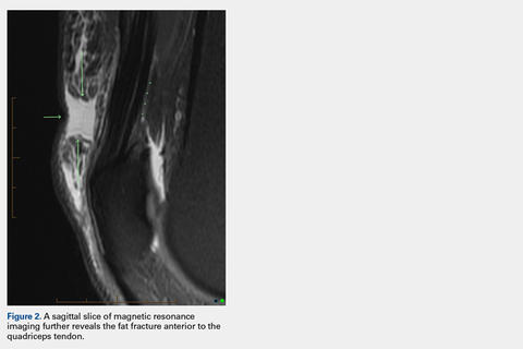

Continue to: These findings explained...
These findings explained the delayed course in resolution of symptoms. Over the next 48 hours, continued conservative management, as outlined above, led to the resolution of symptoms, and the athlete returned to play. At a 2-month follow-up, the athlete described normal function in his knee without any residual symptoms. He returned to play without any symptoms. At 6 months, the athlete underwent MRI of the same knee for an unrelated reason. MRI revealed a healed fat fracture with resolution of the fluid defect in the subcutaneous fat (Figures 4A, 4B).

DISCUSSION
A fat fracture was first described in 1972 in 12 cases of buttock fat fractures after blunt trauma.3 The authors explained that fat lobules are typically arranged in layers and supported by horizontal and vertical fibrous septa. Typical loads flatten the lobules and disperse the forces throughout the layer. However, abnormal loads to a local area disrupt the fat lobules and shear the septa, resulting in decreased integrity of the interface between the epidermis and the fascia.
However, the extremities typically have less adipose tissue than in the buttocks, and the anterior knee is prone to blunt trauma. A previous description of a fat fracture in the knee noted a palpable defect in the quadriceps tendon and an inability to perform a straight leg raise. Our case initially presented with swelling, which concealed any soft-tissue defect. Furthermore, a straight leg raise was always intact despite the fat fracture defect surfacing after anterior swelling subsided. However, the disparity in these 2 cases highlights the spectrum of injury that is possible, as well as the difficulty in diagnosing a fat fracture. The previous report used ultrasound to confirm the diagnosis and assess the integrity of adjacent musculotendinous structures. An ultrasound may be readily available in athletic training rooms.1 Of note, to the best of our knowledge, this is the first case in the literature to report a fat fracture in a professional athlete and in baseball players. Furthermore, this case report describes an athlete who presented with anterior and medial knee pain. The edema from the fat fracture dispersed into the medial prepatellar bursa, which could be confused with edema from an injury to the medial-sided soft tissues.
Although these injuries do not require operative management, conservative measures may not be as effective as those in a patellar contusion or ligamentous sprain, and prolonged treatment may be necessary. Additionally, healthcare providers should be aware of this possible source of injury and counsel on an appropriate recovery time. Ideally, further recognition of such injuries can facilitate improved management and a faster return to activity.
1. Thomas RH, Holt MD, James SH, White PG. 'Fat fracture'—a physical sign mimicking tendon rupture. J Bone Joint Surg Br. 2001;83(2):204-205.
2. Antich T, Randall CC, Westbrook RA, Morrissey MC, Brewster CE. Physical therapy treatment of knee extensor mechanism disorders: comparison of four treatment modalities*. J Orthop Sports Phys Ther. 1986;8(5):255-259.
3. Meggitt BF, Wilson JN. The battered buttock syndrome—fat fractures. A report on a group of traumatic lipomata. Br J Surg. 1972;59(3):165-169.
ABSTRACT
Blunt trauma to the anterior knee typically results in a contusion or fracture of the patella. Additionally, injury to the extensor mechanism may come from a partial or full disruption of the patellar or quadriceps tendon. A professional baseball player suffered an injury to his knee after he collided with an outfield wall. Acute swelling in the suprapatellar soft tissues concealed a palpable defect, which initially was suspected to be an injury to the quadriceps tendon. Magnetic resonance imaging of the knee revealed an intact extensor mechanism; moreover, a fracture of the subcutaneous fat anterior to the quadriceps tendon was evident and diagnosed as a fat fracture.
Fat fracture is a rare diagnosis, and to the best of our knowledge, this is the first reported diagnosis in a professional athlete. Conservative management including, but not limited to, range of motion exercises, hydrotherapy, and iontophoresis effectively treated the athlete’s injury.
Blunt trauma to the anterior knee can result in a contusion or fracture of the patella, subluxation of the patella, and injury to the quadriceps or patellar tendon. Typically, a contusion or non-displaced fracture of the patella clinically presents with a direct anterior effusion and point tenderness. A displaced fracture or tendon deficit typically has an extensor lag or weakness in extension. Fat fracture or traumatic lipomata has been previously described in 1 case of anterior knee pain after blunt injury.1
In this article, we present the case of a 32-year-old professional baseball player who suffered a blunt injury to his left knee after collision with the outfield wall and experienced both anterior and medial knee pain. The patient provided written informed consent for print and electronic publication of this case report.
CASE
A 32-year-old outfielder for a professional baseball team was attempting a catch in the outfield when his left knee collided with the padded outfield wall in a semiflexed position. The player was able to walk off the field in the middle of the inning; however, he then experienced increasing pain and was unable to return to play. He had no prior history of significant knee pain or injury. He complained only of pain, with no instability or sensation of catching or locking.
Continue to: Physical examination of the patient...
Physical examination of the patient revealed a grade 1+ swelling over the anterior aspect of the superior pole of the patella in the prepatellar region, as well as medially over the medial femoral condyle. However, there was no joint effusion. Palpation of the superomedial aspect of the patella elicited pain, but no medial joint line tenderness was elicited. Percussion testing to the patella was negative. There were no gross palpable defects in the extensor mechanism, and the patient was able to perform a straight leg raise against resistance with pain.
Mild coronal laxity of the patella was noted compared with that of the contralateral knee. Hip range of motion (ROM) was intact, but knee ROM was limited to 110° of flexion, with the complaint of anterior tightness at this position. He was able to fully extend his knee without symptoms. The knee was stable to varus and valgus stress at both 0° and 30° of flexion. Lachman and anterior and posterior drawer tests were negative and symmetric to the contralateral knee. The McMurray test for meniscal pathology also was negative. Radiographs of the left knee were completed and were negative for fracture.
OUTCOMES
The initial clinical diagnosis was a patellar contusion and sprain of the medial retinaculum, and the athlete was treated with multiple modalities available in the athletic training room. Rehabilitation included activity modifications, passive and active ROM activities, quadriceps isometric exercises, and neuromuscular control activities. Adjunctive modalities included cryotherapy, hydrotherapy, topical hematoma cream, and iontophoresis.2 This aggressive treatment was continued for 3 days with decreased but persistent pain with running drills and limited knee flexion. Repeat clinical examination revealed a decreased swelling, but there was evidence of a clinically palpable defect anteriorly proximal to the patella. Although the patient could perform a straight leg raise, a partial injury to the quadriceps became plausible. Magnetic resonance imaging (MRI) of the left knee was performed, owing to the persistent pain and limited flexion despite aggressive conservative management, as well as the palpable soft-tissue defect.
MRI was performed using a 3T (Tesla) system (GE Healthcare) with a GE Healthcare Precision 8-channel knee coil. Routine knee protocol imaging was performed to include the distal quadriceps tendon due to clinical concern for a quadriceps tear. Sagittal proton density and proton-density fat-saturated (PD FS), coronal T1 and PD FS, and axial T1 and PD FS sequences were acquired.
An acutely marginated, 1.5 cm × 3 cm, longitudinal and transverse fluid defect “crevasse” was identified at the midline in the prepatellar subcutaneous fat overlying the distal quadriceps tendon and corresponded to a clinically palpable abnormality (Figures 1, 2).
Continue to: These findings explained...
These findings explained the delayed course in resolution of symptoms. Over the next 48 hours, continued conservative management, as outlined above, led to the resolution of symptoms, and the athlete returned to play. At a 2-month follow-up, the athlete described normal function in his knee without any residual symptoms. He returned to play without any symptoms. At 6 months, the athlete underwent MRI of the same knee for an unrelated reason. MRI revealed a healed fat fracture with resolution of the fluid defect in the subcutaneous fat (Figures 4A, 4B).
DISCUSSION
A fat fracture was first described in 1972 in 12 cases of buttock fat fractures after blunt trauma.3 The authors explained that fat lobules are typically arranged in layers and supported by horizontal and vertical fibrous septa. Typical loads flatten the lobules and disperse the forces throughout the layer. However, abnormal loads to a local area disrupt the fat lobules and shear the septa, resulting in decreased integrity of the interface between the epidermis and the fascia.
However, the extremities typically have less adipose tissue than in the buttocks, and the anterior knee is prone to blunt trauma. A previous description of a fat fracture in the knee noted a palpable defect in the quadriceps tendon and an inability to perform a straight leg raise. Our case initially presented with swelling, which concealed any soft-tissue defect. Furthermore, a straight leg raise was always intact despite the fat fracture defect surfacing after anterior swelling subsided. However, the disparity in these 2 cases highlights the spectrum of injury that is possible, as well as the difficulty in diagnosing a fat fracture. The previous report used ultrasound to confirm the diagnosis and assess the integrity of adjacent musculotendinous structures. An ultrasound may be readily available in athletic training rooms.1 Of note, to the best of our knowledge, this is the first case in the literature to report a fat fracture in a professional athlete and in baseball players. Furthermore, this case report describes an athlete who presented with anterior and medial knee pain. The edema from the fat fracture dispersed into the medial prepatellar bursa, which could be confused with edema from an injury to the medial-sided soft tissues.
Although these injuries do not require operative management, conservative measures may not be as effective as those in a patellar contusion or ligamentous sprain, and prolonged treatment may be necessary. Additionally, healthcare providers should be aware of this possible source of injury and counsel on an appropriate recovery time. Ideally, further recognition of such injuries can facilitate improved management and a faster return to activity.
ABSTRACT
Blunt trauma to the anterior knee typically results in a contusion or fracture of the patella. Additionally, injury to the extensor mechanism may come from a partial or full disruption of the patellar or quadriceps tendon. A professional baseball player suffered an injury to his knee after he collided with an outfield wall. Acute swelling in the suprapatellar soft tissues concealed a palpable defect, which initially was suspected to be an injury to the quadriceps tendon. Magnetic resonance imaging of the knee revealed an intact extensor mechanism; moreover, a fracture of the subcutaneous fat anterior to the quadriceps tendon was evident and diagnosed as a fat fracture.
Fat fracture is a rare diagnosis, and to the best of our knowledge, this is the first reported diagnosis in a professional athlete. Conservative management including, but not limited to, range of motion exercises, hydrotherapy, and iontophoresis effectively treated the athlete’s injury.
Blunt trauma to the anterior knee can result in a contusion or fracture of the patella, subluxation of the patella, and injury to the quadriceps or patellar tendon. Typically, a contusion or non-displaced fracture of the patella clinically presents with a direct anterior effusion and point tenderness. A displaced fracture or tendon deficit typically has an extensor lag or weakness in extension. Fat fracture or traumatic lipomata has been previously described in 1 case of anterior knee pain after blunt injury.1
In this article, we present the case of a 32-year-old professional baseball player who suffered a blunt injury to his left knee after collision with the outfield wall and experienced both anterior and medial knee pain. The patient provided written informed consent for print and electronic publication of this case report.
CASE
A 32-year-old outfielder for a professional baseball team was attempting a catch in the outfield when his left knee collided with the padded outfield wall in a semiflexed position. The player was able to walk off the field in the middle of the inning; however, he then experienced increasing pain and was unable to return to play. He had no prior history of significant knee pain or injury. He complained only of pain, with no instability or sensation of catching or locking.
Continue to: Physical examination of the patient...
Physical examination of the patient revealed a grade 1+ swelling over the anterior aspect of the superior pole of the patella in the prepatellar region, as well as medially over the medial femoral condyle. However, there was no joint effusion. Palpation of the superomedial aspect of the patella elicited pain, but no medial joint line tenderness was elicited. Percussion testing to the patella was negative. There were no gross palpable defects in the extensor mechanism, and the patient was able to perform a straight leg raise against resistance with pain.
Mild coronal laxity of the patella was noted compared with that of the contralateral knee. Hip range of motion (ROM) was intact, but knee ROM was limited to 110° of flexion, with the complaint of anterior tightness at this position. He was able to fully extend his knee without symptoms. The knee was stable to varus and valgus stress at both 0° and 30° of flexion. Lachman and anterior and posterior drawer tests were negative and symmetric to the contralateral knee. The McMurray test for meniscal pathology also was negative. Radiographs of the left knee were completed and were negative for fracture.
OUTCOMES
The initial clinical diagnosis was a patellar contusion and sprain of the medial retinaculum, and the athlete was treated with multiple modalities available in the athletic training room. Rehabilitation included activity modifications, passive and active ROM activities, quadriceps isometric exercises, and neuromuscular control activities. Adjunctive modalities included cryotherapy, hydrotherapy, topical hematoma cream, and iontophoresis.2 This aggressive treatment was continued for 3 days with decreased but persistent pain with running drills and limited knee flexion. Repeat clinical examination revealed a decreased swelling, but there was evidence of a clinically palpable defect anteriorly proximal to the patella. Although the patient could perform a straight leg raise, a partial injury to the quadriceps became plausible. Magnetic resonance imaging (MRI) of the left knee was performed, owing to the persistent pain and limited flexion despite aggressive conservative management, as well as the palpable soft-tissue defect.
MRI was performed using a 3T (Tesla) system (GE Healthcare) with a GE Healthcare Precision 8-channel knee coil. Routine knee protocol imaging was performed to include the distal quadriceps tendon due to clinical concern for a quadriceps tear. Sagittal proton density and proton-density fat-saturated (PD FS), coronal T1 and PD FS, and axial T1 and PD FS sequences were acquired.
An acutely marginated, 1.5 cm × 3 cm, longitudinal and transverse fluid defect “crevasse” was identified at the midline in the prepatellar subcutaneous fat overlying the distal quadriceps tendon and corresponded to a clinically palpable abnormality (Figures 1, 2).
Continue to: These findings explained...
These findings explained the delayed course in resolution of symptoms. Over the next 48 hours, continued conservative management, as outlined above, led to the resolution of symptoms, and the athlete returned to play. At a 2-month follow-up, the athlete described normal function in his knee without any residual symptoms. He returned to play without any symptoms. At 6 months, the athlete underwent MRI of the same knee for an unrelated reason. MRI revealed a healed fat fracture with resolution of the fluid defect in the subcutaneous fat (Figures 4A, 4B).
DISCUSSION
A fat fracture was first described in 1972 in 12 cases of buttock fat fractures after blunt trauma.3 The authors explained that fat lobules are typically arranged in layers and supported by horizontal and vertical fibrous septa. Typical loads flatten the lobules and disperse the forces throughout the layer. However, abnormal loads to a local area disrupt the fat lobules and shear the septa, resulting in decreased integrity of the interface between the epidermis and the fascia.
However, the extremities typically have less adipose tissue than in the buttocks, and the anterior knee is prone to blunt trauma. A previous description of a fat fracture in the knee noted a palpable defect in the quadriceps tendon and an inability to perform a straight leg raise. Our case initially presented with swelling, which concealed any soft-tissue defect. Furthermore, a straight leg raise was always intact despite the fat fracture defect surfacing after anterior swelling subsided. However, the disparity in these 2 cases highlights the spectrum of injury that is possible, as well as the difficulty in diagnosing a fat fracture. The previous report used ultrasound to confirm the diagnosis and assess the integrity of adjacent musculotendinous structures. An ultrasound may be readily available in athletic training rooms.1 Of note, to the best of our knowledge, this is the first case in the literature to report a fat fracture in a professional athlete and in baseball players. Furthermore, this case report describes an athlete who presented with anterior and medial knee pain. The edema from the fat fracture dispersed into the medial prepatellar bursa, which could be confused with edema from an injury to the medial-sided soft tissues.
Although these injuries do not require operative management, conservative measures may not be as effective as those in a patellar contusion or ligamentous sprain, and prolonged treatment may be necessary. Additionally, healthcare providers should be aware of this possible source of injury and counsel on an appropriate recovery time. Ideally, further recognition of such injuries can facilitate improved management and a faster return to activity.
1. Thomas RH, Holt MD, James SH, White PG. 'Fat fracture'—a physical sign mimicking tendon rupture. J Bone Joint Surg Br. 2001;83(2):204-205.
2. Antich T, Randall CC, Westbrook RA, Morrissey MC, Brewster CE. Physical therapy treatment of knee extensor mechanism disorders: comparison of four treatment modalities*. J Orthop Sports Phys Ther. 1986;8(5):255-259.
3. Meggitt BF, Wilson JN. The battered buttock syndrome—fat fractures. A report on a group of traumatic lipomata. Br J Surg. 1972;59(3):165-169.
1. Thomas RH, Holt MD, James SH, White PG. 'Fat fracture'—a physical sign mimicking tendon rupture. J Bone Joint Surg Br. 2001;83(2):204-205.
2. Antich T, Randall CC, Westbrook RA, Morrissey MC, Brewster CE. Physical therapy treatment of knee extensor mechanism disorders: comparison of four treatment modalities*. J Orthop Sports Phys Ther. 1986;8(5):255-259.
3. Meggitt BF, Wilson JN. The battered buttock syndrome—fat fractures. A report on a group of traumatic lipomata. Br J Surg. 1972;59(3):165-169.
TAKE-HOME POINTS
- A fat fracture should be considered in the setting of a blunt injury to the anterior knee when a palpable soft-tissue defect is observed and the extensor mechanism is clinically intact.
- An ultrasound or MRI can assist in making the diagnosis, which can aid in guiding the patient with management and in determining the expected duration of symptoms.
- Injuries to the anterior knee that may present as contusions but have a prolonged course of symptoms should not be overlooked.

Patient advocacy groups take in millions from drugmakers. Is there a payback?
Pharmaceutical companies gave at least $116 million to patient advocacy groups in a single year, reveals a new database logging 12,000 donations from large publicly traded drugmakers to such organizations.
Even as these patient groups grow in number and political influence, their funding and their relationships to drugmakers are little understood. Unlike payments to doctors and lobbying expenses, companies do not have to report payments to the groups.
The database, called “Pre$cription for Power,” shows that . The 14 companies that contributed $116 million to patient advocacy groups reported only about $63 million in lobbying activities that same year.
Though their primary missions are to focus attention on the needs of patients with a particular disease – such as arthritis, heart disease or various cancers – some groups effectively supplement the work lobbyists perform, providing patients to testify on Capitol Hill and organizing letter-writing and social media campaigns that are beneficial to pharmaceutical companies.
Six drugmakers, the data show, contributed a million dollars or more to individual groups that represent patients who rely on their drugs. The database identifies more than 1,200 patient groups. Of those, 594 accepted money from the drugmakers in the database.
The financial ties are troubling if they cause even one patient group to act in a way that’s “not fully representing the interest of its constituents,” said Matthew McCoy, a medical ethics professor at the University of Pennsylvania, Philadelphia, who coauthored a 2017 study about patient advocacy groups’ influence and transparency.
Notably, such groups have been silent or slow to complain about high or escalating prices, a prime concern of patients.
“When so many patient organizations are being influenced in this way, it can shift our whole approach to health policy, taking away from the interests of patients and towards the interests of industry,” Mr. McCoy said. “That’s not just a problem for the patients and caregivers that particular patient organizations serve; that’s a problem for everyone.”
Bristol-Myers Squibb provides a stark example of how patient groups are valued. In 2015, it spent more than $20.5 million on patient groups, compared with $2.9 million on federal lobbying and less than $1 million on major trade associations, according to public records and company disclosures. The company said its decisions regarding lobbying and contributions to patient groups are “unrelated.”
“Bristol-Myers Squibb is focused on supporting a health care environment that rewards innovation and ensures access to medicines for patients,” said spokeswoman Laura Hortas. “The company supports patient organizations with this shared objective.”
The first-of-its-kind database, compiled by Kaiser Health News, tallies the money from Big Pharma to patient groups. KHN examined the 20 pharmaceutical firms included in the S&P 500, 14 of which were transparent – in varying degrees – about giving money to patient groups. Pre$cription for Power is based on information contained in charitable giving reports from company websites and federal 990 regulatory filings.
It spotlights donations pharma companies made to patient groups large and small. The recipients include well-known disease groups, like the American Diabetes Association, with revenues of hundreds of millions of dollars; high-profile foundations like Susan G. Komen, a patient group focused on breast cancer; and smaller, lesser-known groups, like the Caring Ambassadors Program, which focuses on lung cancer and hepatitis C.
The data show that 15 patient groups – with annual revenues as large as $3.6 million – relied on the pharmaceutical companies for at least 20% of their revenue, and some relied on them for more than half of their revenue. The database explores only a slice of the pharmaceutical industry’s giving overall and will be expanded with more companies and groups over time.
“It’s clear that more transparency in this space is vitally important,” said Sen. Claire McCaskill (D-Mo.), who has been investigating the links between patient advocates and opioid manufacturers and is considering legislation to track funding. “This database is one step forward in that effort, but we also need Congress to act.”
What drives the money flow
The financial ties between drugmakers and the organizations that represent those who use or prescribe their blockbuster medicines have been of growing concern as drug prices escalate. The Senate investigated conflicts of interest in the run-up to the passage of the 2010 Physician Payments Sunshine Act – a law that required payments to physicians from makers of drugs and devices to be registered on a public website – but patient groups were not addressed in the bill.
Some of the patient groups with ties to trade groups echo industry talking points in media campaigns and letters to federal agencies, and do little else. And patients, supported by pharma, are dispatched to state capitals and Washington to support research funding. Some groups send patients updates on the newest drugs and industry products.
“It’s through groups like this that patients often learn about illnesses and treatments,” said Rick Claypool, a research director for Public Citizen, a consumer advocacy group that says it does not accept pharmaceutical funding.
For the patient group Caring Ambassadors Program, industry funds are needed to make up for a lack of public funding, said the group’s executive director, Lorren Sandt. According to IRS filings and published company reports, in 2015, the group received $413,000, the bulk of which came from one company, AbbVie, which makes a hepatitis C treatment and has been testing a new lung cancer drug, Rova-T, not yet approved. She said the money had no influence on the Caring Ambassadors Program’s priorities.
“There aren’t a lot of large pockets of funding outside of the pharmaceutical money,” Sandt said. “We take it where we can find it.”
Other patient groups such as the National Women’s Health Network, based in Washington, make sacrifices to avoid pharmaceutical funding. That includes operating with a small staff in a “modest” office building with few windows and outdated computers, according to executive director Cindy Pearson. “You can see the effect of our approach to funding as soon as you walk [in] the door.”
Pearson said it’s hard for patient groups not to be influenced by the funder, even if they proclaim independence. Patient groups “build relationships with their funders and feel in sync and have sympathy” for them. “It’s human nature. It’s not evil or weak, but it’s wrong.”
Charity as marketing
Patients newly diagnosed with a disease often turn to patient advocacy groups for advice, but the money flow to such groups may distort patients’ knowledge and public debate over treatment options, said Dr. Adriane Fugh-Berman, the director of PharmedOut, a Georgetown University Medical Center program in Washington that is critical of some pharmaceutical marketing practices.
“[The money flow limits] their advocacy agenda to competing branded products when the best therapy might be generics, over-the-counter drugs or diet and exercise,” she said.
AbbVie – whose specialty drug Humira made up 65% of the company’s net revenue in 2017 and is used to treat patients with autoimmune diseases, including Crohn’s disease and certain kinds of arthritis – gave $2.7 million to the Crohn’s & Colitis Foundation and $1.6 million to the Arthritis Foundation, according to the company’s public disclosures included in the database. The list price for a month’s supply of Humira, a biologic drug, is $4,872, according to Express Scripts, a pharmacy benefits manager.
Even though Humira will face competition from near-copycat drugs called biosimilars, it is expected to remain the highest-grossing drug in the United States through 2022, according to drug industry analysts at EvaluatePharma.
The Arthritis and Crohn’s foundations have been largely silent on the cost of Humira and vocal on safety concerns about biosimilars. The Arthritis Foundation has championed state laws that could add extra steps for consumers to receive biosimilars at the pharmacy counter, potentially keeping more patients on the brand-name drug. Experts say those laws could help protect Humira’s market share from generic competitors.
A coalition of patient groups, Patients for Biologics Safety & Access, opposes the automatic substitution of a cheaper biosimilar when doctors prescribe a biologic. In 2015, members of that coalition, including the Crohn’s & Colitis Foundation, the Arthritis Foundation and the Lupus Foundation of America, accepted about $9.1 million from pharmaceutical companies in the database, according to public disclosures. They include AbbVie and Johnson & Johnson, makers of blockbuster biologics.
The Arthritis Foundation did not deny receiving the money but said the foundation represents patients, not sponsors. It is “optimistic” about biosimilars’ ability to help patients and save them money, said Anna Hyde, vice president of advocacy and access. “The Foundation supports the Food and Drug Administration’s scientific standards in evaluating the safety and efficacy of biosimilars, and we support policies that encourage innovation and foster a competitive marketplace.”
The Crohn’s & Colitis Foundation maintains “more than an arm’s-length distance” from its donors in the pharmaceutical industry, who have no say over the foundation’s strategic objectives, said president and CEO Michael Osso.
He added that the foundation’s position on biosimilars is “evolving.”
Lupus Foundation CEO Sandra Raymond said she could not explain how her group, also based in Washington, was involved in the coalition. She confirmed the Lupus Foundation received $444,000 from Pfizer in 2015 but said the money was not linked to any relationship with Patients for Biologics Safety & Access.
“I never went to a meeting,” Raymond said. “A former employee signed us up for a whole host of coalitions. I think we put our name on something or someone did.”
She said the Lupus Foundation was no longer a member of the coalition. Days after Kaiser Health News reached out to the coalition, its website was updated, excluding the Lupus Foundation.
For its part, AbbVie – which overall donated $24.7 million to patient groups in 2015, according to the new database – stipulates that its grants to nonprofits are “non-promotional” and provide no direct benefit to its business, according to a company statement. The company gives to patient groups because they serve as an “important, unbiased and independent resource for patients and caregivers.”
Insulin and influence
The American Diabetes Association said in an email to KHN that it received $18.3 million in pharmaceutical funding in 2017, accounting for 12.3% of its revenue; that was down from $26.7 million in 2015. The money flowed in as insulin makers continued to hike prices in those years – up to four times per product – leading to hardships for patients.
The only “Big Three” insulin maker in the database, Eli Lilly, gave $2.9 million to the American Diabetes Association in 2015, according to disclosures from the company and its foundation. Sanofi and Novo Nordisk are the other two major insulin makers, but neither was in the S&P 500 and therefore not included in the database. Over the past 20 years, Eli Lilly has repeatedly raised prices on its best-selling insulins, Humalog and Humulin, even though the medicines have been around for decades. The drugmaker faced protests – by people demanding to know the cost of manufacturing a vial of insulin – at its Indianapolis headquarters last fall.
The ADA launched a campaign decrying “skyrocketing” insulin in late 2016 but did not call out any drugmaker in its literature. When legislators in Nevada passed a bill last year requiring insulin makers to disclose their profits to the public, the ADA did not take a public stance.
The American Diabetes Association said it doesn’t confront individual companies because it is seeking action from “all entities in the supply chain” – manufacturers, wholesalers, pharmacy benefit managers and insurers.
“As a public health organization, the ADA’s commitment and focus is on the needs of the more than 30 million people with diabetes,” said Dr. William Cefalu, its chief scientific and medical officer. “The ADA requires support from a diverse set of partners to achieve this objective.”
Eli Lilly said it contributes money to the American Diabetes Association because the two share a “common goal” of helping diabetes patients.
“We provide funding for a wide variety of educational programs and opportunities at ADA, and they design and implement those programs in ways that are aligned with their goals,” Eli Lilly said in a statement. “We’re proud to support the ADA on important work that helps millions of people living with diabetes.”
Most patient groups say that funders have little or no influence in shaping their programs and policies, but their agreements are private.
They weren’t always backed by Pharma
Into the ’80s and early ’90s, patient lobbying was generally limited and self-funded with only one or two affluent patients from an organization traveling to Washington on a given day, said Diana Zuckerman, PhD, president of the nonprofit National Center for Health Research.
But the power of patient-lobbyists became apparent after a successful campaign by AIDS patients led to government action and a national push to find drugs to treat the then-terminal disease. Dr. Zuckerman said she will never forget when two women visited her office and asked how breast cancer patients could be as effective as the AIDS patients.
“At the time, there were no breast cancer patients advocating for money or anything else. It’s hard to believe,” she said. “I still remember that conversation, because it was really a turning point.”
Soon after, breast cancer patients started visiting the Hill more frequently. Patients with other diseases followed. Over time, patients’ voices became a potent force, often with industry support.
Even some wealthy, high-profile organizations take industry money: For example, $459,000 of Susan G. Komen’s $118 million in 2015 revenue came from drugmakers in the database, according to public disclosures. Asked about the pharma money, the foundation said it has institutional processes in place to ensure that “no corporate partner – pharma or otherwise – decides our mission priorities,” including a scientific advisory board – free of sponsor influence – that reviews its research program.
Today, patient advocacy groups flush with more industry dollars fly patients in for testimony and training about how to lobby for their drugs.
Some years ago, as the groups increased in number, Dr. Zuckerman said, she started getting email invitations from advocacy groups to attend so-called lobbying days explicitly sponsored by the pharmaceutical industry. The hosts often promised training and usually some kind of keynote speaker at a luncheon in Washington – plus a potential scholarship to cover travel. Now, lobbying days involving dozens of patients from a single group are part of the landscape.
Dan Boston, president of lobbying firm Health Policy Source, said, “It would be naive to think these people on a Tuesday afternoon just happen to turn up in XYZ places,” adding that the money isn’t necessarily a bad thing. Money tends to flow toward citizen groups that already have the same priorities as their funders, he said.
Marching into the future
Patient groups have been successful at campaigning for drug approvals, at times sparking controversy.
When scientists within the FDA advised against the approval of Exondys 51, a drug to treat Duchenne muscular dystrophy, parents of children with the rare genetic disorder and patients rallied to lobby for it in Washington. They were seen as pivotal to the FDA’s 2016 decision to grant approval for the drug, made by Sarepta Therapeutics. The decision was controversial in part because the FDA noted that clinical benefits of the drug – aimed at a subset of people with Duchenne muscular dystrophy – were not yet established.
Sarepta Therapeutics, which is not featured in the database, has taken measures to support its patient base. In March, it announced an annual scholarship program – 10 grants of up to $10,000 each for students with Duchenne muscular dystrophy to attend university or trade schools. Sarepta is also among the funders of Parent Project Muscular Dystrophy, a patient advocacy group at the forefront of the push for Exondys 51’s approval.
The Pre$cription for Power database will grow to include new disclosures. Not all drugmakers are willing to disclose their company giving. Eleven of the 20 companies examined – Allergan, Baxter International, Biogen, Celgene, Endo International, Gilead Sciences, Mallinckrodt, Mylan, Perrigo Co., Regeneron Pharmaceuticals, and Vertex Pharmaceuticals – declined to disclose their company giving or did not respond to repeated calls.
Paul Thacker, a former investigator for Sen. Chuck Grassley (R-Iowa) who helped draft the Physician Payments Sunshine Act in 2010, said there is reason to question the flow of money to patient advocacy groups. The pharmaceutical industry has fostered relationships in every link of the drug supply chain, including payments to researchers, doctors and professional societies.
“There’s so much money out there, and they’ve created all of these allies, so nobody is clamoring for change,” Mr. Thacker said.
Since the Physician Payments Sunshine Act began requiring the industry to report its payments to physicians, the industry is more reluctant to co-opt them, so “pharma has to find other megaphones,” PharmedOut’s Dr. Fugh-Berman said.
And, in times of public outrage over high drug prices and soaring insurance costs, patients are particularly sympathetic messengers, she said.
“Sick consumers make for good press,” Dr. Fugh-Berman said. “They make for good testimony before Congress. They can be very powerful spokespeople for pharmaceutical companies.”
KHN’s coverage of prescription drug development, costs and pricing is supported by the Laura and John Arnold Foundation. Kaiser Health News is a nonprofit news service covering health issues. It is an editorially independent program of the Kaiser Family Foundation that is not affiliated with Kaiser Permanente.
Pharmaceutical companies gave at least $116 million to patient advocacy groups in a single year, reveals a new database logging 12,000 donations from large publicly traded drugmakers to such organizations.
Even as these patient groups grow in number and political influence, their funding and their relationships to drugmakers are little understood. Unlike payments to doctors and lobbying expenses, companies do not have to report payments to the groups.
The database, called “Pre$cription for Power,” shows that . The 14 companies that contributed $116 million to patient advocacy groups reported only about $63 million in lobbying activities that same year.
Though their primary missions are to focus attention on the needs of patients with a particular disease – such as arthritis, heart disease or various cancers – some groups effectively supplement the work lobbyists perform, providing patients to testify on Capitol Hill and organizing letter-writing and social media campaigns that are beneficial to pharmaceutical companies.
Six drugmakers, the data show, contributed a million dollars or more to individual groups that represent patients who rely on their drugs. The database identifies more than 1,200 patient groups. Of those, 594 accepted money from the drugmakers in the database.
The financial ties are troubling if they cause even one patient group to act in a way that’s “not fully representing the interest of its constituents,” said Matthew McCoy, a medical ethics professor at the University of Pennsylvania, Philadelphia, who coauthored a 2017 study about patient advocacy groups’ influence and transparency.
Notably, such groups have been silent or slow to complain about high or escalating prices, a prime concern of patients.
“When so many patient organizations are being influenced in this way, it can shift our whole approach to health policy, taking away from the interests of patients and towards the interests of industry,” Mr. McCoy said. “That’s not just a problem for the patients and caregivers that particular patient organizations serve; that’s a problem for everyone.”
Bristol-Myers Squibb provides a stark example of how patient groups are valued. In 2015, it spent more than $20.5 million on patient groups, compared with $2.9 million on federal lobbying and less than $1 million on major trade associations, according to public records and company disclosures. The company said its decisions regarding lobbying and contributions to patient groups are “unrelated.”
“Bristol-Myers Squibb is focused on supporting a health care environment that rewards innovation and ensures access to medicines for patients,” said spokeswoman Laura Hortas. “The company supports patient organizations with this shared objective.”
The first-of-its-kind database, compiled by Kaiser Health News, tallies the money from Big Pharma to patient groups. KHN examined the 20 pharmaceutical firms included in the S&P 500, 14 of which were transparent – in varying degrees – about giving money to patient groups. Pre$cription for Power is based on information contained in charitable giving reports from company websites and federal 990 regulatory filings.
It spotlights donations pharma companies made to patient groups large and small. The recipients include well-known disease groups, like the American Diabetes Association, with revenues of hundreds of millions of dollars; high-profile foundations like Susan G. Komen, a patient group focused on breast cancer; and smaller, lesser-known groups, like the Caring Ambassadors Program, which focuses on lung cancer and hepatitis C.
The data show that 15 patient groups – with annual revenues as large as $3.6 million – relied on the pharmaceutical companies for at least 20% of their revenue, and some relied on them for more than half of their revenue. The database explores only a slice of the pharmaceutical industry’s giving overall and will be expanded with more companies and groups over time.
“It’s clear that more transparency in this space is vitally important,” said Sen. Claire McCaskill (D-Mo.), who has been investigating the links between patient advocates and opioid manufacturers and is considering legislation to track funding. “This database is one step forward in that effort, but we also need Congress to act.”
What drives the money flow
The financial ties between drugmakers and the organizations that represent those who use or prescribe their blockbuster medicines have been of growing concern as drug prices escalate. The Senate investigated conflicts of interest in the run-up to the passage of the 2010 Physician Payments Sunshine Act – a law that required payments to physicians from makers of drugs and devices to be registered on a public website – but patient groups were not addressed in the bill.
Some of the patient groups with ties to trade groups echo industry talking points in media campaigns and letters to federal agencies, and do little else. And patients, supported by pharma, are dispatched to state capitals and Washington to support research funding. Some groups send patients updates on the newest drugs and industry products.
“It’s through groups like this that patients often learn about illnesses and treatments,” said Rick Claypool, a research director for Public Citizen, a consumer advocacy group that says it does not accept pharmaceutical funding.
For the patient group Caring Ambassadors Program, industry funds are needed to make up for a lack of public funding, said the group’s executive director, Lorren Sandt. According to IRS filings and published company reports, in 2015, the group received $413,000, the bulk of which came from one company, AbbVie, which makes a hepatitis C treatment and has been testing a new lung cancer drug, Rova-T, not yet approved. She said the money had no influence on the Caring Ambassadors Program’s priorities.
“There aren’t a lot of large pockets of funding outside of the pharmaceutical money,” Sandt said. “We take it where we can find it.”
Other patient groups such as the National Women’s Health Network, based in Washington, make sacrifices to avoid pharmaceutical funding. That includes operating with a small staff in a “modest” office building with few windows and outdated computers, according to executive director Cindy Pearson. “You can see the effect of our approach to funding as soon as you walk [in] the door.”
Pearson said it’s hard for patient groups not to be influenced by the funder, even if they proclaim independence. Patient groups “build relationships with their funders and feel in sync and have sympathy” for them. “It’s human nature. It’s not evil or weak, but it’s wrong.”
Charity as marketing
Patients newly diagnosed with a disease often turn to patient advocacy groups for advice, but the money flow to such groups may distort patients’ knowledge and public debate over treatment options, said Dr. Adriane Fugh-Berman, the director of PharmedOut, a Georgetown University Medical Center program in Washington that is critical of some pharmaceutical marketing practices.
“[The money flow limits] their advocacy agenda to competing branded products when the best therapy might be generics, over-the-counter drugs or diet and exercise,” she said.
AbbVie – whose specialty drug Humira made up 65% of the company’s net revenue in 2017 and is used to treat patients with autoimmune diseases, including Crohn’s disease and certain kinds of arthritis – gave $2.7 million to the Crohn’s & Colitis Foundation and $1.6 million to the Arthritis Foundation, according to the company’s public disclosures included in the database. The list price for a month’s supply of Humira, a biologic drug, is $4,872, according to Express Scripts, a pharmacy benefits manager.
Even though Humira will face competition from near-copycat drugs called biosimilars, it is expected to remain the highest-grossing drug in the United States through 2022, according to drug industry analysts at EvaluatePharma.
The Arthritis and Crohn’s foundations have been largely silent on the cost of Humira and vocal on safety concerns about biosimilars. The Arthritis Foundation has championed state laws that could add extra steps for consumers to receive biosimilars at the pharmacy counter, potentially keeping more patients on the brand-name drug. Experts say those laws could help protect Humira’s market share from generic competitors.
A coalition of patient groups, Patients for Biologics Safety & Access, opposes the automatic substitution of a cheaper biosimilar when doctors prescribe a biologic. In 2015, members of that coalition, including the Crohn’s & Colitis Foundation, the Arthritis Foundation and the Lupus Foundation of America, accepted about $9.1 million from pharmaceutical companies in the database, according to public disclosures. They include AbbVie and Johnson & Johnson, makers of blockbuster biologics.
The Arthritis Foundation did not deny receiving the money but said the foundation represents patients, not sponsors. It is “optimistic” about biosimilars’ ability to help patients and save them money, said Anna Hyde, vice president of advocacy and access. “The Foundation supports the Food and Drug Administration’s scientific standards in evaluating the safety and efficacy of biosimilars, and we support policies that encourage innovation and foster a competitive marketplace.”
The Crohn’s & Colitis Foundation maintains “more than an arm’s-length distance” from its donors in the pharmaceutical industry, who have no say over the foundation’s strategic objectives, said president and CEO Michael Osso.
He added that the foundation’s position on biosimilars is “evolving.”
Lupus Foundation CEO Sandra Raymond said she could not explain how her group, also based in Washington, was involved in the coalition. She confirmed the Lupus Foundation received $444,000 from Pfizer in 2015 but said the money was not linked to any relationship with Patients for Biologics Safety & Access.
“I never went to a meeting,” Raymond said. “A former employee signed us up for a whole host of coalitions. I think we put our name on something or someone did.”
She said the Lupus Foundation was no longer a member of the coalition. Days after Kaiser Health News reached out to the coalition, its website was updated, excluding the Lupus Foundation.
For its part, AbbVie – which overall donated $24.7 million to patient groups in 2015, according to the new database – stipulates that its grants to nonprofits are “non-promotional” and provide no direct benefit to its business, according to a company statement. The company gives to patient groups because they serve as an “important, unbiased and independent resource for patients and caregivers.”
Insulin and influence
The American Diabetes Association said in an email to KHN that it received $18.3 million in pharmaceutical funding in 2017, accounting for 12.3% of its revenue; that was down from $26.7 million in 2015. The money flowed in as insulin makers continued to hike prices in those years – up to four times per product – leading to hardships for patients.
The only “Big Three” insulin maker in the database, Eli Lilly, gave $2.9 million to the American Diabetes Association in 2015, according to disclosures from the company and its foundation. Sanofi and Novo Nordisk are the other two major insulin makers, but neither was in the S&P 500 and therefore not included in the database. Over the past 20 years, Eli Lilly has repeatedly raised prices on its best-selling insulins, Humalog and Humulin, even though the medicines have been around for decades. The drugmaker faced protests – by people demanding to know the cost of manufacturing a vial of insulin – at its Indianapolis headquarters last fall.
The ADA launched a campaign decrying “skyrocketing” insulin in late 2016 but did not call out any drugmaker in its literature. When legislators in Nevada passed a bill last year requiring insulin makers to disclose their profits to the public, the ADA did not take a public stance.
The American Diabetes Association said it doesn’t confront individual companies because it is seeking action from “all entities in the supply chain” – manufacturers, wholesalers, pharmacy benefit managers and insurers.
“As a public health organization, the ADA’s commitment and focus is on the needs of the more than 30 million people with diabetes,” said Dr. William Cefalu, its chief scientific and medical officer. “The ADA requires support from a diverse set of partners to achieve this objective.”
Eli Lilly said it contributes money to the American Diabetes Association because the two share a “common goal” of helping diabetes patients.
“We provide funding for a wide variety of educational programs and opportunities at ADA, and they design and implement those programs in ways that are aligned with their goals,” Eli Lilly said in a statement. “We’re proud to support the ADA on important work that helps millions of people living with diabetes.”
Most patient groups say that funders have little or no influence in shaping their programs and policies, but their agreements are private.
They weren’t always backed by Pharma
Into the ’80s and early ’90s, patient lobbying was generally limited and self-funded with only one or two affluent patients from an organization traveling to Washington on a given day, said Diana Zuckerman, PhD, president of the nonprofit National Center for Health Research.
But the power of patient-lobbyists became apparent after a successful campaign by AIDS patients led to government action and a national push to find drugs to treat the then-terminal disease. Dr. Zuckerman said she will never forget when two women visited her office and asked how breast cancer patients could be as effective as the AIDS patients.
“At the time, there were no breast cancer patients advocating for money or anything else. It’s hard to believe,” she said. “I still remember that conversation, because it was really a turning point.”
Soon after, breast cancer patients started visiting the Hill more frequently. Patients with other diseases followed. Over time, patients’ voices became a potent force, often with industry support.
Even some wealthy, high-profile organizations take industry money: For example, $459,000 of Susan G. Komen’s $118 million in 2015 revenue came from drugmakers in the database, according to public disclosures. Asked about the pharma money, the foundation said it has institutional processes in place to ensure that “no corporate partner – pharma or otherwise – decides our mission priorities,” including a scientific advisory board – free of sponsor influence – that reviews its research program.
Today, patient advocacy groups flush with more industry dollars fly patients in for testimony and training about how to lobby for their drugs.
Some years ago, as the groups increased in number, Dr. Zuckerman said, she started getting email invitations from advocacy groups to attend so-called lobbying days explicitly sponsored by the pharmaceutical industry. The hosts often promised training and usually some kind of keynote speaker at a luncheon in Washington – plus a potential scholarship to cover travel. Now, lobbying days involving dozens of patients from a single group are part of the landscape.
Dan Boston, president of lobbying firm Health Policy Source, said, “It would be naive to think these people on a Tuesday afternoon just happen to turn up in XYZ places,” adding that the money isn’t necessarily a bad thing. Money tends to flow toward citizen groups that already have the same priorities as their funders, he said.
Marching into the future
Patient groups have been successful at campaigning for drug approvals, at times sparking controversy.
When scientists within the FDA advised against the approval of Exondys 51, a drug to treat Duchenne muscular dystrophy, parents of children with the rare genetic disorder and patients rallied to lobby for it in Washington. They were seen as pivotal to the FDA’s 2016 decision to grant approval for the drug, made by Sarepta Therapeutics. The decision was controversial in part because the FDA noted that clinical benefits of the drug – aimed at a subset of people with Duchenne muscular dystrophy – were not yet established.
Sarepta Therapeutics, which is not featured in the database, has taken measures to support its patient base. In March, it announced an annual scholarship program – 10 grants of up to $10,000 each for students with Duchenne muscular dystrophy to attend university or trade schools. Sarepta is also among the funders of Parent Project Muscular Dystrophy, a patient advocacy group at the forefront of the push for Exondys 51’s approval.
The Pre$cription for Power database will grow to include new disclosures. Not all drugmakers are willing to disclose their company giving. Eleven of the 20 companies examined – Allergan, Baxter International, Biogen, Celgene, Endo International, Gilead Sciences, Mallinckrodt, Mylan, Perrigo Co., Regeneron Pharmaceuticals, and Vertex Pharmaceuticals – declined to disclose their company giving or did not respond to repeated calls.
Paul Thacker, a former investigator for Sen. Chuck Grassley (R-Iowa) who helped draft the Physician Payments Sunshine Act in 2010, said there is reason to question the flow of money to patient advocacy groups. The pharmaceutical industry has fostered relationships in every link of the drug supply chain, including payments to researchers, doctors and professional societies.
“There’s so much money out there, and they’ve created all of these allies, so nobody is clamoring for change,” Mr. Thacker said.
Since the Physician Payments Sunshine Act began requiring the industry to report its payments to physicians, the industry is more reluctant to co-opt them, so “pharma has to find other megaphones,” PharmedOut’s Dr. Fugh-Berman said.
And, in times of public outrage over high drug prices and soaring insurance costs, patients are particularly sympathetic messengers, she said.
“Sick consumers make for good press,” Dr. Fugh-Berman said. “They make for good testimony before Congress. They can be very powerful spokespeople for pharmaceutical companies.”
KHN’s coverage of prescription drug development, costs and pricing is supported by the Laura and John Arnold Foundation. Kaiser Health News is a nonprofit news service covering health issues. It is an editorially independent program of the Kaiser Family Foundation that is not affiliated with Kaiser Permanente.
Pharmaceutical companies gave at least $116 million to patient advocacy groups in a single year, reveals a new database logging 12,000 donations from large publicly traded drugmakers to such organizations.
Even as these patient groups grow in number and political influence, their funding and their relationships to drugmakers are little understood. Unlike payments to doctors and lobbying expenses, companies do not have to report payments to the groups.
The database, called “Pre$cription for Power,” shows that . The 14 companies that contributed $116 million to patient advocacy groups reported only about $63 million in lobbying activities that same year.
Though their primary missions are to focus attention on the needs of patients with a particular disease – such as arthritis, heart disease or various cancers – some groups effectively supplement the work lobbyists perform, providing patients to testify on Capitol Hill and organizing letter-writing and social media campaigns that are beneficial to pharmaceutical companies.
Six drugmakers, the data show, contributed a million dollars or more to individual groups that represent patients who rely on their drugs. The database identifies more than 1,200 patient groups. Of those, 594 accepted money from the drugmakers in the database.
The financial ties are troubling if they cause even one patient group to act in a way that’s “not fully representing the interest of its constituents,” said Matthew McCoy, a medical ethics professor at the University of Pennsylvania, Philadelphia, who coauthored a 2017 study about patient advocacy groups’ influence and transparency.
Notably, such groups have been silent or slow to complain about high or escalating prices, a prime concern of patients.
“When so many patient organizations are being influenced in this way, it can shift our whole approach to health policy, taking away from the interests of patients and towards the interests of industry,” Mr. McCoy said. “That’s not just a problem for the patients and caregivers that particular patient organizations serve; that’s a problem for everyone.”
Bristol-Myers Squibb provides a stark example of how patient groups are valued. In 2015, it spent more than $20.5 million on patient groups, compared with $2.9 million on federal lobbying and less than $1 million on major trade associations, according to public records and company disclosures. The company said its decisions regarding lobbying and contributions to patient groups are “unrelated.”
“Bristol-Myers Squibb is focused on supporting a health care environment that rewards innovation and ensures access to medicines for patients,” said spokeswoman Laura Hortas. “The company supports patient organizations with this shared objective.”
The first-of-its-kind database, compiled by Kaiser Health News, tallies the money from Big Pharma to patient groups. KHN examined the 20 pharmaceutical firms included in the S&P 500, 14 of which were transparent – in varying degrees – about giving money to patient groups. Pre$cription for Power is based on information contained in charitable giving reports from company websites and federal 990 regulatory filings.
It spotlights donations pharma companies made to patient groups large and small. The recipients include well-known disease groups, like the American Diabetes Association, with revenues of hundreds of millions of dollars; high-profile foundations like Susan G. Komen, a patient group focused on breast cancer; and smaller, lesser-known groups, like the Caring Ambassadors Program, which focuses on lung cancer and hepatitis C.
The data show that 15 patient groups – with annual revenues as large as $3.6 million – relied on the pharmaceutical companies for at least 20% of their revenue, and some relied on them for more than half of their revenue. The database explores only a slice of the pharmaceutical industry’s giving overall and will be expanded with more companies and groups over time.
“It’s clear that more transparency in this space is vitally important,” said Sen. Claire McCaskill (D-Mo.), who has been investigating the links between patient advocates and opioid manufacturers and is considering legislation to track funding. “This database is one step forward in that effort, but we also need Congress to act.”
What drives the money flow
The financial ties between drugmakers and the organizations that represent those who use or prescribe their blockbuster medicines have been of growing concern as drug prices escalate. The Senate investigated conflicts of interest in the run-up to the passage of the 2010 Physician Payments Sunshine Act – a law that required payments to physicians from makers of drugs and devices to be registered on a public website – but patient groups were not addressed in the bill.
Some of the patient groups with ties to trade groups echo industry talking points in media campaigns and letters to federal agencies, and do little else. And patients, supported by pharma, are dispatched to state capitals and Washington to support research funding. Some groups send patients updates on the newest drugs and industry products.
“It’s through groups like this that patients often learn about illnesses and treatments,” said Rick Claypool, a research director for Public Citizen, a consumer advocacy group that says it does not accept pharmaceutical funding.
For the patient group Caring Ambassadors Program, industry funds are needed to make up for a lack of public funding, said the group’s executive director, Lorren Sandt. According to IRS filings and published company reports, in 2015, the group received $413,000, the bulk of which came from one company, AbbVie, which makes a hepatitis C treatment and has been testing a new lung cancer drug, Rova-T, not yet approved. She said the money had no influence on the Caring Ambassadors Program’s priorities.
“There aren’t a lot of large pockets of funding outside of the pharmaceutical money,” Sandt said. “We take it where we can find it.”
Other patient groups such as the National Women’s Health Network, based in Washington, make sacrifices to avoid pharmaceutical funding. That includes operating with a small staff in a “modest” office building with few windows and outdated computers, according to executive director Cindy Pearson. “You can see the effect of our approach to funding as soon as you walk [in] the door.”
Pearson said it’s hard for patient groups not to be influenced by the funder, even if they proclaim independence. Patient groups “build relationships with their funders and feel in sync and have sympathy” for them. “It’s human nature. It’s not evil or weak, but it’s wrong.”
Charity as marketing
Patients newly diagnosed with a disease often turn to patient advocacy groups for advice, but the money flow to such groups may distort patients’ knowledge and public debate over treatment options, said Dr. Adriane Fugh-Berman, the director of PharmedOut, a Georgetown University Medical Center program in Washington that is critical of some pharmaceutical marketing practices.
“[The money flow limits] their advocacy agenda to competing branded products when the best therapy might be generics, over-the-counter drugs or diet and exercise,” she said.
AbbVie – whose specialty drug Humira made up 65% of the company’s net revenue in 2017 and is used to treat patients with autoimmune diseases, including Crohn’s disease and certain kinds of arthritis – gave $2.7 million to the Crohn’s & Colitis Foundation and $1.6 million to the Arthritis Foundation, according to the company’s public disclosures included in the database. The list price for a month’s supply of Humira, a biologic drug, is $4,872, according to Express Scripts, a pharmacy benefits manager.
Even though Humira will face competition from near-copycat drugs called biosimilars, it is expected to remain the highest-grossing drug in the United States through 2022, according to drug industry analysts at EvaluatePharma.
The Arthritis and Crohn’s foundations have been largely silent on the cost of Humira and vocal on safety concerns about biosimilars. The Arthritis Foundation has championed state laws that could add extra steps for consumers to receive biosimilars at the pharmacy counter, potentially keeping more patients on the brand-name drug. Experts say those laws could help protect Humira’s market share from generic competitors.
A coalition of patient groups, Patients for Biologics Safety & Access, opposes the automatic substitution of a cheaper biosimilar when doctors prescribe a biologic. In 2015, members of that coalition, including the Crohn’s & Colitis Foundation, the Arthritis Foundation and the Lupus Foundation of America, accepted about $9.1 million from pharmaceutical companies in the database, according to public disclosures. They include AbbVie and Johnson & Johnson, makers of blockbuster biologics.
The Arthritis Foundation did not deny receiving the money but said the foundation represents patients, not sponsors. It is “optimistic” about biosimilars’ ability to help patients and save them money, said Anna Hyde, vice president of advocacy and access. “The Foundation supports the Food and Drug Administration’s scientific standards in evaluating the safety and efficacy of biosimilars, and we support policies that encourage innovation and foster a competitive marketplace.”
The Crohn’s & Colitis Foundation maintains “more than an arm’s-length distance” from its donors in the pharmaceutical industry, who have no say over the foundation’s strategic objectives, said president and CEO Michael Osso.
He added that the foundation’s position on biosimilars is “evolving.”
Lupus Foundation CEO Sandra Raymond said she could not explain how her group, also based in Washington, was involved in the coalition. She confirmed the Lupus Foundation received $444,000 from Pfizer in 2015 but said the money was not linked to any relationship with Patients for Biologics Safety & Access.
“I never went to a meeting,” Raymond said. “A former employee signed us up for a whole host of coalitions. I think we put our name on something or someone did.”
She said the Lupus Foundation was no longer a member of the coalition. Days after Kaiser Health News reached out to the coalition, its website was updated, excluding the Lupus Foundation.
For its part, AbbVie – which overall donated $24.7 million to patient groups in 2015, according to the new database – stipulates that its grants to nonprofits are “non-promotional” and provide no direct benefit to its business, according to a company statement. The company gives to patient groups because they serve as an “important, unbiased and independent resource for patients and caregivers.”
Insulin and influence
The American Diabetes Association said in an email to KHN that it received $18.3 million in pharmaceutical funding in 2017, accounting for 12.3% of its revenue; that was down from $26.7 million in 2015. The money flowed in as insulin makers continued to hike prices in those years – up to four times per product – leading to hardships for patients.
The only “Big Three” insulin maker in the database, Eli Lilly, gave $2.9 million to the American Diabetes Association in 2015, according to disclosures from the company and its foundation. Sanofi and Novo Nordisk are the other two major insulin makers, but neither was in the S&P 500 and therefore not included in the database. Over the past 20 years, Eli Lilly has repeatedly raised prices on its best-selling insulins, Humalog and Humulin, even though the medicines have been around for decades. The drugmaker faced protests – by people demanding to know the cost of manufacturing a vial of insulin – at its Indianapolis headquarters last fall.
The ADA launched a campaign decrying “skyrocketing” insulin in late 2016 but did not call out any drugmaker in its literature. When legislators in Nevada passed a bill last year requiring insulin makers to disclose their profits to the public, the ADA did not take a public stance.
The American Diabetes Association said it doesn’t confront individual companies because it is seeking action from “all entities in the supply chain” – manufacturers, wholesalers, pharmacy benefit managers and insurers.
“As a public health organization, the ADA’s commitment and focus is on the needs of the more than 30 million people with diabetes,” said Dr. William Cefalu, its chief scientific and medical officer. “The ADA requires support from a diverse set of partners to achieve this objective.”
Eli Lilly said it contributes money to the American Diabetes Association because the two share a “common goal” of helping diabetes patients.
“We provide funding for a wide variety of educational programs and opportunities at ADA, and they design and implement those programs in ways that are aligned with their goals,” Eli Lilly said in a statement. “We’re proud to support the ADA on important work that helps millions of people living with diabetes.”
Most patient groups say that funders have little or no influence in shaping their programs and policies, but their agreements are private.
They weren’t always backed by Pharma
Into the ’80s and early ’90s, patient lobbying was generally limited and self-funded with only one or two affluent patients from an organization traveling to Washington on a given day, said Diana Zuckerman, PhD, president of the nonprofit National Center for Health Research.
But the power of patient-lobbyists became apparent after a successful campaign by AIDS patients led to government action and a national push to find drugs to treat the then-terminal disease. Dr. Zuckerman said she will never forget when two women visited her office and asked how breast cancer patients could be as effective as the AIDS patients.
“At the time, there were no breast cancer patients advocating for money or anything else. It’s hard to believe,” she said. “I still remember that conversation, because it was really a turning point.”
Soon after, breast cancer patients started visiting the Hill more frequently. Patients with other diseases followed. Over time, patients’ voices became a potent force, often with industry support.
Even some wealthy, high-profile organizations take industry money: For example, $459,000 of Susan G. Komen’s $118 million in 2015 revenue came from drugmakers in the database, according to public disclosures. Asked about the pharma money, the foundation said it has institutional processes in place to ensure that “no corporate partner – pharma or otherwise – decides our mission priorities,” including a scientific advisory board – free of sponsor influence – that reviews its research program.
Today, patient advocacy groups flush with more industry dollars fly patients in for testimony and training about how to lobby for their drugs.
Some years ago, as the groups increased in number, Dr. Zuckerman said, she started getting email invitations from advocacy groups to attend so-called lobbying days explicitly sponsored by the pharmaceutical industry. The hosts often promised training and usually some kind of keynote speaker at a luncheon in Washington – plus a potential scholarship to cover travel. Now, lobbying days involving dozens of patients from a single group are part of the landscape.
Dan Boston, president of lobbying firm Health Policy Source, said, “It would be naive to think these people on a Tuesday afternoon just happen to turn up in XYZ places,” adding that the money isn’t necessarily a bad thing. Money tends to flow toward citizen groups that already have the same priorities as their funders, he said.
Marching into the future
Patient groups have been successful at campaigning for drug approvals, at times sparking controversy.
When scientists within the FDA advised against the approval of Exondys 51, a drug to treat Duchenne muscular dystrophy, parents of children with the rare genetic disorder and patients rallied to lobby for it in Washington. They were seen as pivotal to the FDA’s 2016 decision to grant approval for the drug, made by Sarepta Therapeutics. The decision was controversial in part because the FDA noted that clinical benefits of the drug – aimed at a subset of people with Duchenne muscular dystrophy – were not yet established.
Sarepta Therapeutics, which is not featured in the database, has taken measures to support its patient base. In March, it announced an annual scholarship program – 10 grants of up to $10,000 each for students with Duchenne muscular dystrophy to attend university or trade schools. Sarepta is also among the funders of Parent Project Muscular Dystrophy, a patient advocacy group at the forefront of the push for Exondys 51’s approval.
The Pre$cription for Power database will grow to include new disclosures. Not all drugmakers are willing to disclose their company giving. Eleven of the 20 companies examined – Allergan, Baxter International, Biogen, Celgene, Endo International, Gilead Sciences, Mallinckrodt, Mylan, Perrigo Co., Regeneron Pharmaceuticals, and Vertex Pharmaceuticals – declined to disclose their company giving or did not respond to repeated calls.
Paul Thacker, a former investigator for Sen. Chuck Grassley (R-Iowa) who helped draft the Physician Payments Sunshine Act in 2010, said there is reason to question the flow of money to patient advocacy groups. The pharmaceutical industry has fostered relationships in every link of the drug supply chain, including payments to researchers, doctors and professional societies.
“There’s so much money out there, and they’ve created all of these allies, so nobody is clamoring for change,” Mr. Thacker said.
Since the Physician Payments Sunshine Act began requiring the industry to report its payments to physicians, the industry is more reluctant to co-opt them, so “pharma has to find other megaphones,” PharmedOut’s Dr. Fugh-Berman said.
And, in times of public outrage over high drug prices and soaring insurance costs, patients are particularly sympathetic messengers, she said.
“Sick consumers make for good press,” Dr. Fugh-Berman said. “They make for good testimony before Congress. They can be very powerful spokespeople for pharmaceutical companies.”
KHN’s coverage of prescription drug development, costs and pricing is supported by the Laura and John Arnold Foundation. Kaiser Health News is a nonprofit news service covering health issues. It is an editorially independent program of the Kaiser Family Foundation that is not affiliated with Kaiser Permanente.