User login
Studies provide insight into link between cancer immunotherapy and autoimmune disease
MADRID – Rheumatologists all over the world are beginning to find that the new class of anticancer immune checkpoint inhibitor therapies have the potential to elicit symptoms of rheumatoid arthritis (RA) and other rheumatic diseases in patients with no previous history of them, and two reports from the European Congress of Rheumatology provide typical examples.
These immune checkpoint inhibitor (ICI) agents, which include ipilimumab (Yervoy), nivolumab (Opdivo), and pembrolizumab (Keytruda), target regulatory pathways in T cells to boost antitumor immune responses, leading to improved survival for many cancer patients, but the induction of rheumatic disease can sometimes lead to the suspension of the agents, according to investigators.
“This phenomenon was unknown to me and my group before [February 2016], when we started noting referrals of patients from oncology,” Dr. Calabrese said. “We were seeing symptoms of everything from Sjögren’s syndrome to inflammatory arthritis and myositis in patients being treated with these drugs for their cancer.” The same year, Dr. Calabrese and her team began coordinating an ongoing study to assess these patients.
Dr. Calabrese said that the cohort has shown so far that patients who develop autoimmune disease after immune checkpoint inhibitors “require much higher doses – of steroids in particular – to treat their symptoms,” and this can all too often result in being taken out of a clinical trial or having to stop cancer treatment.
Most of the patients in the cohort were treated with steroids only, while three patients received biologic agents, and four received methotrexate or antimalarials.
Dr. Calabrese said that the serology results were available for all the patients in the cohort and “were largely unremarkable.”
She noted that the rheumatic symptoms did not always resolve after pausing or stopping the cancer treatment. “We have some patients that have been off their checkpoint inhibitors for over a year and still have symptoms, so it’s looking like it might be a more long-term effect,” she said.
“In my unit, we also manage patients with myeloma, and I developed a weekly consultation with a cancer center,” Dr. Belkhir said. In 2015, she saw her first patient with RA and no previous history who had been treated with checkpoint inhibitors. That patient’s symptoms resolved after treatment with nonsteroidal anti-inflammatory drugs alone.
Dr. Belkhir is sharing results from this and five other patients presenting with symptoms of RA after their cancer treatment with immune checkpoint inhibitors, taken from a larger cohort of patients (n = 13) with a spectrum of rheumatic disease–like adverse effects. None of the six patients in this study had a previous clinical history of RA. They manifested their RA symptoms after a median of 1 month on cancer immunotherapy.
Some were able to continue their checkpoint inhibitors and be treated simultaneously for RA with steroids, antimalarials, methotrexate, and NSAIDs, Dr. Belkhir said. None received biologic agents, and each medication strategy, she said, was arrived at in consultation with the treating oncologist.
Dr. Belkhir’s team also looked closely at serology and found all six patients to be at least weakly, and mostly strongly, seropositive for RA. Three patients underwent testing for anticyclic citrullinated protein antibodies prior to starting cancer immunotherapy and two of these three were anti-CCP positive. Now, she said, the oncologists she’s working with are testing for anticyclic citrullinated peptides and rheumatoid factor prior to initiating cancer immunotherapy, so that this relationship is better understood.
“It is possible that antibodies were already present and that the anti-PD1 immunotherapy,” one type of immune checkpoint inhibitor, “acted as a trigger for the disease.” Animal studies have suggested a role for PD1 in the development of autoimmune disease, “but it’s not well investigated,” Dr. Belkhir said.
Dr. Belkhir and Dr. Calabrese both acknowledged that the understanding of checkpoint inhibitor–induced autoimmune disease is in its infancy. Clinical trials largely missed the phenomenon, the researchers said, because the trials were not designed to capture musculoskeletal adverse effects with the same granularity as other serious adverse events.
“This will be a long discussion in the months and the years ahead with oncologists,” Dr. Belkhir said.
Neither Dr. Calabrese nor Dr. Belkhir reported having any relevant conflicts of interest.
AT THE EULAR 2017 CONGRESS
Key clinical point:
Major finding: Rheumatic symptoms did not always resolve after pausing or stopping the cancer treatment, and some were able to continue their checkpoint inhibitors and be treated simultaneously for RA.
Data source: Two retrospective cohort reviews of patients on immune checkpoint inhibitors.
Disclosures: Neither Dr. Calabrese nor Dr. Belkhir reported having any relevant conflicts of interest.
All isn’t well with HIV-exposed uninfected infants
MADRID – Children who were HIV-exposed antenatally but not infected are at double the risk of hospitalization for infectious diseases during their first year of life, compared with HIV-unexposed controls, according to what’s believed to be the first prospective study examining the issue in a Western industrialized country.
That’s one key take-away message from the study conducted in Brussels. Another key finding was that the sharply increased risk of hospitalization for infection during infancy was erased if HIV-infected mothers started antiretroviral therapy prior to, rather than during, pregnancy, Catherine Adler, MD, reported at the annual meeting of the European Society for Paediatric Infectious Diseases.
She presented a prospective study of 125 HIV-positive and 119 HIV-negative pregnant Belgian women of comparable ethnic and sociodemographic backgrounds. All of the HIV-positive mothers were on antiretroviral therapy, which they started either prior to or during pregnancy.
The two groups of women gave birth to 132 HEU and 123 HIV-unexposed babies, all born after 35 weeks’ gestation. The babies didn’t differ in terms of gender, prematurity rate, mode of delivery, or the use of antibiotics at delivery. However, 17% of the HEU babies had a birth weight below 2,500 g, compared with just 3% of the HIV-unexposed controls. Also, as a matter of policy, none of the HEU babies were breastfed, while 95% of the controls were, Dr. Adler explained.
The primary outcome in the study was the rate of hospitalization for infection during the first 12 months of life. The rate was 21% in the HEU babies, significantly greater than the 11% rate in HIV-unexposed babies. In a multivariate analysis adjusted for preterm birth, low birth weight, literacy, and maternal age, HEU status was associated with twofold increased risk of hospitalization for infection in infancy.
“The increased susceptibility of HEU infants to infectious disease is not restricted to children born in developing countries,” she declared.
The disparity in hospitalization rates was driven by hospitalization for viral infections, which occurred at a rate of 20% in the HEU group, versus 9% in controls. Particularly notable were the 10 hospitalizations for respiratory syncytial virus infection in the HEU patients, compared with just 1 in the controls.
Dr. Adler and her coinvestigators will continue following the children out to about 3 years of age. After age 12 months, the two groups no longer differed significantly in their risk of hospitalization for infection.
“The first year is a vulnerable period. Our data highlight the importance of a close follow-up of these infants,” she said.
The biggest risk factor for hospitalization for infectious illness in the HEU group was initiation of antiretroviral therapy during pregnancy. The hospitalization rate in HEU infants whose mothers began therapy prior to pregnancy was the same as in HIV-unexposed infants. The inference is that it’s not in utero exposure to antiretroviral drugs that is responsible for the increased risk of hospitalization during infancy.
“This observation supports the notion that it’s the activity of the maternal HIV infection – the exposure to a strongly proinflammatory state in the mother – that contributes to the risk of severe infection in HEU infants, probably by causing changes in innate immunity cells,” according to Dr. Adler.
Even though the increased risk of hospitalization for infectious illnesses in HEU children falls off after age 12 months, she continued, her group is following them out to about age 3 years because “we have the impression that they are at risk for neurodevelopmental problems, including language delay.”
Other researchers in the audience confirmed this risk, reporting that, as they follow HEU children through adolescence, they see an increased rate of attention deficits and associated comorbidities.
Dr. Adler called the administration of antiretroviral therapy to pregnant HIV-infected women in order to prevent maternal-to-child transmission of the disease “one of the major successes of the 21st century.”
“The number of new HIV infections among children has collapsed, leading to an increasing number of HIV-exposed but uninfected children. One million of them are born each year,” she said.
Dr. Adler reported having no financial conflicts of interest regarding her study.
*The article was updated 6/15/17.
AT ESPID 2017
Key clinical point:
Major finding: The rate of hospitalization for a serious infectious illness during the first 12 months of life was 21% in HIV-exposed uninfected children, significantly greater than the 11% rate in HIV-unexposed babies.
Data source: This prospective observational study included 125 HIV-positive and 119 HIV-negative pregnant Belgian women of comparable ethnic and sociodemographic backgrounds and their offspring, followed to date through the infants’ first birthday.
Disclosures: Dr. Adler reported having no financial conflicts of interest.
Prophylaxis prevents PCP in rheumatic disease patients
MADRID – The benefits of primary prophylaxis for pneumocystis pneumonia (PCP) outweighed the risks of treatment in patients taking prolonged, high-dose corticosteroids for various rheumatic diseases in a study presented at the European Congress of Rheumatology.
In a single-center, retrospective cohort study of 1,522 corticosteroid treatment episodes in 1,092 patients with a variety of rheumatic conditions given over a 12-year follow-up period, the estimated incidence of PCP was 2.37 per 100 person-years.
Significantly fewer cases of PCP occurred at 1 year, however, in the 262 patients who were cotreated with the antibiotic combination of trimethoprim and sulfamethoxazole (TMP-SMX), than in the 1,260 patients who received no such antibiotic prophylaxis in addition to their steroid therapy.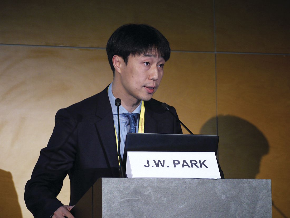
The adjusted hazard ratio (HR) for no PCP at 1 year of follow-up in the prophylaxis group, versus the no prophylaxis group, was 0.096 (P = .022).
The TMP-SMX combination also significantly reduced the mortality associated with PCP, with an adjusted HR of 0.09, versus no prophylaxis (P = .023).
“Pneumocystis pneumonia is a major opportunistic infection in immunocompromised patients associated with high morbidity and mortality,” explained the presenting study investigator Jun Won Park, MD, of Seoul National University Hospital in the Republic of Korea.
Dr. Park added that corticosteroid therapy was an important risk factor for PCP but that the risk-benefit ratio had not been evaluated sufficiently in patients with rheumatic diseases and that there was “different opinion among rheumatologists regarding [the value of] PCP prophylaxis.”
The current study aimed to see if primary antibiotic prophylaxis could prevent PCP in patients with rheumatic diseases, which included patients with systemic lupus erythematosus (SLE), dermatomyositis, rheumatoid arthritis, and Behçet’s disease.
For inclusion, patients had to have been treated with prednisolone at a dose of 30 mg/day or more (or its equivalent) for at least 4 weeks and observed for 1 year. Patients with a prior history of PCP or conditions associated with this opportunistic infection, such as HIV, cancer, or solid organ or hematopoietic stem cell transplantation, were excluded.
Dr. Park reported that PCP prophylaxis was given at the discretion of the treating physician, and the mean duration of TMP-SMX was 230 days.
In the prophylaxis group, 34 adverse drug reactions occurred. Two of these reactions were serious – one case of pancytopenia and one case of Steven’s Johnson syndrome – but both resolved after the antibiotic treatment was discontinued.
A sensitivity analysis was performed, giving consistent results, and a risk-benefit analysis showed that the number needed to treat to prevent one case of PCP was 52, considering all rheumatic disease studied, while the number needed to cause one serious adverse drug reaction was 131.
Taken together, these results suggest a role for TMP-SMX as primary prophylaxis for PCP in patients with rheumatic diseases who need prolonged treatment with high-dose corticosteroids, Dr. Park said.
Dr. Park reported having no relevant financial disclosures.
AT THE EULAR 2017 CONGRESS
Key clinical point:
Major finding: The adjusted hazard ratio (HR) for no PCP at 1 year of follow-up in the prophylaxis group, versus the no prophylaxis group, was 0.096 (P = .022).
Data source: A single-center, retrospective cohort study of 1,522 episodes of prolonged, high-dose steroid use in 1,092 patients with various rheumatic diseases.
Disclosures: Dr. Park reported having no relevant financial disclosures.
Romosozumab cuts new vertebral fracture risk by 73%, but safety data are concerning
MADRID – Romosozumab, an investigational bone-building agent, reduced new vertebral fractures by 73% among postmenopausal women with osteoporosis.
Compared with placebo, the monoclonal antibody also reduced the risk of a clinical fracture by 36% after 12 months of treatment, Piet Geusens, MD, said at the European Congress of Rheumatology. The effect was maintained at 24 months, with a 50% reduction in fracture risk, said Dr. Geusens of Maastricht University, the Netherlands.
Romosozumab also significantly reduced clinical and nonvertebral fractures and increased bone mineral density at the total hip, femoral neck, and lumbar spine in the phase III FRAME study, cosponsored by Amgen and UCB Pharma.
But recently, the finding of increased cardiovascular events in another highly anticipated phase III study of romosozumab cast a cloud of doubt over its rising star. During an interview at EULAR, a UCB company spokesman said the company no longer anticipates a 2017 Food and Drug Administration approval.
FRAME randomized 7,180 postmenopausal women with osteoporosis to monthly injections of romosozumab 210 mg or placebo for 12 months; after that, patients who had been taking placebo switched to denosumab. Dr. Geusens presented only the first year’s placebo-controlled portion. The full FRAME study was published in the New England Journal of Medicine last September (doi: 10.1056/NEJMoa1607948).
At baseline, the women had a mean bone mineral density T score of –2.7 at the lumbar spine, –2.4 at the total hip, and –2.7 at the femoral neck. Mean age was 71 years. About 20% of the women had a previous vertebral fracture, and 22% a previous nonvertebral fracture. The mean FRAX (fracture risk assessment tool) score was 13.4 in both groups.
After 12 months, a new vertebral fracture had occurred in 59 women taking placebo and 16 taking romosozumab (1.8% vs. 0.5%). This amounted to a 73% risk reduction, Dr. Geusens said. Although he did not present 24-month data, the published article cited the antibody’s sustained effect, with a 50% risk reduction evident after the 12-month comparison with denosumab.
The drug also exerted its benefit quickly, Dr. Geusens said. Most of the fractures in the active group occurred in the first 6 months of treatment, with only two additional fractures later.
Romosozumab also was associated with a 36% decrease in the risk of clinical fracture by 12 months (1.6% vs. 2.5% placebo). There was also a positive effect on nonvertebral fractures, which constituted more than 85% of the clinical fractures. Nonvertebral fractures occurred in 56 of those taking the antibody and 75 of those taking placebo (1.6% vs. 2.1%; hazard ratio [HR], .75).
By 12 months, bone mineral density had increased in the romosozumab group by 13% more than in the placebo group at the lumbar spine, by 7% more at the total hip, and by 6% more at the femoral neck.
Dr. Geusens did not address the adverse event profile during his talk. However, according to the published study, romosozumab was generally well tolerated. Serious hypersensitivity events occurred in seven romosozumab patients. Injection site reactions were mostly mild and occurred in 5% of the active group and 3% of the placebo group.
Two patients taking romosozumab experienced osteonecrosis of the jaw; both incidences occurred during the second 12 months and in conjunction with dental issues (tooth extraction and poorly fitted dentures). Anti-romosozumab antibodies developed in 18% and neutralizing antibodies in 0.7%.
Serious cardiovascular events occurred in about 1% of each treatment group, with 17 among those taking romosozumab and15 cardiovascular deaths among those taking placebo – not a significant difference.
UCB and Amgen were pleased with FRAME’s results and, last July, submitted a Biologics License Application to the FDA based on the positive data. A 2017 approval was anticipated, UCB spokesman Scott Fleming said in an interview. But in May, the primary safety analysis of another phase III study, ARCH, threw a monkey wrench in the works.
ARCH compared romosozumab to alendronate in 4,100 postmenopausal women with osteoporosis. ARCH met its primary and secondary endpoints, reducing the incidence of new vertebral fractures by 50%, clinical fractures by 27%, and nonvertebral fractures by 19%. But significantly more women taking the antibody experienced an adjudicated serious cardiovascular event (2.5% vs. 1.9% on alendronate).
On May 21, the companies said these new data would delay romosozumab’s progress toward approval, despite the fact that the submission was based on FRAMES’s positive safety and efficacy data.
Mr. Fleming confirmed this in an interview.
“Amgen has agreed with the FDA that the ARCH data should be considered in the regulatory review prior to the initial marketing authorization, and as a result we do not expect approval of romosozumab in the U.S. to occur in 2017,” he said. “Patient safety is of utmost importance and whilst the cardiac imbalance observed in ARCH was not seen in FRAME, it is important and our responsibility to better understand this imbalance. Further analysis of the ARCH study data is ongoing and will be submitted to a future medical conference and for publication.”
Dr. Geusens refused to comment on the cardiovascular adverse events, saying he had not seen the ARCH data; nor did he explain the cardiovascular events that did occur in FRAME.
Romosozumab also is being reviewed in Canada and Japan; those processes are still underway. Mr. Fleming said the companies are preparing a European Medicines Agency application as well. “The preparation for the European regulatory submission will continue as planned – second half of 2017,” he said.
Dr. Geusens has received research support from Amgen and other pharmaceutical companies. He is a consultant for Amgen and a member of its speakers bureau.
msullivan@frontlinemedcom.com
On Twitter @Alz_gal
AT THE EULAR 2017 CONGRESS
Key clinical point:
Major finding: Compared with placebo, the antibody reduced the risk of a new-onset vertebral fracture by 73% over 1 year.
Data source: FRAME, which randomized 7,180 women to monthly injections of 210 mg romosozumab or placebo for 12 months; for an additional 12 months, those taking placebo switched to denosumab.
Disclosures: Amgen and UCB Pharma are codeveloping the drug. Dr. Geusens is a consultant and speaker for Amgen, and has received research from the company.
VIDEO: No cancer risk found from biological DMARDs
MADRID – Additional real-world evidence confirmed that biological disease modifying drugs used to treat rheumatoid arthritis produced no spikes in new cancers or in cancer recurrences in registry data from tens of thousands of Swedish patients.
Among rheumatoid arthritis patients with a history of cancer, patients treated with a tumor necrosis factor inhibitor (TNFi) were not at an increased risk for cancer recurrence, Johan Askling, MD, said at the European Congress of Rheumatology. In a second study, patients with rheumatoid arthritis (RA) treated with a non-TNFi, biological, disease-modifying drug, specifically abatacept, rituximab, or tocilizumab, had no significantly different rate of new cancer onset when compared with RA patients who never received a biological disease modifying drug nor when compared with the general Swedish adult population, said Dr. Askling, a professor of clinical epidemiology at the Karolinska Institute in Stockholm.
“Five-year data are a good start, but we need data on 30-year risk,” Dr. Askling said in an interview.
The cancer-recurrence risk study with TNFi treatment used data collected by the Swedish national outpatient care registry on nearly 62,000 people, the Swedish cancer registry, and a rheumatology treatment registry called ARTIS. It also included patients treated during 2001-2014. From these sources, the researchers identified 446 RA patients with a history of at least one cancer who then began treatment with any type of TNFi and matched these cases with 1,278 similar RA patients with a cancer history who had never received a biologic drug. On average, the patients were nearly 10 years removed from their initial cancer diagnoses, and the average duration on TNFi treatment was nearly 5 years.
The adjusted hazard ratio for cancer recurrence among the TNFi recipients was reduced by a nominal 30%, compared with that of the controls, a difference that was not statistically significant, Dr. Askling reported.
The second study used data from similar sources for patients treated during 2006-2014 and included nearly 100,000 Swedes from the general population, more than 42,000 RA patients who did not receive a biological drug, more than 14,000 treated with either a first or second TNFi drug, and 1,693 patients treated with tocilizumab (Actemra), 1,894 on abatacept (Orencia), and 3,119 on rituximab (Rituxan).
The rates of new onset cancer in any of these treatment groups, including the patients on tocilizumab, abatacept, or rituximab, was not significantly different from the rate among RA patients who never received a biologic drug, nor from the general Swedish population rate, Dr. Askling said.
This is “one of the first large-scale assessments” of the cancer risk posed by non-TNFi biological drugs, aside from what was reported from the pivotal trials for these drugs, Dr. Askling said.
Dr. Askling has received research support from AbbVie, Lilly, MSD, Pfizer, Roche, and UCB.
mzoler@frontlinemedcom.com
On Twitter @mitchelzoler
Rheumatologists began having concerns about the possible impact of biological drugs on cancer when these types of drugs first became available 20 or more years ago. Registries have allowed us to follow these patients, and, so far, we have consistently seen that the risk for cancer is very low. The major adverse effect from treatment with biological drugs is infection.
The most confirmed finding has been that biologic drugs do not cause new cancers. We have known less about the risk patients with a history of cancer face for recurrence by taking a biological drug. The data on this have so far been scarce. Most guidelines advise that, when patients have had cancer, the possible use of a biologic drug should be the subject of a shared-decision discussion with the patient. The new data reported by Dr. Askling add to the risk information we have available to discuss with patients.
The risk that biologic drugs poses for infections is more complex. The infection risk also depends on a patient’s use of glucocorticoids, their age, and their comorbidities. The infection risk faced by a patient from treatment with a biological drug requires an individualized discussion that takes into account the severity of all the relevant risk factors.
The video associated with this article is no longer available on this site. Please view all of our videos on the MDedge YouTube channel
João E. Fonseca, MD is a professor of rheumatology at the University of Lisbon. He has been a speaker for or has received research funding from Abbvie, MSD, Pfizer, Roche, and UCB. He made these comments in a video interview.
MADRID – Additional real-world evidence confirmed that biological disease modifying drugs used to treat rheumatoid arthritis produced no spikes in new cancers or in cancer recurrences in registry data from tens of thousands of Swedish patients.
Among rheumatoid arthritis patients with a history of cancer, patients treated with a tumor necrosis factor inhibitor (TNFi) were not at an increased risk for cancer recurrence, Johan Askling, MD, said at the European Congress of Rheumatology. In a second study, patients with rheumatoid arthritis (RA) treated with a non-TNFi, biological, disease-modifying drug, specifically abatacept, rituximab, or tocilizumab, had no significantly different rate of new cancer onset when compared with RA patients who never received a biological disease modifying drug nor when compared with the general Swedish adult population, said Dr. Askling, a professor of clinical epidemiology at the Karolinska Institute in Stockholm.
“Five-year data are a good start, but we need data on 30-year risk,” Dr. Askling said in an interview.
The cancer-recurrence risk study with TNFi treatment used data collected by the Swedish national outpatient care registry on nearly 62,000 people, the Swedish cancer registry, and a rheumatology treatment registry called ARTIS. It also included patients treated during 2001-2014. From these sources, the researchers identified 446 RA patients with a history of at least one cancer who then began treatment with any type of TNFi and matched these cases with 1,278 similar RA patients with a cancer history who had never received a biologic drug. On average, the patients were nearly 10 years removed from their initial cancer diagnoses, and the average duration on TNFi treatment was nearly 5 years.
The adjusted hazard ratio for cancer recurrence among the TNFi recipients was reduced by a nominal 30%, compared with that of the controls, a difference that was not statistically significant, Dr. Askling reported.
The second study used data from similar sources for patients treated during 2006-2014 and included nearly 100,000 Swedes from the general population, more than 42,000 RA patients who did not receive a biological drug, more than 14,000 treated with either a first or second TNFi drug, and 1,693 patients treated with tocilizumab (Actemra), 1,894 on abatacept (Orencia), and 3,119 on rituximab (Rituxan).
The rates of new onset cancer in any of these treatment groups, including the patients on tocilizumab, abatacept, or rituximab, was not significantly different from the rate among RA patients who never received a biologic drug, nor from the general Swedish population rate, Dr. Askling said.
This is “one of the first large-scale assessments” of the cancer risk posed by non-TNFi biological drugs, aside from what was reported from the pivotal trials for these drugs, Dr. Askling said.
Dr. Askling has received research support from AbbVie, Lilly, MSD, Pfizer, Roche, and UCB.
mzoler@frontlinemedcom.com
On Twitter @mitchelzoler
AT THE EULAR 2017 CONGRESS
Key clinical point:
Major finding: Former cancer patients on a TNFi had no increased cancer recurrences, compared with patients on other rheumatic treatments.
Data source: Data from the Swedish national registries.
Disclosures: Dr. Askling has received research support from AbbVie, Lilly, MSD, Pfizer, Roche, and UCB.
Hexavalent DTaP5-IPV-HB-Hib is immunogenic in series with pentavalent vaccine
against all the hexavalent antigens, said Federico Martinón-Torres, MD, of Hospital Clinico Universitario de Santiago de Compostela, Spain, and his associates.
DTaP5-IPV-HB-Hib combination vaccine has a five-antigen pertussis component and contains polyribosylribitol phosphate–Neisseria meningitidis serogroup B outer membrane protein [PRP-OMP] Hib conjugate, while DTaP5-IPV-Hib contains polyribosylribitol phosphate-tetanus toxoid (PRP-T) Hib conjugate.
The phase III, open-label, single-arm study in 12 Spanish hospitals and health centers included 384 infants who had received only one dose of monovalent hepatitis B vaccine within 3 days of birth prior to starting the study. They then received the hexa/penta/hexa combo vaccines concomitantly with the MCC vaccine. The response rate was 100% against Hib and 98.9% against hepatitis B.
Also, a “robust immune response was induced against all other disease antigens,” said Dr. Martinón-Torres and his colleagues. Coadministration of the combo vaccines with the meningococcal vaccine likewise did not appear to compromise immunogenicity of any of the vaccine antigens.
Use of the hexa/penta/hexa combo vaccine series was mostly safe and well tolerated, the researchers said.
Read more in the journal Vaccine (2017 Jun 17;35[30];3764-72).
FROM VACCINE
Two SNPs linked to survival in R-CHOP–treated DLBCL
Two variations of the BCL2 gene are linked with the survival prospects of patients with diffuse large B-cell lymphoma (DLBCL) who are treated with the R-CHOP regimen, based on a study published in Haematologica.
In the population-based, case-control study of patients with non-Hodgkin’s lymphoma across the British Columbia province, Morteza Bashash, PhD, of the Dalla Lana School of Public Health, Toronto, and researchers at the British Columbia Cancer Agency analyzed 217 germline DLBCL samples, excluding those with primary mediastinal large B-cell lymphoma, specifically looking at nine single nucleotide polymorphisms (SNPs).
Patients receiving R-CHOP who had the AA genotype at rs7226979 had a risk of death that was four times higher than that of those with a G allele, researchers said (P less than .01). The same pattern was seen for PFS, with AA carriers having twice the risk of an event, compared with the other genotypes (P less than .05).
For those with rs4456611, patients with the GG genotype had a risk of death that was 3 times greater than that of those with an A allele (P less than .01), but there was no association with PFS for that SNP.
In an analysis of an independent cohort, only the associations that were seen with rs7226979 – and not those with rs4456611 – were able to be replicated.
The researchers noted that, while most predictive markers that are used to guide clinical treatment are drawn from actual tumor material, host-related factors could also be important.
“Compared to genetic analysis of the tumor, the patient’s constitutional genetic profile is relatively easy to obtain and can be assessed before treatment is started,” they wrote. “Our result has the potential to be useful as a complementary tool to predict the outcome of patients treated with R-CHOP and enhance clinical decision-making after confirmation by further studies.”
FROM HAEMATOLOGICA
Key clinical point: Two SNPs were found to be linked to survival prospects in patients with diffuse large B-cell lymphoma who receive primary R-CHOP therapy.
Major finding: For the rs7226979 SNP, those with the AA genotype had a four times higher risk of death than those with a G allele.
Data source: A population-based, case-control study of patients with non-Hodgkin’s lymphoma in British Columbia, with DNA samples analyzed for 9nine SNPs among the DLBCL patients, excluding those with primary mediastinal large B-cell lymphoma.
Disclosures: Some of the study authors reported institutional research funding from Roche; honoraria from Roche/Genentech, Janssen Pharmaceuticals and Celgene; and/or consultant or advisory roles with Roche/Genentech, Janssen Pharmaceuticals, Celgene, and NanoString Technologies.
Liposomes boost bortezomib efficacy
Bortezomib treatment using liposome nanocarriers leads to decreased cell viability and greater apoptosis in vitro, compared with treatment with free bortezomib, according to a study in the Journal of Pharmaceutical Sciences and Pharmacology.
Liposomes are lipid sacs with a watery compartment, which can be used to encapsulate and deliver a therapeutic cargo. The delivery method has been found to have improved efficacy with lesser side effects.
Ceramide liposomes are an attractive drug-delivery vehicle, the researchers said, because of their cell-permeability and because they’ve been found, on their own, to mediate apoptosis. Researchers said they believed this was the first time results have been reported on combining ceramide liposomes with an anticancer drug such as bortezomib. Cationic liposomes were picked because they’re known to destabilize cell membranes, helping with intracellular delivery of the drug.
Free bortezomib and bortezomib loaded into liposomes were tested for efficacy on mouse preosteoclast calvaria MC3T3 cells, mouse macrophage-like RAW 264.7 cells, and human osteosarcoma U2OS cells.
On the RAW 264.7 cells, researchers found a significant difference in cell viability between free bortezomib and ceramide liposomes after 24 hours (P less than .01) and 48 hours (P less than .05) and between free bortezomib and cationic liposomes at 24 hours (P less than .01). They also reported a significant difference with cationic liposomes on MC3T3 cells and U2OS cells at 48 hours (both P less than .01).
One nanomolar (nM) of ceramide-loaded bortezomib induced significantly more apoptosis than did 1 nM of free bortezomib (P less than .01), and 10 nM of ceramide-loaded bortezomib brought about more cell death and apoptosis than did 10 nM of free bortezomib (P less than .05). These effects were likely the result of increased expression of proteins involved in apoptosis.
Liposomes might be able to boost the efficacy of bortezomib, according to the researchers, who are now studying the localization of these liposomes with confocal microscopes to better understand the mechanism of action.
“Such improvements,” they wrote, “offer the potential to reduce side effects known to occur with this chemotherapy, such as peripheral neuropathy, as well as to target Bort-resistant cancers.”
FROM THE JOURNAL OF PHARMACEUTICAL SCIENCES AND PHARMACOLOGY
Key clinical point: Ceramide and cationic liposomes loaded with bortezomib decreased cell viability and increased apoptosis in vitro, compared with bortezomib alone.
Major finding: One nanomolar (nM) of ceramide-loaded bortezomib induced significantly more apoptosis than did 1 nM of free bortezomib (P less than .01), and 10 nM of ceramide-loaded bortezomib brought about more cell death and apoptosis than did 10 nM of free bortezomib (P less than .05).
Data source: An in vitro study conducted at Midwestern University.
Disclosures: Researchers reported no conflicts of interest.
Treatment response rates for psychotic bipolar depression similar to those without
MIAMI – Response rates for psychotic and nonpsychotic depression in bipolar disorder were statistically similar, regardless of treatment, an ad hoc analysis has shown.
Over a 6 month period, results from the multisite, randomized, controlled Bipolar CHOICE (Clinical Health Outcomes Initiative in Comparative Effectiveness for Bipolar Disorder) study showed that 482 patients anywhere on the bipolar spectrum, given either lithium or quetiapine, had similar treatment response rates over 6 months.
“When you look at the course of the improvement for those with psychosis, they had more severe disorder at baseline,and presented with these symptoms throughout the study. But, when we compare curves of improvement, those with severe disorder responded to treatment at the same pace [as those without psychosis],” Dr. Caldieraro said.
The overall scores for the Bipolar Inventory of Symptoms Scale (BISS) at baseline were 75.2 plus or minus 17.6 percentage points for those with psychosis, vs. 54.9 plus or minus 16.3 for those without (P less than .001). At 6 months, the scores were more in range with one another: 37.2 plus or minus 19.7 for those with psychosis and 26.3 plus or minus 18.0 for those without (P = .003). The BISS depression scores at baseline for those with psychosis were 29.5 plus or minus 7.0, compared with 24.9 plus or minus 8.0 for those without (P = .002). At study end, the scores were 13.0 plus or minus 8.6, vs. 10.9 plus or minus 9.5 (P = .253).
Overall Clinical Global Impressions (CGI) scores for bipolar disorder at baseline in the group with psychosis were 5.1 plus or minus 0.9, compared with 4.5 plus or minus 0.8 in those without (P less than .001). At 6 months, the scores were 3.4 plus or minus 1.3, vs. 2.8 plus or minus 1.3 (P = .032). The CGI scores for depression in the psychosis group at baseline were 4.9 plus or minus 0.9, compared with 4.4 plus or minus 0.9 in the nonpsychosis group (P = .006). At 6 months, the psychosis groups’ scores were 3.1 plus or minus 1.4, compared with 2.6 plus or minus 1.3 in the nonpsychosis group (P = .07).
In addition to either lithium or quetiapine, patients in the CHOICE study also received adjunctive personalized treatment. Patients who received lithium plus APT were not given second-generation antipsychotics, while those given quetiapine plus APT were not given lithium or any other second-generation antipsychotic.
In the quetiapine group, 21 people had psychotic depression at baseline. In the lithium group, there were 11. The time to remission was numerically, although not statistically, similar between the patients with psychosis in the lithium and the quetiapine groups.
Compared with the CHOICE study participants without psychosis, the subanalysis showed that the 32 people with psychotic features were far more likely to be single or never married (P = .036), employed at half the rate (P = .035), twice as likely to suffer from generalized anxiety disorder (P = .028), and more likely to have social phobias (P = .018). People with psychotic depression in the study also were more likely to suffer from agoraphobia.
One reason for his interest in the study, Dr. Caldieraro said, was that, despite the worse prognosis for people on the bipolar spectrum with psychotic depression, the literature on treatment outcomes for this cohort is scant.
“Ours is a small sample, so you could say that we didn’t have enough power, but we have some interesting results,” he said during his presentation. “The results need replication, but the study suggests that maybe, if we make the patient better, it doesn’t matter which medication we use.”
Dr. Caldieraro had no relevant disclosures. The Agency for Healthcare Research and Quality funded the CHOICE study, NCT01331304.
AT THE ASCP ANNUAL MEETING
Key clinical point:
Major finding: Patients with psychotic or nonpsychotic bipolar depression responded equally well to lithium and quetiapine when compared with response rates in patients without bipolar depression.
Data source: A secondary analysis of 32 patients from a multisite, randomized, controlled trial of 482 patients with bipolar disorder I or bipolar II and psychosis, assigned to receive either lithium or quetiapine for 6 months.
Disclosures: Dr. Caldieraro had no relevant disclosures. The Agency for Healthcare Research and Quality funded the CHOICE study, NCT01331304.
Intra-amniotic sludge: Does its presence rule out cerclage for short cervix?
CASE: Woman with short cervix, intra-amniotic sludge, and prior preterm delivery
An asymptomatic 32-year-old woman with a prior preterm delivery, presently pregnant with a singleton at 17 weeks of gestation, underwent transvaginal ultrasonography and was found to have a cervical length of 22 mm and dense intra-amniotic sludge. She received one dose of 17α-hydroxyprogesterone caproate (17P) at 16 weeks of gestation. What are your next steps in management?
Intra-amniotic sludge is a conundrum
Intra-amniotic sludge is a sonographic finding of free-floating, hyperechoic, particulate matter in the amniotic fluid close to the internal os. The precise nature of this material varies, and it may include blood, meconium, or vernix and may signal inflammation or infection. In a retrospective case-control study, 27% of asymptomatic women with sludge and a short cervix had positive amniotic fluid cultures, and 27% had evidence of inflammation in the amniotic fluid (>50 white blood cells/mm3).1 In a separate report, the authors proposed that "the detection of amniotic fluid 'sludge' represents a sign that microbial invasion of the amniotic cavity and an inflammatory process are in progress."2
Benefit of cerclage in high-risk women. Several systematic reviews have highlighted the benefit of cerclage for women with a singleton pregnancy, short cervix, and previous preterm birth or second-trimester loss (ultrasound-indicated cerclage for high-risk women).3 Cerclage is presumed to work by providing some degree of structural support and by maintaining a barrier to protect the fetal membranes against exposure to ascending pathogens.4
Since dense intra-amniotic sludge may represent chronic intra-amniotic infection, can cerclage still be expected to be beneficial when microbiologic invasion of the amniotic cavity already has occurred? Furthermore, intra-amniotic infection has been cited as a possible complication of ultrasound-indicated cerclage, with a rate of 10%.5 The traditional view is that the presence of subclinical intra-amniotic infection may further increase this risk and therefore should be considered a contraindication to cerclage.6
Evaluating the patient for cerclage placement
The patient history and physical examination should focus on the signs and symptoms of labor, vaginal bleeding, amniotic membrane rupture, and intra-amniotic infection. Particular attention should be paid to maternal temperature, pulse, and the presence of uterine tenderness or foul-smelling vaginal discharge. A sterile speculum examination followed by digital examination would complement the ultrasonography evaluation in assessing cervical dilation and effacement. The ultrasonography evaluation should be completed to confirm a viable pregnancy with accurate dating and the absence of detectable fetal anomalies.
Currently, evidence is insufficient for recommending routine amniocentesis to exclude intra-amniotic infection in an asymptomatic woman prior to ultrasound-indicated cerclage, even in the presence of intra-amniotic sludge, as there are no data demonstrating improved outcomes.4 In addition, intra-amniotic sludge has been associated with intra-amniotic infection and/or inflammation in the form of microbial biofilms, which may prevent detection of infection by routine culture techniques.7
Related Article:
Universal cervical length screening–saving babies lives
Study results offer limited guidance
Data are limited on the clinical implications of intra-amniotic sludge in women with cervical cerclage. In a retrospective cohort of 177 patients with cerclage, 60 had evidence of sludge and 46 of those with sludge underwent ultrasound-indicated cerclage.8 There were no significant differences in the mean gestational age at delivery, neonatal outcomes, rate of preterm delivery, preterm premature rupture of membranes, or intra-amniotic infection between women with or without intra-amniotic sludge. A subanalysis was performed comparing women with sludge detected before or after cerclage and, again, no difference was found in measured outcomes.
Similarly, in a small (N = 20) retrospective review of the Arabin pessary used as a noninvasive intervention for short cervix, the presence of intra-amniotic sludge in 5 cases did not appear to impact outcomes.9
Case patient: How would you manage her care?
Based on her obstetric history and ultrasonography findings, the patient described in the case vignette is at high risk for preterm delivery. The presence of both intra-amniotic sludge and short cervix is associated with an increased risk for spontaneous preterm delivery. After evaluating for clinical intra-amniotic infection and performing a work-up for other contraindications to cerclage placement, cerclage placement may be offered--even in the presence of intra-amniotic sludge.
The next practical question is whether 17P, already started, should be continued after cerclage placement. From the literature on 17P, it is unclear whether progesterone provides additional benefit. One randomized, placebo-controlled study in women with at least 2 preterm deliveries or mid-trimester losses and cerclage in place showed that the 17P-treated women had a significant reduction in preterm delivery compared with the control group, from 37.8% to 16.1%.10
By contrast, in a secondary analysis of a randomized trial evaluating cerclage in high-risk women with short cervix in the current pregnancy, addition of 17P to cerclage was not beneficial.11 Results of 2 retrospective cohort studies showed the same lack of difference on preterm delivery rates with the addition of 17P.12,13
Accepting that the interpretation of these data is challenging, in our practice we would choose to continue the progesterone supplementation, siding with other recently expressed expert opinions.14
The bottom line
While clinical intra-amniotic infection is a contraindication to cerclage, there is no evidence to support withholding cerclage from eligible women due to the presence of intra-amniotic fluid sludge alone.
Share your thoughts! Send your Letter to the Editor to rbarbieri@frontlinemedcom.com. Please include your name and the city and state in which you practice.
- Kusanovic JP, Espinoza J, Romero R, et al. Clinical significance of the presence of amniotic fluid "sludge" in asymptomatic patients at high risk for spontaneous preterm delivery. Ultrasound Obstet Gynecol. 2007;30(5):706-714.
- Romero R, Kusanovic JP, Espinoza J, et al. What is amniotic fluid "sludge"? Ultrasound Obstet Gynecol. 2007;30(5):793-798.
- Alfirevic Z, Stampalija T, Roberts D, Jorgensen AL. Cervical stitch (cerclage) for preventing preterm birth in singleton pregnancy. Cochrane Database Syst Rev. 2012;4:CD008991.
- Abbott D, To M, Shennan A. Cervical cerclage: a review of current evidence. Aust N Z J Obstet Gynaecol. 2012;52(3):220-223.
- Drassinower D, Poggi SH, Landy HJ, Gilo N, Benson JE, Ghidini A. Perioperative complications of history-indicated and ultrasound-indicated cervical cerclage. Am J Obstet Gynecol. 2011;205(1):53.e1-e5.
- Mays JK, Figueroa R, Shah J, Khakoo H, Kaminsky S, Tejani N. Amniocentesis for selection before rescue cerclage. Obstet Gynecol. 2000;95(5):652-655.
- Vaisbuch E, Romero R, Erez IO, et al. Clinical significance of early (<20 weeks) vs late (20-24 weeks) detection of sonographic short cervix in asymptomatic women in the mid-trimester. Ultrasound Obstet Gynecol. 2010;36(4):471-481.
- Gorski LA, Huang WH, Iriye BK, Hancock J. Clinical implication of intra-amniotic sludge on ultrasound in patients with cervical cerclage. Ultrasound Obstet Gynecol. 2010;36(4):482-485.
- Ting YH, Lao TT, Wa Law LW, et al. Arabin cerclage pessary in the management of cervical insufficiency. J Matern Fetal Neonatal Med. 2012;25(12):2693-2695.
- Yemini M, Borenstein R, Dreazen E, et al. Prevention of premature labor by 17 alpha-hydroxyprogesterone caproate. Am J Obstet Gynecol. 1985;151(5):574-577.
- Berghella V, Figueroa D, Szychowski JM, et al; Vaginal Ultrasound Trial Consortium. 17-alpha-hydroxyprogesterone caproate for the prevention of preterm birth in women with prior preterm birth and a short cervical length. Am J Obstet Gynecol. 2010;202(4):351.e1-e6.
- Rebarber A, Cleary-Goldman J, Istwan NB, et al. The use of 17 alpha-hydroxyprogesterone caproate (17P) in women with cervical cerclage. Am J Perinatol. 2008;25(5):271-275.
- Stetson B, Hibbard JU, Wilkins I, Leftwich H. Outcomes with cerclage alone compared with cerclage plus 17 α-hydroxyprogesterone caproate. Obstet Gynecol. 2016;128(5):983-988.
- Iams JD. Identification of candidates for progesterone: why, who, how, and when? Obstet Gynecol. 2014;123(6):1317-1326.

Dr. Tolcher is a Fellow in Maternal-Fetal Medicine at Baylor College of Medicine and Texas Children's Hospital, Pavilion for Women, Houston, Texas.

Dr. Vidaeff is Professor and Fellowship Program Director, Maternal-Fetal Medicine, at Baylor College of Medicine and Texas Children's Hospital, Pavilion for Women.
The authors report no financial relationships relevant to this article.
Dr. Tolcher is a Fellow in Maternal-Fetal Medicine at Baylor College of Medicine and Texas Children's Hospital, Pavilion for Women, Houston, Texas.
Dr. Vidaeff is Professor and Fellowship Program Director, Maternal-Fetal Medicine, at Baylor College of Medicine and Texas Children's Hospital, Pavilion for Women.
The authors report no financial relationships relevant to this article.
CASE: Woman with short cervix, intra-amniotic sludge, and prior preterm delivery
An asymptomatic 32-year-old woman with a prior preterm delivery, presently pregnant with a singleton at 17 weeks of gestation, underwent transvaginal ultrasonography and was found to have a cervical length of 22 mm and dense intra-amniotic sludge. She received one dose of 17α-hydroxyprogesterone caproate (17P) at 16 weeks of gestation. What are your next steps in management?
Intra-amniotic sludge is a conundrum
Intra-amniotic sludge is a sonographic finding of free-floating, hyperechoic, particulate matter in the amniotic fluid close to the internal os. The precise nature of this material varies, and it may include blood, meconium, or vernix and may signal inflammation or infection. In a retrospective case-control study, 27% of asymptomatic women with sludge and a short cervix had positive amniotic fluid cultures, and 27% had evidence of inflammation in the amniotic fluid (>50 white blood cells/mm3).1 In a separate report, the authors proposed that "the detection of amniotic fluid 'sludge' represents a sign that microbial invasion of the amniotic cavity and an inflammatory process are in progress."2
Benefit of cerclage in high-risk women. Several systematic reviews have highlighted the benefit of cerclage for women with a singleton pregnancy, short cervix, and previous preterm birth or second-trimester loss (ultrasound-indicated cerclage for high-risk women).3 Cerclage is presumed to work by providing some degree of structural support and by maintaining a barrier to protect the fetal membranes against exposure to ascending pathogens.4
Since dense intra-amniotic sludge may represent chronic intra-amniotic infection, can cerclage still be expected to be beneficial when microbiologic invasion of the amniotic cavity already has occurred? Furthermore, intra-amniotic infection has been cited as a possible complication of ultrasound-indicated cerclage, with a rate of 10%.5 The traditional view is that the presence of subclinical intra-amniotic infection may further increase this risk and therefore should be considered a contraindication to cerclage.6
Evaluating the patient for cerclage placement
The patient history and physical examination should focus on the signs and symptoms of labor, vaginal bleeding, amniotic membrane rupture, and intra-amniotic infection. Particular attention should be paid to maternal temperature, pulse, and the presence of uterine tenderness or foul-smelling vaginal discharge. A sterile speculum examination followed by digital examination would complement the ultrasonography evaluation in assessing cervical dilation and effacement. The ultrasonography evaluation should be completed to confirm a viable pregnancy with accurate dating and the absence of detectable fetal anomalies.
Currently, evidence is insufficient for recommending routine amniocentesis to exclude intra-amniotic infection in an asymptomatic woman prior to ultrasound-indicated cerclage, even in the presence of intra-amniotic sludge, as there are no data demonstrating improved outcomes.4 In addition, intra-amniotic sludge has been associated with intra-amniotic infection and/or inflammation in the form of microbial biofilms, which may prevent detection of infection by routine culture techniques.7
Related Article:
Universal cervical length screening–saving babies lives
Study results offer limited guidance
Data are limited on the clinical implications of intra-amniotic sludge in women with cervical cerclage. In a retrospective cohort of 177 patients with cerclage, 60 had evidence of sludge and 46 of those with sludge underwent ultrasound-indicated cerclage.8 There were no significant differences in the mean gestational age at delivery, neonatal outcomes, rate of preterm delivery, preterm premature rupture of membranes, or intra-amniotic infection between women with or without intra-amniotic sludge. A subanalysis was performed comparing women with sludge detected before or after cerclage and, again, no difference was found in measured outcomes.
Similarly, in a small (N = 20) retrospective review of the Arabin pessary used as a noninvasive intervention for short cervix, the presence of intra-amniotic sludge in 5 cases did not appear to impact outcomes.9
Case patient: How would you manage her care?
Based on her obstetric history and ultrasonography findings, the patient described in the case vignette is at high risk for preterm delivery. The presence of both intra-amniotic sludge and short cervix is associated with an increased risk for spontaneous preterm delivery. After evaluating for clinical intra-amniotic infection and performing a work-up for other contraindications to cerclage placement, cerclage placement may be offered--even in the presence of intra-amniotic sludge.
The next practical question is whether 17P, already started, should be continued after cerclage placement. From the literature on 17P, it is unclear whether progesterone provides additional benefit. One randomized, placebo-controlled study in women with at least 2 preterm deliveries or mid-trimester losses and cerclage in place showed that the 17P-treated women had a significant reduction in preterm delivery compared with the control group, from 37.8% to 16.1%.10
By contrast, in a secondary analysis of a randomized trial evaluating cerclage in high-risk women with short cervix in the current pregnancy, addition of 17P to cerclage was not beneficial.11 Results of 2 retrospective cohort studies showed the same lack of difference on preterm delivery rates with the addition of 17P.12,13
Accepting that the interpretation of these data is challenging, in our practice we would choose to continue the progesterone supplementation, siding with other recently expressed expert opinions.14
The bottom line
While clinical intra-amniotic infection is a contraindication to cerclage, there is no evidence to support withholding cerclage from eligible women due to the presence of intra-amniotic fluid sludge alone.
Share your thoughts! Send your Letter to the Editor to rbarbieri@frontlinemedcom.com. Please include your name and the city and state in which you practice.
- Kusanovic JP, Espinoza J, Romero R, et al. Clinical significance of the presence of amniotic fluid "sludge" in asymptomatic patients at high risk for spontaneous preterm delivery. Ultrasound Obstet Gynecol. 2007;30(5):706-714.
- Romero R, Kusanovic JP, Espinoza J, et al. What is amniotic fluid "sludge"? Ultrasound Obstet Gynecol. 2007;30(5):793-798.
- Alfirevic Z, Stampalija T, Roberts D, Jorgensen AL. Cervical stitch (cerclage) for preventing preterm birth in singleton pregnancy. Cochrane Database Syst Rev. 2012;4:CD008991.
- Abbott D, To M, Shennan A. Cervical cerclage: a review of current evidence. Aust N Z J Obstet Gynaecol. 2012;52(3):220-223.
- Drassinower D, Poggi SH, Landy HJ, Gilo N, Benson JE, Ghidini A. Perioperative complications of history-indicated and ultrasound-indicated cervical cerclage. Am J Obstet Gynecol. 2011;205(1):53.e1-e5.
- Mays JK, Figueroa R, Shah J, Khakoo H, Kaminsky S, Tejani N. Amniocentesis for selection before rescue cerclage. Obstet Gynecol. 2000;95(5):652-655.
- Vaisbuch E, Romero R, Erez IO, et al. Clinical significance of early (<20 weeks) vs late (20-24 weeks) detection of sonographic short cervix in asymptomatic women in the mid-trimester. Ultrasound Obstet Gynecol. 2010;36(4):471-481.
- Gorski LA, Huang WH, Iriye BK, Hancock J. Clinical implication of intra-amniotic sludge on ultrasound in patients with cervical cerclage. Ultrasound Obstet Gynecol. 2010;36(4):482-485.
- Ting YH, Lao TT, Wa Law LW, et al. Arabin cerclage pessary in the management of cervical insufficiency. J Matern Fetal Neonatal Med. 2012;25(12):2693-2695.
- Yemini M, Borenstein R, Dreazen E, et al. Prevention of premature labor by 17 alpha-hydroxyprogesterone caproate. Am J Obstet Gynecol. 1985;151(5):574-577.
- Berghella V, Figueroa D, Szychowski JM, et al; Vaginal Ultrasound Trial Consortium. 17-alpha-hydroxyprogesterone caproate for the prevention of preterm birth in women with prior preterm birth and a short cervical length. Am J Obstet Gynecol. 2010;202(4):351.e1-e6.
- Rebarber A, Cleary-Goldman J, Istwan NB, et al. The use of 17 alpha-hydroxyprogesterone caproate (17P) in women with cervical cerclage. Am J Perinatol. 2008;25(5):271-275.
- Stetson B, Hibbard JU, Wilkins I, Leftwich H. Outcomes with cerclage alone compared with cerclage plus 17 α-hydroxyprogesterone caproate. Obstet Gynecol. 2016;128(5):983-988.
- Iams JD. Identification of candidates for progesterone: why, who, how, and when? Obstet Gynecol. 2014;123(6):1317-1326.