User login
Children with ASD less likely to get vision screening
Children with autism spectrum disorder (ASD) are significantly less likely to have vision screening at well visits for 3- to 5-year-olds than are typically developing children, researchers have found.
The report, by Kimberly Hoover, MD, of Thomas Jefferson University in Philadelphia, and colleagues, was published online in Pediatrics.
While 59.9% of children without ASD got vision screening in these visits, only 36.5% of children with ASD got the screening. Both screening rates miss the mark set by American Academy of Pediatrics guidelines.
The AAP recommends “annual instrument-based vision screening, if available, at well visits for children starting at age 12 months to 3 years, and direct visual acuity testing beginning at 4 years of age. However, in children with developmental delays, the AAP recommends instrument-based screening, such as photoscreening, as a useful alternative at any age.”
Racial, age disparities as well
Racial disparities were evident in the data as well. Of the children who had ASD, Black children had the lowest rates of screening (27.6%), while the rate for White children was 39.7%. The rate for other/multiracial children with ASD was 39.8%.
The lowest rates of screening occurred in the youngest children, at the 3-year visit.
The researchers analyzed data from 63,829 well-child visits between January 2016 and December 2019, collected from the large primary care database PEDSnet.
Photoscreening vs. acuity screening
The authors pointed out that children with ASD are less likely to complete a vision test, which can be problematic in a busy primary care office.
“Children with ASD were significantly less likely to have at least one completed vision screening (43.2%) compared with children without ASD (72.1%; P <. 01),” the authors wrote, “with only 6.9% of children with ASD having had two or more vision screenings compared with 22.3% of children without ASD.”
The researchers saw higher vision test completion rates with photoscreening, using a sophisticated camera, compared with acuity screening, which uses a wall chart and requires responses.
Less patient participation is required for photoscreening and it can be done in less than 2 minutes.
If ability to complete the vision tests is a concern, the authors wrote, photoscreening may be a better solution.
Photoscreening takes 90 seconds
“Photoscreening has high sensitivity in detecting ocular conditions in children with ASD and has an average screening time of 90 seconds, and [it has] been validated in both children with ASD and developmental delays,” the authors wrote.
Andrew Adesman, MD, chief of developmental and behavioral pediatrics at Cohen Children’s Medical Center in New Hyde Park, N.Y., said the authors of this study quantify the gap between need and reality for vision tests for those with ASD.
“Other studies have shown that children on the autism spectrum have more than three times greater risk of having eye disease or vision problems,” he said in an interview. “You’ve got a high-risk population in need of assessment and the likelihood of them getting an assessment is much reduced.”
He said in addition to attention problems in taking the test, vision screening may get lost in the plethora of concerns parents want to talk about in well-child visits.
“If you’re the parent of a child with developmental delays, language delays, poor social engagement, there are a multitude of things the visit could be focused on and it may be that vision screening possibly gets compromised or not done,” Dr. Adesman said.
That, he said, may be a focus area for improving the screening numbers.
Neither parents nor providers should forget that vision screening is important, despite the myriad other issues to address, he said. “They don’t have to take a long time.”
When it comes to vision problems and children, “the earlier they’re identified the better,” Dr. Adesman says, particularly to identify the need for eye muscle surgery or corrective lenses, the two major interventions for strabismus or refractive error.
“If those problems are significant and go untreated, there’s a risk of loss of vision in the affected eye,” he said.
Reimbursement concerns for photoscreening
This study strongly supports the use of routine photoscreening to help eliminate the vision screening gap in children with ASD, the authors wrote.
They noted, however, that would require insurance reimbursement for primary care practices to effectively use that screening.
The researchers advised, “Providers treating patients with race, ethnicity, region, or age categories that reduce the adjusted odds of photoscreening can take steps in their practices to address these disparities, particularly in children with ASD.”
The study authors and Dr. Adesman reported no relevant financial relationships.
FROM PEDIATRICS
CLL and surgery are more compatible than ever
In the past decade, as targeted therapies have permitted better management of CLL, a new realm of possibilities has opened up for patients with this blood cancer.
“Previously, patients may not have been candidates for elective surgeries, such as hip replacements,” said hematologist-oncologist Helen Ma, MD, of the University of Irvine (Calif.) and VA Long Beach Healthcare System. She is the lead author of the report, which appeared in the British Journal of Hematology.
“Now that targeted therapies are controlling CLL well, patients may elect to have procedures that they may not have considered if their blood counts were very low or they felt too unwell to go through such invasive surgeries,” said Dr. Ma in an interview. In fact, the study authors noted that, “with currently available treatments, many patients with CLL are living considerably longer than the 1-year life expectancy threshold that proceduralists require.”
But extra surgical risks persist. “Both CLL and its treatment can increase the risk of complications during and after procedures, though available data are not consistently stratified by stage and whether patients are undergoing treatment,” the report authors noted.
Research has linked CLL to higher rates of blood transfusions in cardiac surgeries: One study, conducted partially in the era of targeted therapy, found that 87% of these surgery patients with CLL needed blood products vs. 65% of those who didn’t have CLL (P = .01). Studies didn’t find any extra risk of infections in patients with CLL, however, and there are conflicting findings about whether hospital mortality is higher.
Another study, also conducted partially in the era of targeted therapy, found that patients with CLL who had percutaneous coronary intervention procedures “developed higher rates of in-hospital mortality, any complication, bleeding and postoperative stroke compared to those seen in patients without leukemia.”
The authors of the new report noted that “patients with more advanced stage are at increased risk of bleeding and thromboembolic events relevant to their disease and invasive procedures.” Patients at more than minimal risk should undergo electrocardiograms prior to cardiac procedures, they wrote. Stress tests, coronary angiography, and percutaneous coronary intervention may also be warranted.
“To optimize evaluation and perioperative management, we strongly recommend the prospective collaborative inclusion of a multidisciplinary team including hematologists/oncologists, cardiologists (ideally cardio-oncologists), surgeons and anesthetists, as well as their ongoing involvement during the postoperative period,” the authors wrote.
As for medications, the researchers said that “generally, antibody therapy has no impact on surgery.” They added, “There is no evidence to hold treatment with anti-CD20 monoclonal antibodies prior to procedures unless the patient has cytopenias that may be a contra-indication. If that is the case, we recommend holding until counts recover to the parameters required for the procedure.”
In regard to Bruton’s tyrosine kinase inhibitors such as ibrutinib, “patients undergoing major surgeries with high risk of bleeding should hold Bruton’s tyrosine kinase inhibitors for a week prior to surgery to ensure adequate platelet function recovery given the disruption between collagen and platelet aggregation. Medications can be resumed 3-7 days after achieving postoperative hemostasis, depending on the type of surgery and risk of bleeding.”
As for venetoclax, “prior to surgery, patients should receive granulocyte colony-stimulating factor for neutropenia, blood transfusions for anemia, and platelet transfusions for thrombocytopenia to maintain procedural parameters.”
In the big picture, study lead author Dr. Ma said, “patients with CLL are doing well on continuous targeted treatments, and if there are otherwise no contraindications, they should be considered for procedures to improve their quality of life.”
In an interview, Stanford (Calif.) University surgeon Joe Forrester MD, MSc, who’s familiar with the report findings, said its conclusions are valid. “The nice thing is that a lot of the [CLL] therapies don’t have a lot of surgical side effects. Most should not preclude a patient from going to surgery.”
He advised colleagues to make sure to be open with patients about the heightened surgical risks due to CLL, such when they need emergency procedures. And it’s important to be realistic about whether patients will live long enough to benefit from the rare surgeries – such as weight-loss procedures – that won’t show major benefits for 5-10 years, he said.
The Lymphoma Research Foundation supported the study. Dr. Ma, several coauthors, and Dr. Forrester report no disclosures. One coauthor reports multiple relationships with industry.
FROM THE BRITISH JOURNAL OF HEMATOLOGY
NSCLC- Clinical Presentation
Studies validate IL-17 as hidradenitis suppurativa drug target
NEW ORLEANS – In two phase 3 trials, bimekizumab, a monoclonal antibody targeting two types of interleukin-17 — IL-17A and IL-17F — reduced the abscess and inflammatory nodule count better than placebo in the chronic inflammatory skin condition hidradenitis suppurativa (HS), according to results presented together during a late-breaker session at the annual meeting of the American Academy of Dermatology.
“We are very excited to add this data to what we already have around IL-17 inhibition. This clearly validates this target for the control of HS,” reported lead investigator Alexa B. Kimball, MD, MPH, professor of dermatology at Harvard Medical School and Beth Israel Deaconess Medical Center, both in Boston.

The trials, called BE HEARD I and BE HEARD II, enrolled 505 and 509 patients with HS, respectively. About 50% of patients in BE HEARD I and 60% of patients in BE HEARD II had Hurley stage 3 disease, which is the most severe of the three stratifications. The remainder were in Hurley stage 2. The mean duration of HS was 8.3 and 7.1 years, respectively.
Patients in both studies were randomized to one of four groups – either to a dosing regimen of 320 mg of bimekizumab administered by subcutaneous injection or to a placebo group. Both trials comprised double-blind 16-week initial and 32-week maintenance treatment periods.
In one experimental group, bimekizumab was given once every 2 weeks for the full course of the 48-week study (Q2W/Q2W). In another, patients started on the every-2-week schedule for 16 weeks and then were switched to every-4-week dosing (Q2W/Q4W). In the third group, patients started and remained on the every-4-week schedule (Q4W/Q4W). Patients in a fourth group started on placebo and switched at 16 weeks to the every-2-week bimekizumab schedule (placebo/Q2W).
Results at primary endpoint
The primary endpoint was HiSCR50, signifying a 50% reduction from baseline in abscess and inflammatory nodule count on the Hidradenitis Suppurativa Clinical Response (HiSCR) assessment tool. At 16 weeks, the initial Q2W dose in two of the groups outperformed the placebo in both BE HEARD I (47.8% vs. 28.7%) and BE HEARD II (52.0% vs. 32.2%). The response rates in the Q4W arm in BE HEARD I (45.3%) and BE HEARD II (53.8%) were also higher than the placebo, but the difference was only significant in BE HEARD II.
At 48 weeks, the proportion of patients with an HiSCR50 response climbed in all groups in both trials. The patterns were generally the same with slightly higher numerical responses among the groups that received the every-2-week dosing schedule relative to the every-4-week schedule.
In BE HEARD I at 48 weeks, the HiSCR50 response rate was about 60% for those who started and remained on every-2-week bimekizumab (Q2W/Q2W) or were switched at 16 weeks to every-4-week bimekizumab (Q2W/Q4W). For those who started and remained on every-4-week bimekizumab and the group started on placebo and switched to every-2-week bimekizumab, the response rates were 52.7% and 45.3%, respectively.
In BE HEARD II, the HiSCR50 response rates were higher in all groups, including the placebo, and the patterns of response were similar at 48 weeks. Most patients reached the HiSCR50 response – 79.8% (Q2W/Q2W), 78.4% (Q2W/Q4W), 76.7% (Q4W/Q4W), and 65.9 % (placebo/Q2W) of patients.
It is notable that, although there was rapid increase in the proportion of placebo patients reaching HiSCR50 after the switch at 16 weeks, there appeared to be an advantage at 48 weeks for starting on full-dose bimekizumab over starting on placebo.
In this trial, patients were listed as nonresponders if they received antibiotics at any time and for any reason after randomization. This might have concealed an even greater benefit of bimekizumab, Dr. Kimball said, but the study design element was considered necessary to isolate the activity of the study drug.
“In future HS trials, it will be helpful to address the difficulty of handling the impact of antibiotics and pain medications [in assessing results],” Dr. Kimball said.
Clinically meaningful secondary endpoint
For HS patients, the secondary endpoint of HiSCR75 might be considered the most meaningful, according to Dr. Kimball. She said that this higher bar not only documents a higher level of efficacy but correlates with meaningful improvement in quality of life. In the two trials combined, more than 55% of patients on continuous bimekizumab achieved HiSCR75 at week 48 in the observed case analysis, according to a news release from biopharmaceutical company UCB, developer of bimekizumab.
In BE HEARD I, the HiSCR75 rates were 33.4% and 24.7% for the every-2-week and every-4-week bimekizumab doses, respectively. The 33.4% response was statistically superior to placebo (18.4%). In BE HEARD II, both the every-2-week dose (35.7%) and the every-4-week dose (33.7%) were superior to the 15.6% response in placebo patients.
The improvements in quality of life as measured with the Dermatology Life Quality Index (DLQI), reflected the changes in disease activity. Relative to about a 3-point reduction from baseline in the placebo groups of the two trials, the 5-point reduction for either the 2-week or 4-week bimekizumab groups in each clinical trial were highly significant, Dr. Kimball said.
Bimekizumab was relatively well tolerated, although it shares the increased risk for candidiasis observed with this agent when used in psoriasis and with other IL-17 inhibitors, such as secukinumab (Cosentyx), in general. The risk of candidiasis appeared to be dose related, but cases were generally mild and easily managed, according to Dr. Kimball. She noted that only three patients discontinued treatment for this reason. Discontinuations for a treatment-related adverse event overall was less than 4% at 16 weeks.
This is only the third phase 3 trial ever completed in patients with HS. In fact, Dr. Kimball has led all of the phase 3 trials so far, including clinical studies of adalimumab (Humira), published in 2016, and of secukinumab, published earlier this year. All were positive studies.
“This is amazing news for our patients,” Dr. Kimball said. HS remains a challenging disease, even with a growing number of options showing benefit in large studies, she said, and the high rate of response, particularly at the level of HiSCR75, “is a huge milestone for what we can achieve.”
Multiple treatment options important
Her assessment was echoed by other experts, including Christopher J. Sayed, MD, an associate professor of dermatology at the University of North Carolina at Chapel Hill, who publishes frequently about this disease.

“It is incredibly exciting to see the strong phase 3 data on bimekizumab, particularly the deep responses at the HiSCR75 in a majority of patients after the first year,” he said.
Importantly, he does not see the growing array of treatment options as necessarily competitive for a disease with heterogeneous manifestations and variable responses to any one agent.
“While this may be a major step forward, it will still be critical to see more drugs come along for those who do not respond fully enough or have comorbidities that prevent the use of IL-17 and TNF [tumor necrosis factor] antagonists,” he said.
Bimekizumab is not approved for any indication in the United States; it is approved for treating moderate to severe plaque psoriasis in adults who are candidates for systemic therapy in the EU/EEA, where it is marketed as Bimzelx, according to UCB. Dr. Kimball reports financial relationships with AbbVie, Janssen, Kymera, Lilly, Novartis, Pfizer, and UCB. Dr. Sayed reports financial relationships with AbbVie, InflaRx, and UCB.
A version of this article first appeared on Medscape.com.
NEW ORLEANS – In two phase 3 trials, bimekizumab, a monoclonal antibody targeting two types of interleukin-17 — IL-17A and IL-17F — reduced the abscess and inflammatory nodule count better than placebo in the chronic inflammatory skin condition hidradenitis suppurativa (HS), according to results presented together during a late-breaker session at the annual meeting of the American Academy of Dermatology.
“We are very excited to add this data to what we already have around IL-17 inhibition. This clearly validates this target for the control of HS,” reported lead investigator Alexa B. Kimball, MD, MPH, professor of dermatology at Harvard Medical School and Beth Israel Deaconess Medical Center, both in Boston.
The trials, called BE HEARD I and BE HEARD II, enrolled 505 and 509 patients with HS, respectively. About 50% of patients in BE HEARD I and 60% of patients in BE HEARD II had Hurley stage 3 disease, which is the most severe of the three stratifications. The remainder were in Hurley stage 2. The mean duration of HS was 8.3 and 7.1 years, respectively.
Patients in both studies were randomized to one of four groups – either to a dosing regimen of 320 mg of bimekizumab administered by subcutaneous injection or to a placebo group. Both trials comprised double-blind 16-week initial and 32-week maintenance treatment periods.
In one experimental group, bimekizumab was given once every 2 weeks for the full course of the 48-week study (Q2W/Q2W). In another, patients started on the every-2-week schedule for 16 weeks and then were switched to every-4-week dosing (Q2W/Q4W). In the third group, patients started and remained on the every-4-week schedule (Q4W/Q4W). Patients in a fourth group started on placebo and switched at 16 weeks to the every-2-week bimekizumab schedule (placebo/Q2W).
Results at primary endpoint
The primary endpoint was HiSCR50, signifying a 50% reduction from baseline in abscess and inflammatory nodule count on the Hidradenitis Suppurativa Clinical Response (HiSCR) assessment tool. At 16 weeks, the initial Q2W dose in two of the groups outperformed the placebo in both BE HEARD I (47.8% vs. 28.7%) and BE HEARD II (52.0% vs. 32.2%). The response rates in the Q4W arm in BE HEARD I (45.3%) and BE HEARD II (53.8%) were also higher than the placebo, but the difference was only significant in BE HEARD II.
At 48 weeks, the proportion of patients with an HiSCR50 response climbed in all groups in both trials. The patterns were generally the same with slightly higher numerical responses among the groups that received the every-2-week dosing schedule relative to the every-4-week schedule.
In BE HEARD I at 48 weeks, the HiSCR50 response rate was about 60% for those who started and remained on every-2-week bimekizumab (Q2W/Q2W) or were switched at 16 weeks to every-4-week bimekizumab (Q2W/Q4W). For those who started and remained on every-4-week bimekizumab and the group started on placebo and switched to every-2-week bimekizumab, the response rates were 52.7% and 45.3%, respectively.
In BE HEARD II, the HiSCR50 response rates were higher in all groups, including the placebo, and the patterns of response were similar at 48 weeks. Most patients reached the HiSCR50 response – 79.8% (Q2W/Q2W), 78.4% (Q2W/Q4W), 76.7% (Q4W/Q4W), and 65.9 % (placebo/Q2W) of patients.
It is notable that, although there was rapid increase in the proportion of placebo patients reaching HiSCR50 after the switch at 16 weeks, there appeared to be an advantage at 48 weeks for starting on full-dose bimekizumab over starting on placebo.
In this trial, patients were listed as nonresponders if they received antibiotics at any time and for any reason after randomization. This might have concealed an even greater benefit of bimekizumab, Dr. Kimball said, but the study design element was considered necessary to isolate the activity of the study drug.
“In future HS trials, it will be helpful to address the difficulty of handling the impact of antibiotics and pain medications [in assessing results],” Dr. Kimball said.
Clinically meaningful secondary endpoint
For HS patients, the secondary endpoint of HiSCR75 might be considered the most meaningful, according to Dr. Kimball. She said that this higher bar not only documents a higher level of efficacy but correlates with meaningful improvement in quality of life. In the two trials combined, more than 55% of patients on continuous bimekizumab achieved HiSCR75 at week 48 in the observed case analysis, according to a news release from biopharmaceutical company UCB, developer of bimekizumab.
In BE HEARD I, the HiSCR75 rates were 33.4% and 24.7% for the every-2-week and every-4-week bimekizumab doses, respectively. The 33.4% response was statistically superior to placebo (18.4%). In BE HEARD II, both the every-2-week dose (35.7%) and the every-4-week dose (33.7%) were superior to the 15.6% response in placebo patients.
The improvements in quality of life as measured with the Dermatology Life Quality Index (DLQI), reflected the changes in disease activity. Relative to about a 3-point reduction from baseline in the placebo groups of the two trials, the 5-point reduction for either the 2-week or 4-week bimekizumab groups in each clinical trial were highly significant, Dr. Kimball said.
Bimekizumab was relatively well tolerated, although it shares the increased risk for candidiasis observed with this agent when used in psoriasis and with other IL-17 inhibitors, such as secukinumab (Cosentyx), in general. The risk of candidiasis appeared to be dose related, but cases were generally mild and easily managed, according to Dr. Kimball. She noted that only three patients discontinued treatment for this reason. Discontinuations for a treatment-related adverse event overall was less than 4% at 16 weeks.
This is only the third phase 3 trial ever completed in patients with HS. In fact, Dr. Kimball has led all of the phase 3 trials so far, including clinical studies of adalimumab (Humira), published in 2016, and of secukinumab, published earlier this year. All were positive studies.
“This is amazing news for our patients,” Dr. Kimball said. HS remains a challenging disease, even with a growing number of options showing benefit in large studies, she said, and the high rate of response, particularly at the level of HiSCR75, “is a huge milestone for what we can achieve.”
Multiple treatment options important
Her assessment was echoed by other experts, including Christopher J. Sayed, MD, an associate professor of dermatology at the University of North Carolina at Chapel Hill, who publishes frequently about this disease.
“It is incredibly exciting to see the strong phase 3 data on bimekizumab, particularly the deep responses at the HiSCR75 in a majority of patients after the first year,” he said.
Importantly, he does not see the growing array of treatment options as necessarily competitive for a disease with heterogeneous manifestations and variable responses to any one agent.
“While this may be a major step forward, it will still be critical to see more drugs come along for those who do not respond fully enough or have comorbidities that prevent the use of IL-17 and TNF [tumor necrosis factor] antagonists,” he said.
Bimekizumab is not approved for any indication in the United States; it is approved for treating moderate to severe plaque psoriasis in adults who are candidates for systemic therapy in the EU/EEA, where it is marketed as Bimzelx, according to UCB. Dr. Kimball reports financial relationships with AbbVie, Janssen, Kymera, Lilly, Novartis, Pfizer, and UCB. Dr. Sayed reports financial relationships with AbbVie, InflaRx, and UCB.
A version of this article first appeared on Medscape.com.
AT AAD 2023
Early exercise intervention improves knee osteoarthritis
DENVER – Initiating exercise therapy early on in people who develop symptoms of knee osteoarthritis – even within their first year of pain or reduced function – is associated with modestly lower pain scores and modestly better function than in those whose symptoms have lasted longer, according to a study presented at the OARSI 2023 World Congress.
Although the benefits of exercise therapy for advanced knee osteoarthritis had already been well established, this study looked specifically at benefits from exercise therapy earlier on, in patients with a shorter duration of symptoms.
“Exercise indeed seems especially beneficial in patients with shorter symptom duration and should therefore be encouraged at first symptom presentation,” Marienke van Middelkoop, PhD, of Erasmus MC Medical University in Rotterdam, the Netherlands, told attendees at the meeting, sponsored by Osteoarthritis Research Society International. “It is, however, still a challenge how we can identify patients but also how we can motivate these patients with early symptoms of osteoarthritis.” She noted that a separate pilot study had experienced difficulty recruiting people with short-term symptom duration.
The researchers compared the effect of exercise therapy and no exercise among adults at least 45 years old with knee osteoarthritis, relying on individual participant data from the STEER OA study, a meta-analysis of 31 studies that involved 4,241 participants. After excluding studies that didn’t report symptom duration, lacked a control group or consent, or focused on hip osteoarthritis, the researchers ended up with 10 studies involving 1,895 participants. These participants were stratified based on the duration of their symptoms: up to 1 year (14.4%), 1-2 years (11%), and 2 years or longer (74%).
About two-thirds of the participants were women (65.9%), with an average age of 65 years and an average body mass index (BMI) of 30.7 kg/m2. Any land-based or water-based therapeutic exercise counted for the 62% of participants in the intervention group, while the control group had no exercise. Outcomes were assessed based on self-reported pain or physical function at short-term and long-term follow-up, which were as close as possible to 3 months for short-term and the closest date to 12 months for longer term. At baseline, the participants reported an average pain score of 41.7 on a 0-to-100 scale and an average physical function score of 37.4 on a 0-to-100 scale where lower scores indicate better function.
Among those doing exercise therapy, average pain scores dropped 4.56 points in the short term and 7.43 points in the long term. Short-term and long-term pain scores were lower among those whose symptom durations were shorter. For example, those with symptoms for less than a year reported a short-term pain score of 29, compared with 30 for those with 1-2 years of pain and 32 for those with at least 2 years of pain. Results were similar for long-term pain (a score of 26, compared with 28 and 33, respectively).
Participants engaging in exercise therapy also improved average function scores, with a pattern of improvement that was similar to pain scores based on patients’ symptom duration. The average short-term function score was 26 among those with less than a year of symptoms, compared with 28 for those with symptoms for 1-2 years, and 30 for those with symptoms for at least 2 years. Longer-term function scores were 21, 24, and 29, respectively, based on increasing symptom durations.
Chris Yun Lane, PT, DPT, a physical therapist and a fourth-year PhD student at the University of North Carolina at Chapel Hill, was not surprised at the exercise benefit given the extensive evidence already showing that exercise is beneficial for patients with osteoarthritis whose symptoms have lasted longer.
“Just spending a little bit of time on education, designing kind of simple exercise programs, such as walking programs, can be very helpful,” Dr. Lane said in an interview. “Of course, some of it is dependent on the patient itself, but strengthening range of motion is often very helpful.” Dr. Lane said it’s particularly important for physicians and physical therapists to emphasize the importance of exercise to their patients because that guidance doesn’t always occur as often as it should.
Ron Ellis Jr., DO, MBA, chief strategy officer of Pacira BioSciences in Tampa, Fla., noted that a lot of patients with knee osteoarthritis have weakness in their quads, so quad strengthening is “a typical part of our improvement program for patients with osteoarthritis,” he said in an interview. Dr. Ellis also referenced a session he attended the previous day that showed exercise results in reduced inflammation.
“So you may not have weight loss, but you can lower the inflammatory state of the overall body and of the specific joints,” Dr. Ellis said, “so that would support [this study’s] conclusion.”
The STEER OA study was funded by the Chartered Society of Physiotherapy Charitable Trust and the National Institute for Health Research School of Primary Care Research. Dr. van Middelkoop and Dr. Lane both reported having no relevant financial relationships.
AT OARSI 2023
Complaints of cough and fatigue
The history and findings in this case are suggestive of non–small cell lung cancer (NSCLC) large cell carcinoma.
Lung cancer is the most common cancer worldwide and has the highest mortality rate of all cancers. It comprises two major subtypes: NSCLC and small cell lung cancer (SCLC). Histologically, NSCLC is further classified as adenocarcinoma, squamous cell carcinoma, and large cell carcinoma with or without neuroendocrine features. Large cell carcinoma accounts for 9% of all cases and is frequently associated with poor prognosis. Most patients with NSCLC large cell carcinoma are older than 60 years and are diagnosed with stage III or IV disease. NSCLC large cell carcinoma appears to occur more commonly in men than in women and in patients with a history of smoking. It often presents as a large mass with central necrosis.
NSCLC is often asymptomatic in its early stages. The most frequently reported signs and symptoms of lung cancer include:
• Cough
• Chest pain
• Shortness of breath
• Coughing up blood
• Wheezing
• Hoarseness
• Recurring infections, such as bronchitis and pneumonia
• Weight loss and loss of appetite
• Fatigue
Signs and symptoms of metastatic disease may include bone pain, spinal cord impingement, or neurologic problems, such as headache, weakness or numbness of limbs, dizziness, and seizures.
All patients with NSCLC require a complete staging workup to evaluate the extent of disease because stage plays a central role in treatment selection. After physical examination and a complete blood count, a chest radiograph is often the first test performed. Chest radiographs may show a pulmonary nodule, mass, or infiltrate; mediastinal widening; atelectasis; hilar enlargement; and/or pleural effusion.
Various methods are available to confirm the diagnosis, and the method chosen may be determined at least in part by lesion location. These include:
• Bronchoscopy
• Sputum cytology
• Mediastinoscopy
• Thoracentesis
• Thoracoscopy
• Transthoracic needle biopsy (CT- or fluoroscopy-guided)
According to 2023 guidelines from the National Comprehensive Cancer Network (NCCN), the diagnosis of NSCLC large cell carcinoma requires a thoroughly sampled resected tumor with immunohistochemical stains that exclude adenocarcinoma (TTF-1, napsin A) and squamous cell (p40, p63) carcinoma. Nonresected specimens or cytology specimens are insufficient for its diagnosis. NSCLC large cell carcinoma lacks the cytologic, architectural, and histochemical features of small cell carcinoma, adenocarcinoma, or squamous cell carcinoma and is undifferentiated.
When the NSCLC histologic subtype is determined, molecular testing should be performed as part of broad molecular profiling with the goal of identifying rare driver mutations for which effective drugs may already be available or to appropriately counsel patients regarding the availability of clinical trials. NSCLC diagnostic standards include the detection of EGFR, BRAF, and MET mutations, ERBB2 (HER2) expression, and the analysis of ALK, ROS1, RET, and NTRK translocations. In addition, analysis of programmed death-ligand 1 expression is necessary to identify patients who may benefit from the use of immune checkpoint inhibitors.
Surgery combined with chemotherapy has been shown to improve the prognosis of patients with NSCLC large cell carcinoma. Preferred regimens in various lines of treatment and according to molecular characteristics can be found in the NCCN guidelines.
Karl J. D'Silva, MD, Clinical Assistant Professor, Department of Medicine, Tufts University School of Medicine, Boston; Medical Director, Department of Oncology and Hematology, Lahey Hospital and Medical Center, Peabody, Massachusetts.
Karl J. D'Silva, MD, has disclosed no relevant financial relationships.
Image Quizzes are fictional or fictionalized clinical scenarios intended to provide evidence-based educational takeaways.

A 67-year-old White man presents to the emergency department with reports of cough, dyspnea, fatigue, hoarseness, and unintentional weight loss. The patient states that his symptoms began approximately 3 weeks earlier and have progressively worsened. In the past year, he has been treated twice for respiratory infections (bronchitis and pneumonia approximately 6 and 9 months before the current presentation, respectively). He has a 45-year history of smoking (45 pack-years). The patient's vital signs include temperature of 100.4 °F, blood pressure of 142/80 mm Hg, and pulse ox of 95%. Physical examination reveals rales in the left side of the chest and decreased breath sounds in bilateral bases of the lungs. The patient appears cachexic. He is 6 ft 2 in and weighs 163 lb.
A chest radiograph reveals a large mass in the left lung field. A subsequent CT of the chest reveals encasement of the left upper and lower lobe bronchus with extensive mediastinal lymphadenopathy and areas of necrosis. Immunohistochemical analysis of the resected tumor reveals a malignant, poorly differentiated epithelial neoplasm composed of large, atypical cells. There is no morphologic or immunohistochemical evidence of glandular, squamous, or neuroendocrine differentiation.
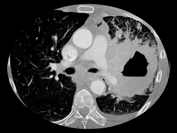
Holy smoke: Air pollution link to bone damage confirmed
Air pollution appears to contribute independently to bone damage in postmenopausal women, new data suggest.
The findings come from a new analysis of data from the Women’s Health Initiative (WHI) and location-specific air particulate information from the U.S. Environmental Protection Agency.
“Our findings confirm that poor air quality may be a risk factor for bone loss, independent of socioeconomic or demographic factors, and expands previous findings to postmenopausal women. Indeed, to our knowledge, this is the first study of the impact of criteria air pollutants on bone health in postmenopausal women,” Diddier Prada, MD, PhD, Columbia University, New York, and colleagues wrote.
The results are also the first to show that “nitrogen oxides contribute the most to bone damage and that the lumbar spine is one of the most susceptible sites,” they added.
Public health policies should aim to reduce air pollution in general, they wrote, and reducing nitrogen oxides, in particular, will reduce bone damage in postmenopausal women, prevent bone fractures, and reduce the health cost burden associated with osteoporosis in this population.
The findings were recently published in eClinicalMedicine.
Asked to comment, Giovanni Adami, MD, PhD, said in an interview that the study “adds to the body of literature on air pollution and bone health. The study confirms and provides further evidence linking air pollution exposure and osteoporosis.”
Dr. Adami, of the University of Verona (Italy), who also studies this topic, said that these new findings align with those from his group and others.
“The scientific literature in the field is clearly pointing toward a negative effect of chronic pollution exposure on bone health.”

He pointed to one study from his group that found chronic exposure to ultrafine particulate matter is associated with low BMD, and consequently, bone fragility, and another study that showed acute exposure to high levels of pollutants could actually cause fractures.
As for what might be done clinically, Dr. Adami said: “It is difficult to extrapolate direct and immediate recommendations for patients.
“However, it might be acceptable to say that patients at risk of osteoporosis, such as older women or those with prior bone fractures, should avoid chronic exposure to air pollution, perhaps using masks when walking in traffic or using air filters for indoor ventilation.”
Dr. Adami also said that this evidence so far might spur the future inclusion of chronic exposure to air pollution in fracture risk assessment tools, although this isn’t likely to come about in the near future.
Particulates linked to whole-body, hip, lumbar, and femoral neck BMD
The prospective observational study included 9,041 WHI participants seen over 32,663 visits who were an average of 63 years old at baseline. More than 70% were White, and just under half were college graduates.
With geocoded address data used to estimate particulate matter concentrations, mean levels of particulate matter of 10 mcm or less, nitrogen oxide nitrogen dioxide, and sulfur dioxide over 1, 3, and 5 years were all negatively associated with whole-body, total hip, femoral neck, and lumbar spine BMD.
In the multivariate analysis, the highest correlations were found between nitrogen oxide and nitrogen dioxide. For example, lumbar spine BMD decreased by 0.026 g/cm2 per year per 10% increase in 3-year mean nitrogen dioxide concentration.
“Our findings show that both particulate matter and gases may adversely impact BMD and that nitrogen oxides may play a critical role in bone damage and osteoporosis risk,” Dr. Prada and colleagues wrote.
Dr. Adami added: “We need more data to understand the precise magnitude of effect of air pollution on fractures, which might depend on levels of exposure but also on genetics and lifestyle.”
The study was funded by the National Institutes of Health. The authors reported no relevant financial relationships. Dr. Adami reported receiving fees from Amgen, Eli Lilly, UCB, Fresenius Kabi, Galapagos, and Theramex.
A version of this article originally appeared on Medscape.com.
Air pollution appears to contribute independently to bone damage in postmenopausal women, new data suggest.
The findings come from a new analysis of data from the Women’s Health Initiative (WHI) and location-specific air particulate information from the U.S. Environmental Protection Agency.
“Our findings confirm that poor air quality may be a risk factor for bone loss, independent of socioeconomic or demographic factors, and expands previous findings to postmenopausal women. Indeed, to our knowledge, this is the first study of the impact of criteria air pollutants on bone health in postmenopausal women,” Diddier Prada, MD, PhD, Columbia University, New York, and colleagues wrote.
The results are also the first to show that “nitrogen oxides contribute the most to bone damage and that the lumbar spine is one of the most susceptible sites,” they added.
Public health policies should aim to reduce air pollution in general, they wrote, and reducing nitrogen oxides, in particular, will reduce bone damage in postmenopausal women, prevent bone fractures, and reduce the health cost burden associated with osteoporosis in this population.
The findings were recently published in eClinicalMedicine.
Asked to comment, Giovanni Adami, MD, PhD, said in an interview that the study “adds to the body of literature on air pollution and bone health. The study confirms and provides further evidence linking air pollution exposure and osteoporosis.”
Dr. Adami, of the University of Verona (Italy), who also studies this topic, said that these new findings align with those from his group and others.
“The scientific literature in the field is clearly pointing toward a negative effect of chronic pollution exposure on bone health.”
He pointed to one study from his group that found chronic exposure to ultrafine particulate matter is associated with low BMD, and consequently, bone fragility, and another study that showed acute exposure to high levels of pollutants could actually cause fractures.
As for what might be done clinically, Dr. Adami said: “It is difficult to extrapolate direct and immediate recommendations for patients.
“However, it might be acceptable to say that patients at risk of osteoporosis, such as older women or those with prior bone fractures, should avoid chronic exposure to air pollution, perhaps using masks when walking in traffic or using air filters for indoor ventilation.”
Dr. Adami also said that this evidence so far might spur the future inclusion of chronic exposure to air pollution in fracture risk assessment tools, although this isn’t likely to come about in the near future.
Particulates linked to whole-body, hip, lumbar, and femoral neck BMD
The prospective observational study included 9,041 WHI participants seen over 32,663 visits who were an average of 63 years old at baseline. More than 70% were White, and just under half were college graduates.
With geocoded address data used to estimate particulate matter concentrations, mean levels of particulate matter of 10 mcm or less, nitrogen oxide nitrogen dioxide, and sulfur dioxide over 1, 3, and 5 years were all negatively associated with whole-body, total hip, femoral neck, and lumbar spine BMD.
In the multivariate analysis, the highest correlations were found between nitrogen oxide and nitrogen dioxide. For example, lumbar spine BMD decreased by 0.026 g/cm2 per year per 10% increase in 3-year mean nitrogen dioxide concentration.
“Our findings show that both particulate matter and gases may adversely impact BMD and that nitrogen oxides may play a critical role in bone damage and osteoporosis risk,” Dr. Prada and colleagues wrote.
Dr. Adami added: “We need more data to understand the precise magnitude of effect of air pollution on fractures, which might depend on levels of exposure but also on genetics and lifestyle.”
The study was funded by the National Institutes of Health. The authors reported no relevant financial relationships. Dr. Adami reported receiving fees from Amgen, Eli Lilly, UCB, Fresenius Kabi, Galapagos, and Theramex.
A version of this article originally appeared on Medscape.com.
FROM ECLINICALMEDICINE
Rheumatology fellows learn about career opportunities
SAN FRANCISCO – Various career paths open to newly board-certified rheumatologists – and some of the pros and cons for each – were explored at the 2023 Fellows Conference of the Coalition for State Rheumatology Organizations.
CSRO’s annual Fellows Conference aims to helps rheumatology fellows-in-training transition into future roles as practicing physicians, said Christopher Sonntag, MD, a 2nd-year rheumatology fellow at Roger Williams Medical Center, Providence, R.I., and the Fellow-At-Large representative on CSRO’s Board of Directors. He will launch his own career at Washington Regional Medical Center in Fayetteville, Ark., close to where he grew up, when his fellowship winds up in June.
“I started going to CSRO meetings in 2019, when I was still a resident,” said Dr. Sonntag, who fell in love with rheumatology in medical school. “This conference is a great opportunity for fellows. They can learn a lot of critical issues and skills that we just don’t get enough information about in our training, basic things we ought to know about: how insurance works, medical benefits, and the like.”
Job seekers in a specialty in short supply like rheumatology have some competitive advantages, but that varies by locality in a volatile health care market. “The job I ended up taking was not one where they were initially looking to hire another rheumatologist,” Dr. Sonntag told this news organization. The Fayetteville hospital already had two busy rheumatologists, but after Dr. Sonntag had unsatisfying interviews at six other groups, he called them back and they decided to go ahead and hire him. He said the position provides an acceptable work-life balance, as well as opportunities to teach. He hopes eventually to create a rheumatology fellowship program.
Models and Career Paths
Decisions about one’s career path are very important, said CSRO’s president, Gary Feldman, MD, a rheumatologist at Pacific Arthritis in Los Angeles. “We want your choice to work for you,” he told attendees. “We need you to be happy [in your jobs] for the next 30 years. You are the future.”
Dr. Feldman cited a recent Medscape salary survey of 13,000 full-time physicians from 29 specialties, which ranked rheumatologists 22nd in average annual income at $289,000. Total income may not be the first consideration in pursuing rheumatology as a career, Dr. Feldman noted. The same Medscape survey revealed that 60% of rheumatologists believe they are fairly compensated. “Something else is going on, something to do with work/life balance, which is complicated,” he said.
Other contributors to their career fulfillment may include the in-depth, long-term therapeutic relationships rheumatologists develop with their patients who have chronic, incurable illnesses; the ability with new treatments to make such a difference in managing their pain and discomfort; and engagement with giving good medical care that is centered on the patient’s experience.
“We have drugs that work to make our patients feel better. Patients come to us with no idea what’s going on, and we can turn their lives around,” Dr. Sonntag noted.
Other important career-oriented questions to ask, Dr. Feldman said, include:
What is important to you?
Who are you going to be working alongside?
How much autonomy, agency, security, or risk are you comfortable with?
What is your best balance between being a physician and an entrepreneur?
Finding your niche
Presenter Aaron Broadwell, MD, a rheumatologist in a private specialty practice of five physicians and five advanced practice providers in Shreveport, La., discussed the prospects for a career in private practice at the Fellows Conference. Private practice is not dead as a career choice, he observed, “despite what I continue to hear.” Data show that 70% of rheumatologists currently are in employed positions, but he sees signs of a movement back toward private practice.
Other basic career paths outlined by Dr. Broadwell include:
- Academic medicine, which offers opportunities to teach future physicians (although it’s also possible for rheumatologists practicing outside of academia to teach as well).
- Hospital employment, which has a higher starting salary but also a greater emphasis on RVUs (relative value units) and productivity, with less job security than it used to enjoy.
- Military/Veterans Administration positions, which may have antiquated office systems and salary caps.
- Other paths, including corporate medical director positions with pharmaceutical companies and insurance companies.
Newer options include concierge and direct specialty care models where physician-operated practices partner with their patients to provide specialty care services under a flat or periodic membership fee, and joining one of the large, multistate, rheumatology care management groups like United Rheumatology, LLC, and American Arthritis and Rheumatology Associates.
Private practice medical groups are both single specialty and multispecialty, both large and small – as well as solo rheumatology practices, Dr. Broadwell said. “People launch solo private practices all the time. It is good for some doctors. It has the highest risk and the highest potential reward.”
Becoming profitable in solo practice may take a year or two, while the doctor remains responsible 24/7, including the need to arrange for vacation and sick leave coverage. Solo practitioners need to be up to date on billing, coding, revenue cycles, bundled payments and the like, and eventually need to hire and supervise a team the doctor can trust.
What can young rheumatologists do to learn more of the nuances of these approaches? Dr. Broadwell recommended joining their state rheumatology society as well as the American College of Rheumatology. “The National Organization of Rheumatology Management is a phenomenal source of information, not just for your office manager but also for you,” he said. He also recommended linking up with colleagues through social media outlets such as the Rheumatology Private Practice Group on Facebook.
No relevant financial relationships were reported by the conference speakers.
A version of this article originally appeared on Medscape.com.
AT CSRO 2023
Ozempic: The latest weight loss craze and how over-prescribing is harming patients
Social media and mainstream media websites are full of stories on the new wonder weight loss drug: Ozempic. Even Hollywood stars are talking about it.

Recently, the zealous prescribing of this diabetes medication fueled a 6-month shortage making it difficult for anyone to get it. Part of the problem stems from digital access to these medications where a patient can get a prescription online or via a telemedicine platform. Additionally, certain weight loss programs contributed to promoting the weight loss benefits.
Ozempic is a glucagon-like peptide-1 (GLP-1) agonist, with the generic name semaglutide, that lowers hemoglobin A1c in patients with diabetes and lowers the risk of cardiovascular events. Semaglutide is also sold as Wegovy, which is indicated for weight loss. Both Ozempic and Wegovy are sold in multiple doses, but the target dose for Wegovy is higher.
Weight loss with Wegovy is, on average, higher than that seen with Ozempic. However, it is often more difficult to get Wegovy covered by health insurance companies.
As doctors, we must be stewards of the medications we are prescribing. Clearly, the Internet should not be driving our prescribing habits. Prescribing Ozempic for weight loss can make it more difficult for patients with diabetes to receive it, and we should consider other options until it is more available and/or receives FDA approval for treating obesity.
Most of us have seen our patients with diabetes having difficulty getting a prescription for Ozempic filled, either because it is on back-order or because of a lack of coverage. Insurance companies have no incentive to lower the cost when it is in such high demand at its current rate. For these patients, lowering their A1c can be life-saving and prevent complications of diabetes, such as kidney failure and heart disease. In our current environment, we should reserve prescribing Ozempic for our patients with diabetes who need it more. Wegovy is available and can be prescribed for patients wishing to lose weight.
Many patients are looking for a magic cure. Neither medication is that. Patients need to start with making lifestyle changes first. In primary care, advising on and helping patients implement those are often our most difficult tasks. However, no medication is going to work unless the patient makes adjustments to their diet and amount and type of movement they are doing. In patients who have a hard time changing their diet, lowering carbohydrate intake may be a good first step. Exercising, or being more active if a patient is unable to formally exercise, is an important therapy.
As we all know, metformin is the usual preferred method for the treatment of type 2 diabetes unless contraindicated in a given patient. There are many oral diabetes medications available, and which of these and how these are prescribed need to be tailored to the individual patient. Ozempic can be used when a patient is failing on metformin, or other oral meds, or if they would rather do a weekly injection rather than remembering to take daily pills, for example.
Obesity has reached epidemic proportions in the United States. According to the CDC, more than 40% of the U.S. population is obese. Additionally, millions of children between the ages of 2 and 19 are now considered obese, and the medical complications for these individuals ares yet to be seen. Plus, many of us are seeing higher frequencies of diabetes, hypertension, and other chronic medical conditions in adolescents in our daily practices.
Our war against obesity is a fight for future lives and having more tools available is definitely a help. Like with patients with diabetes, all treatment regimens should start off with lifestyle modifications. Fad diets rarely result in long-term weight loss.
There are several medications now available to help with weight loss, Wegovy being just one of them. Patients often come to us with their own personal preferences, and it is our job to guide them on the best course to take. Some people may prefer a weekly injection. There are oral medications available, such as Contrave and Phentermine, and the best one should be decided upon by the patient and doctor after a discussion of the risks.
Let’s stop prescribing Ozempic for weight loss because nonphysicians say we should. Leave it for our patients with diabetes, those whose lives may depend on taking it. If we didn’t have other medications available, it would be a very different story. But, we do, and we need to resist the pressure others place on us and do the right thing for all of our patients.
*This article was updated on 3/23/2023.
Dr. Girgis practices family medicine in South River, N.J., and is a clinical assistant professor of family medicine at Robert Wood Johnson Medical School, New Brunswick, N.J. She has no conflicts related to this piece. You can contact her at fpnews@mdedge.com.
Social media and mainstream media websites are full of stories on the new wonder weight loss drug: Ozempic. Even Hollywood stars are talking about it.
Recently, the zealous prescribing of this diabetes medication fueled a 6-month shortage making it difficult for anyone to get it. Part of the problem stems from digital access to these medications where a patient can get a prescription online or via a telemedicine platform. Additionally, certain weight loss programs contributed to promoting the weight loss benefits.
Ozempic is a glucagon-like peptide-1 (GLP-1) agonist, with the generic name semaglutide, that lowers hemoglobin A1c in patients with diabetes and lowers the risk of cardiovascular events. Semaglutide is also sold as Wegovy, which is indicated for weight loss. Both Ozempic and Wegovy are sold in multiple doses, but the target dose for Wegovy is higher.
Weight loss with Wegovy is, on average, higher than that seen with Ozempic. However, it is often more difficult to get Wegovy covered by health insurance companies.
As doctors, we must be stewards of the medications we are prescribing. Clearly, the Internet should not be driving our prescribing habits. Prescribing Ozempic for weight loss can make it more difficult for patients with diabetes to receive it, and we should consider other options until it is more available and/or receives FDA approval for treating obesity.
Most of us have seen our patients with diabetes having difficulty getting a prescription for Ozempic filled, either because it is on back-order or because of a lack of coverage. Insurance companies have no incentive to lower the cost when it is in such high demand at its current rate. For these patients, lowering their A1c can be life-saving and prevent complications of diabetes, such as kidney failure and heart disease. In our current environment, we should reserve prescribing Ozempic for our patients with diabetes who need it more. Wegovy is available and can be prescribed for patients wishing to lose weight.
Many patients are looking for a magic cure. Neither medication is that. Patients need to start with making lifestyle changes first. In primary care, advising on and helping patients implement those are often our most difficult tasks. However, no medication is going to work unless the patient makes adjustments to their diet and amount and type of movement they are doing. In patients who have a hard time changing their diet, lowering carbohydrate intake may be a good first step. Exercising, or being more active if a patient is unable to formally exercise, is an important therapy.
As we all know, metformin is the usual preferred method for the treatment of type 2 diabetes unless contraindicated in a given patient. There are many oral diabetes medications available, and which of these and how these are prescribed need to be tailored to the individual patient. Ozempic can be used when a patient is failing on metformin, or other oral meds, or if they would rather do a weekly injection rather than remembering to take daily pills, for example.
Obesity has reached epidemic proportions in the United States. According to the CDC, more than 40% of the U.S. population is obese. Additionally, millions of children between the ages of 2 and 19 are now considered obese, and the medical complications for these individuals ares yet to be seen. Plus, many of us are seeing higher frequencies of diabetes, hypertension, and other chronic medical conditions in adolescents in our daily practices.
Our war against obesity is a fight for future lives and having more tools available is definitely a help. Like with patients with diabetes, all treatment regimens should start off with lifestyle modifications. Fad diets rarely result in long-term weight loss.
There are several medications now available to help with weight loss, Wegovy being just one of them. Patients often come to us with their own personal preferences, and it is our job to guide them on the best course to take. Some people may prefer a weekly injection. There are oral medications available, such as Contrave and Phentermine, and the best one should be decided upon by the patient and doctor after a discussion of the risks.
Let’s stop prescribing Ozempic for weight loss because nonphysicians say we should. Leave it for our patients with diabetes, those whose lives may depend on taking it. If we didn’t have other medications available, it would be a very different story. But, we do, and we need to resist the pressure others place on us and do the right thing for all of our patients.
*This article was updated on 3/23/2023.
Dr. Girgis practices family medicine in South River, N.J., and is a clinical assistant professor of family medicine at Robert Wood Johnson Medical School, New Brunswick, N.J. She has no conflicts related to this piece. You can contact her at fpnews@mdedge.com.
Fat Necrosis of the Breast Mimicking Breast Cancer in a Male Patient Following Wax Hair Removal
To the Editor:
Fat necrosis of the breast is a benign inflammatory disease of adipose tissue commonly observed after trauma in the female breast during the perimenopausal period.1 Fat necrosis of the male breast is rare, first described by Silverstone2 in 1949; the condition usually presents with unilateral, painful or asymptomatic, firm nodules, which in rare cases are observed as skin retraction and thickening, ecchymosis, erythematous plaque–like cellulitis, local depression, and/or discoloration of the breast skin.3-5
Diagnosis of fat necrosis of the male breast may need to be confirmed via biopsy in conjunction with clinical and radiologic findings because the condition can mimic breast cancer.1 We report a case of bilateral fat necrosis of the breast mimicking breast cancer following wax hair removal.
A 42-year-old man presented to our outpatient dermatology clinic for evaluation of redness, swelling, and hardness of the skin of both breasts of 3 weeks’ duration. The patient had a history of wax hair removal of the entire anterior aspect of the body. He reported an erythematous, edematous, warm plaque that developed on the breasts 2 days after waxing. The plaque did not respond to antibiotics. The swelling and induration progressed over the 2 weeks after the patient was waxed. The patient had no family history of breast cancer. He had a standing diagnosis of gynecomastia. He denied any history of fat or filler injection in the affected area.
Dermatologic examination revealed erythematous, edematous, indurated, asymptomatic plaques with a peau d’orange appearance on the bilateral pectoral and presternal region. Minimal retraction of the right areola was noted (Figure 1). The bilateral axillary lymph nodes were palpable.

Laboratory results including erythrocyte sedimentation rate (108 mm/h [reference range, 2–20 mm/h]), C-reactive protein (9.2 mg/dL [reference range, >0.5 mg/dL]), and ferritin levels (645
Mammography of both breasts revealed a Breast Imaging Reporting and Data System (BI-RADS) score of 4 with a suspicious abnormality (ie, diffuse edema of the breast, multiple calcifications in a nonspecific pattern, oil cysts with calcifications, and bilateral axillary lymphadenopathy with a diameter of 2.5 cm and a thick and irregular cortex)(Figure 2A). Ultrasonography of both breasts revealed an inflammatory breast. Magnetic resonance imaging showed similar findings with diffuse edema and a heterogeneous appearance. Contrast-enhanced magnetic resonance imaging showed diffuse contrast enhancement in both breasts extending to the pectoral muscles and axillary regions, consistent with inflammatory changes (Figure 2B).

Because of difficulty differentiating inflammation and an infiltrating tumor, histopathologic examination was recommended by radiology. Results from a 5-mm punch biopsy from the right breast yielded the following differential diagnoses: cellulitis, panniculitis, inflammatory breast cancer, subcutaneous fat necrosis, and paraffinoma. Histopathologic examination of the skin revealed a normal epidermis and a dense inflammatory cell infiltrate comprising lymphocytes and monocytes in the dermis and subcutaneous tissue. Marked fibrosis also was noted in the dermis and subcutaneous tissue. Lipophagic fat necrosis accompanied by a variable inflammatory cell infiltrate consisted of histiocytes and neutrophils (Figure 3A). Pankeratin immunostaining was negative. Fat necrosis was present in a biopsy specimen obtained from the right breast; no signs of malignancy were present (Figure 3B). Fine-needle aspiration of the axillary lymph nodes was benign. Given these histopathologic findings, malignancy was excluded from the differential diagnosis. Paraffinoma also was ruled out because the patient insistently denied any history of fat or filler injection.

Based on the clinical, histopathologic, and radiologic findings, as well as the history of minor trauma due to wax hair removal, a diagnosis of fat necrosis of the breast was made. Intervention was not recommended by the plastic surgeons who subsequently evaluated the patient, because the additional trauma may aggravate the lesion. He was treated with nonsteroidal anti-inflammatory drugs.
At 6-month follow-up, there was marked reduction in the erythema and edema but no notable improvement of the induration. A potent topical steroid was added to the treatment, but only slight regression of the induration was observed.
The normal male breast is comprised of fat and a few secretory ducts.6 Gynecomastia and breast cancer are the 2 most common conditions of the male breast; fat necrosis of the male breast is rare. In a study of 236 male patients with breast disease, only 5 had fat necrosis.7
Fat necrosis of the breast can be observed with various clinical and radiological presentations. Subcutaneous nodules, skin retraction and thickening, local skin depression, and ecchymosis are the more common presentations of fat necrosis.3-5 In our case, the first symptoms of disease were similar to those seen in cellulitis. The presentation of fat necrosis–like cellulitis has been described only rarely in the medical literature. Haikin et al5 reported a case of fat necrosis of the leg in a child that presented with cellulitis followed by induration, which did not respond to antibiotics, as was the case with our patient.5
Blunt trauma, breast reduction surgery, and breast augmentation surgery can cause fat necrosis of the breast1,4; in some cases, the cause cannot be determined.8 The only pertinent history in our patient was wax hair removal. Fat necrosis was an unexpected complication, but hair removal can be considered minor trauma; however, this is not commonly reported in the literature following hair removal with wax. In a study that reviewed diseases of the male breast, the investigators observed that all male patients with fat necrosis had pseudogynecomastia (adipomastia).7 Although our patient’s entire anterior trunk was epilated, only the breast was affected. This situation might be explained by underlying gynecomastia because fat necrosis is common in areas of the body where subcutaneous fat tissue is dense.
Fat necrosis of the breast can be mistaken—both clinically and radiologically—for malignancy, such as in our case. Diagnosis of fat necrosis of the breast should be a diagnosis of exclusion; therefore, histopathologic confirmation of the lesion is imperative.9
In conclusion, fat necrosis of the male breast is rare. The condition can present as cellulitis. Hair removal with wax might be a cause of fat necrosis. Because breast cancer and fat necrosis can exhibit clinical and radiologic similarities, the diagnosis of fat necrosis should be confirmed by histopathologic analysis in conjunction with clinical and radiologic findings.
- Tan PH, Lai LM, Carrington EV, et al. Fat necrosis of the breast—a review. Breast. 2006;15:313-318. doi:10.1016/j.breast.2005.07.003
- Silverstone M. Fat necrosis of the breast with report of a case in a male. Br J Surg. 1949;37:49-52. doi:10.1002/bjs.18003714508
- Akyol M, Kayali A, Yildirim N. Traumatic fat necrosis of male breast. Clin Imaging. 2013;37:954-956. doi:10.1016/j.clinimag.2013.05.009
- Crawford EA, King JJ, Fox EJ, et al. Symptomatic fat necrosis and lipoatrophy of the posterior pelvis following trauma. Orthopedics. 2009;32:444. doi:10.3928/01477447-20090511-25
- Haikin Herzberger E, Aviner S, Cherniavsky E. Posttraumatic fat necrosis presented as cellulitis of the leg. Case Rep Pediatr. 2012;2012:672397. doi:10.1155/2012/672397
- Michels LG, Gold RH, Arndt RD. Radiography of gynecomastia and other disorders of the male breast. Radiology. 1977;122:117-122. doi:10.1148/122.1.117
- Günhan-Bilgen I, Bozkaya H, Ustün E, et al. Male breast disease: clinical, mammographic, and ultrasonographic features. Eur J Radiol. 2002;43:246-255. doi:10.1016/s0720-048x(01)00483-1
- Chala LF, de Barros N, de Camargo Moraes P, et al. Fat necrosis of the breast: mammographic, sonographic, computed tomography, and magnetic resonance imaging findings. Curr Probl Diagn Radiol. 2004;33:106-126. doi:10.1067/j.cpradiol.2004.01.001
- Pullyblank AM, Davies JD, Basten J, et al. Fat necrosis of the female breast—Hadfield re-visited. Breast. 2001;10:388-391. doi:10.1054/brst.2000.0287
To the Editor:
Fat necrosis of the breast is a benign inflammatory disease of adipose tissue commonly observed after trauma in the female breast during the perimenopausal period.1 Fat necrosis of the male breast is rare, first described by Silverstone2 in 1949; the condition usually presents with unilateral, painful or asymptomatic, firm nodules, which in rare cases are observed as skin retraction and thickening, ecchymosis, erythematous plaque–like cellulitis, local depression, and/or discoloration of the breast skin.3-5
Diagnosis of fat necrosis of the male breast may need to be confirmed via biopsy in conjunction with clinical and radiologic findings because the condition can mimic breast cancer.1 We report a case of bilateral fat necrosis of the breast mimicking breast cancer following wax hair removal.
A 42-year-old man presented to our outpatient dermatology clinic for evaluation of redness, swelling, and hardness of the skin of both breasts of 3 weeks’ duration. The patient had a history of wax hair removal of the entire anterior aspect of the body. He reported an erythematous, edematous, warm plaque that developed on the breasts 2 days after waxing. The plaque did not respond to antibiotics. The swelling and induration progressed over the 2 weeks after the patient was waxed. The patient had no family history of breast cancer. He had a standing diagnosis of gynecomastia. He denied any history of fat or filler injection in the affected area.
Dermatologic examination revealed erythematous, edematous, indurated, asymptomatic plaques with a peau d’orange appearance on the bilateral pectoral and presternal region. Minimal retraction of the right areola was noted (Figure 1). The bilateral axillary lymph nodes were palpable.
Laboratory results including erythrocyte sedimentation rate (108 mm/h [reference range, 2–20 mm/h]), C-reactive protein (9.2 mg/dL [reference range, >0.5 mg/dL]), and ferritin levels (645
Mammography of both breasts revealed a Breast Imaging Reporting and Data System (BI-RADS) score of 4 with a suspicious abnormality (ie, diffuse edema of the breast, multiple calcifications in a nonspecific pattern, oil cysts with calcifications, and bilateral axillary lymphadenopathy with a diameter of 2.5 cm and a thick and irregular cortex)(Figure 2A). Ultrasonography of both breasts revealed an inflammatory breast. Magnetic resonance imaging showed similar findings with diffuse edema and a heterogeneous appearance. Contrast-enhanced magnetic resonance imaging showed diffuse contrast enhancement in both breasts extending to the pectoral muscles and axillary regions, consistent with inflammatory changes (Figure 2B).
Because of difficulty differentiating inflammation and an infiltrating tumor, histopathologic examination was recommended by radiology. Results from a 5-mm punch biopsy from the right breast yielded the following differential diagnoses: cellulitis, panniculitis, inflammatory breast cancer, subcutaneous fat necrosis, and paraffinoma. Histopathologic examination of the skin revealed a normal epidermis and a dense inflammatory cell infiltrate comprising lymphocytes and monocytes in the dermis and subcutaneous tissue. Marked fibrosis also was noted in the dermis and subcutaneous tissue. Lipophagic fat necrosis accompanied by a variable inflammatory cell infiltrate consisted of histiocytes and neutrophils (Figure 3A). Pankeratin immunostaining was negative. Fat necrosis was present in a biopsy specimen obtained from the right breast; no signs of malignancy were present (Figure 3B). Fine-needle aspiration of the axillary lymph nodes was benign. Given these histopathologic findings, malignancy was excluded from the differential diagnosis. Paraffinoma also was ruled out because the patient insistently denied any history of fat or filler injection.
Based on the clinical, histopathologic, and radiologic findings, as well as the history of minor trauma due to wax hair removal, a diagnosis of fat necrosis of the breast was made. Intervention was not recommended by the plastic surgeons who subsequently evaluated the patient, because the additional trauma may aggravate the lesion. He was treated with nonsteroidal anti-inflammatory drugs.
At 6-month follow-up, there was marked reduction in the erythema and edema but no notable improvement of the induration. A potent topical steroid was added to the treatment, but only slight regression of the induration was observed.
The normal male breast is comprised of fat and a few secretory ducts.6 Gynecomastia and breast cancer are the 2 most common conditions of the male breast; fat necrosis of the male breast is rare. In a study of 236 male patients with breast disease, only 5 had fat necrosis.7
Fat necrosis of the breast can be observed with various clinical and radiological presentations. Subcutaneous nodules, skin retraction and thickening, local skin depression, and ecchymosis are the more common presentations of fat necrosis.3-5 In our case, the first symptoms of disease were similar to those seen in cellulitis. The presentation of fat necrosis–like cellulitis has been described only rarely in the medical literature. Haikin et al5 reported a case of fat necrosis of the leg in a child that presented with cellulitis followed by induration, which did not respond to antibiotics, as was the case with our patient.5
Blunt trauma, breast reduction surgery, and breast augmentation surgery can cause fat necrosis of the breast1,4; in some cases, the cause cannot be determined.8 The only pertinent history in our patient was wax hair removal. Fat necrosis was an unexpected complication, but hair removal can be considered minor trauma; however, this is not commonly reported in the literature following hair removal with wax. In a study that reviewed diseases of the male breast, the investigators observed that all male patients with fat necrosis had pseudogynecomastia (adipomastia).7 Although our patient’s entire anterior trunk was epilated, only the breast was affected. This situation might be explained by underlying gynecomastia because fat necrosis is common in areas of the body where subcutaneous fat tissue is dense.
Fat necrosis of the breast can be mistaken—both clinically and radiologically—for malignancy, such as in our case. Diagnosis of fat necrosis of the breast should be a diagnosis of exclusion; therefore, histopathologic confirmation of the lesion is imperative.9
In conclusion, fat necrosis of the male breast is rare. The condition can present as cellulitis. Hair removal with wax might be a cause of fat necrosis. Because breast cancer and fat necrosis can exhibit clinical and radiologic similarities, the diagnosis of fat necrosis should be confirmed by histopathologic analysis in conjunction with clinical and radiologic findings.
- Tan PH, Lai LM, Carrington EV, et al. Fat necrosis of the breast—a review. Breast. 2006;15:313-318. doi:10.1016/j.breast.2005.07.003
- Silverstone M. Fat necrosis of the breast with report of a case in a male. Br J Surg. 1949;37:49-52. doi:10.1002/bjs.18003714508
- Akyol M, Kayali A, Yildirim N. Traumatic fat necrosis of male breast. Clin Imaging. 2013;37:954-956. doi:10.1016/j.clinimag.2013.05.009
- Crawford EA, King JJ, Fox EJ, et al. Symptomatic fat necrosis and lipoatrophy of the posterior pelvis following trauma. Orthopedics. 2009;32:444. doi:10.3928/01477447-20090511-25
- Haikin Herzberger E, Aviner S, Cherniavsky E. Posttraumatic fat necrosis presented as cellulitis of the leg. Case Rep Pediatr. 2012;2012:672397. doi:10.1155/2012/672397
- Michels LG, Gold RH, Arndt RD. Radiography of gynecomastia and other disorders of the male breast. Radiology. 1977;122:117-122. doi:10.1148/122.1.117
- Günhan-Bilgen I, Bozkaya H, Ustün E, et al. Male breast disease: clinical, mammographic, and ultrasonographic features. Eur J Radiol. 2002;43:246-255. doi:10.1016/s0720-048x(01)00483-1
- Chala LF, de Barros N, de Camargo Moraes P, et al. Fat necrosis of the breast: mammographic, sonographic, computed tomography, and magnetic resonance imaging findings. Curr Probl Diagn Radiol. 2004;33:106-126. doi:10.1067/j.cpradiol.2004.01.001
- Pullyblank AM, Davies JD, Basten J, et al. Fat necrosis of the female breast—Hadfield re-visited. Breast. 2001;10:388-391. doi:10.1054/brst.2000.0287
Practice Points
- Fat necrosis of the breast can be mistaken—both clinically and radiologically—for malignancy; therefore, diagnosis should be confirmed by histopathology in conjunction with clinical and radiologic findings.
- Fat necrosis of the male breast is rare, and hair removal with wax may be a rare cause of the disease.
