User login
Cardiology News is an independent news source that provides cardiologists with timely and relevant news and commentary about clinical developments and the impact of health care policy on cardiology and the cardiologist's practice. Cardiology News Digital Network is the online destination and multimedia properties of Cardiology News, the independent news publication for cardiologists. Cardiology news is the leading source of news and commentary about clinical developments in cardiology as well as health care policy and regulations that affect the cardiologist's practice. Cardiology News Digital Network is owned by Frontline Medical Communications.
When intravascular imaging guides complex PCI, MACE risk is lowered
NEW ORLEANS – In patients undergoing percutaneous intervention (PCI) for complex coronary lesions, intravascular imaging is superior to angiography for reducing the risk of target lesion failure (TLF), according to results of a randomized trial.
Previous studies have produced the same conclusion, but the advantage was demonstrated this time in a multicenter well-powered randomized trial, principal investigator Joo Yong Hahn, MD, PhD, said at the joint scientific sessions of the American College of Cardiology and the World Heart Federation.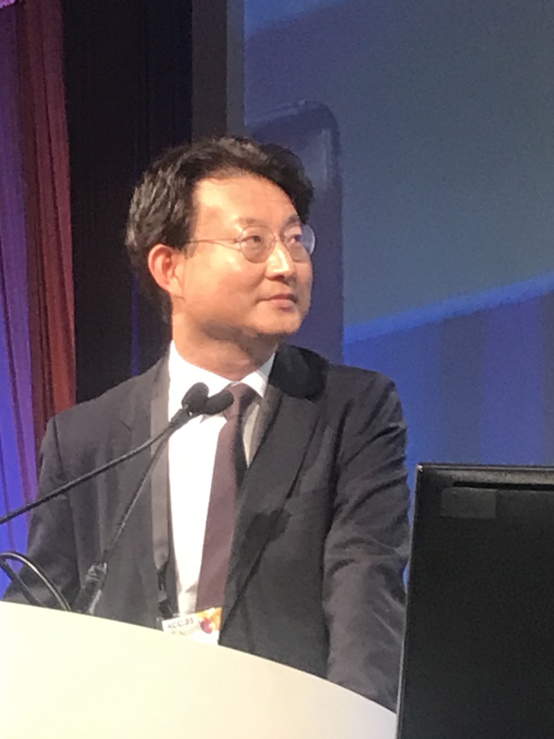
The earlier studies “were not definitive,” said Dr. Hahn, pointing out that even those that were randomized lacked sufficient duration of follow-up or were not inclusive of a broad array of types of complex PCI.
In this clinical outcomes–driven study, called RENOVATE-COMPLEX-PCI, 1,639 patients undergoing complex PCI in 20 South Korean treatment centers were randomized in a 2:1 ratio to PCI guided by intravascular imaging or angiography alone. There were nine types of complex PCI eligible for trial entry, including bifurcated lesions, long lesions (expected stent length ≥ 38 mm), total coronary occlusions, lesions requiring multiple stents, severely calcified lesions, and lesions in multiple vessels.
Intravascular imaging in the experimental arm could be performed with either intravascular ultrasound (IVUS) or optical coherence tomography (OCT), according to Dr. Hahn. Because one might be better than the other for specific patient and lesions characteristics, the type of intravascular imaging in the experimental group was selected at the discretion of the treating investigator, reported Dr. Hahn, of the Heart Vascular Stroke Institute, Samsung Medical Center, Sungkyunkwan University, Seoul.
The primary TLF endpoint was defined as death from cardiovascular causes, target-vessel-related MI, and target-vessel revascularization.
Risk reduction of > 35% observed
After a median of 2.1 years of follow-up, the lower TLF incidence in the group with PCI guided by intravascular imaging (7.7% vs. 12.3%) translated into a 36% reduction in risk (hazard ratio, 0.64; P = .008).
Intravascular imaging was associated with a numerical reduction of each component of TLF. In the case of death from cardiovascular causes, the confidence interval remained below the line of unity (HR 0.47; 95% CI, 0.24-0.93).
Although this was not true for target vessel–related MI (HR, 0.74, 95% CI, 0.45-1.22) or target vessel revascularization (HR, 0.66; 95% CI, 0.36-1.22), it was also true of TLF without procedural-related MI (HR, 0.59; 95% CI, 0.39-0.90) and cardiac death or target vessel–related MI (HR, 0.63; 95% CI, 0.42-0.93).
With few exceptions, all of the secondary outcomes “moved in the right direction” to favor intravascular imaging, including death from any cause (HR 0.71, 95% CI, 0.44-1.15), reported Dr. Hahn, who noted that the results were simultaneously published in the New England Journal of Medicine.
When compared, there were no major baseline differences in the 1,092 patients with PCI guided by intravascular imaging relative to the 547 guided by angiography. The median age was 65.5 years. Most (79%) were male. About half (51%) had an acute coronary syndrome and the remainder had stable ischemic heart disease. The proportions of patients with hypertension (61%), dyslipidemia (51%), and diabetes (38%) were substantial. About 18% of patients were current smokers, 24% had a previous PCI, and 7% had a previous MI.
Stent types were similar in the two groups, and they were delivered by radial access. Procedural success was achieved in about 98% of both groups. Almost all patients were discharged on a statin, aspirin, and a P2Y12 inhibitor, and the other specific postprocedural medications were comparable in the two groups.
Advantage of intravascular imaging consistent
Of the complex lesions, most (55%) had diffuse long coronary artery lesions, but other types of complex PCI, including bifurcated lesions (22%), chronic total occlusions (20%), severely calcified lesions (14%), and ostial lesions of a major coronary artery (15%) were represented. Across these lesion types, intravascular imaging was favored over angiography for TLF at least numerically. The potential exceptions were lesions requiring at least three stents (HR, 1.24; 95% CI, 0.49-3.18), but confidence intervals were wide.
The trial was unblinded, but Dr. Hahn reported that imaging analyses were performed at a core laboratory and events were adjudicated by a committee with members unaware of trial group assignments.
One unanswered question is cost. Because intravascular imaging adds cost to PCI relative to angiography, cost-effectiveness analyses are needed to provide context for the decision to use this approach in all complex PCI patients. These analyses are planned.
Based on the consistency of these trial results with previous studies, almost all of which showed the same thing, “the intravascular imaging world has spoken,” said Wayne B. Batchelor, MD, director of interventional cardiology, Inova Heart and Vascular Institute, Fairfax, Va. “The only question now is when will the interventional community is going to listen.”
Dr. Batchelor predicted that these data will change the mindset of many practitioners “to shift the debate to why not do it [intravascular imaging] from why do it.”
“Only about 15% of PCI is performed with intravascular imaging in the United States, and these [results] argue that this number needs to go up,” Dr. Batchelor said. Although he said there are technical reasons, such as diffuse lesions or small vessels, that prevent intravascular imaging from being used in every complex patient, he suggested the data are compelling.
“If you apply this to the one million patients undergoing PCI in the United States, this will translate potentially into tens of thousands of patients protected from the TVF endpoint,” Dr. Batchelor said.
Dr. Hahn reports no potential conflicts of interest, but this investigator-initiated trial received funding from Boston Scientific and Abbott Vascular. Dr. Batchelor reports financial relationships with Abbott Vascular, Boston Scientific, Idorsia, Medtronic, and V-Wave Medical.
AT ACC 2023
Viability-guided PCI doubted in stable severe CAD: REVIVED-BCIS2
There is no magical amount of viable ventricular myocardium that makes percutaneous coronary intervention (PCI) an effective addition to optimal medical therapy (OMT) in stable patients with coronary disease and poor ventricular function, suggests an analysis from a major trial.
The REVIVED-BCIS2 trial recently made waves when it showed no clinical advantage from adding PCI to OMT in stable patients with severe ischemic left ventricular (LV) dysfunction. All the patients had shown viable but dysfunctional myocardium that could potentially be revascularized.
But in a secondary analysis, extent of such hibernating heart muscle was not a good predictor of clinical outcomes, which in the trial meant death from any cause or hospitalization for heart failure (HHF).
Burden of myocardial scar tissue, however, turned out to be a potent predictor of clinical risk regardless of coronary disease severity or even LV ejection fraction (LVEF).
Because myocardial viability tracks poorly with outcomes in patients like those enrolled in the trial, as the new analysis suggests, conventional viability testing isn’t an effective guide for deciding who among them should get PCI, Divaka Perera, MD, said in an interview.
Dr. Perera, of King’s College London and the trial’s principal investigator, presented the REVIVED-BCIS2 secondary results at the joint scientific sessions of the American College of Cardiology and the World Heart Federation..
Viability testing for ischemia, he noted, is often used in practice to aid revascularization decisions. As the extent of myocardial viability can vary, it’s been asked – ever since the trial’s primary publication – whether there could be “a sweet spot or Goldilocks zone of viability that would allow prediction of which patients will do better with PCI compared to medical therapy,” Dr. Perera said. “The trial conclusively shows that is not the case.”
That the extent of hibernating myocardium, which is viable but dysfunctional, didn’t predict clinical outcomes or LV functional recovery “is disruptive of current practice and challenges a view that’s been held for decades.”
The trial’s 700 patients receiving OMT had been randomly assigned to undergo PCI or not, 347 and 353 patients, respectively. About 12% of the total were women.
About 70% of patients underwent baseline and follow-up myocardial viability testing using cardiac magnetic resonance (CMR) imaging with late gadolinium enhancement for estimation of scar burden; the remainder underwent dobutamine-stress echocardiography. All imaging assessments were conducted at independent core laboratories, Dr. Perera reported.
Extent of myocardial viability was defined three ways: volume of hibernating heart muscle, total volume of viable myocardium, and scar burden – all expressed as a percentage of total LV volume.
Every 10% increment in LV volume found to be hibernating related to a hazard ratio of 0.98 (95% confidence interval, 0.93-1.04; P = .56) for all-cause mortality or HHF at a median of 3.3 years. The analysis was adjusted for age, sex, diabetes, previous HHF, chronic renal failure, extent of CAD, type of viability testing, and baseline LVEF.
The adjusted HR for the same percentage increment in total viable myocardium was marginally significantly reduced at 0.93 (95% CI, 0.87-1.00; P = .048).
The correlation with scar burden was stronger. The adjusted composite-endpoint HR per 10% increment in scar burden was significantly increased at 1.18 (95% CI, 1.04-1.33; P = .009).
Extent of myocardial viability by tertiles, regardless of viability definition, did not highlight any group with a reduced risk for death or HHF, or group with better LV functional recovery, from OMT plus PCI, compared with OMT alone.
The findings appear to suggest that scar burden, but not extent of viability as it’s usually measured, may effectively guide PCI decisions in such patients, Dr. Perera said.
“I would say that viability testing as we understand it now, based on the paradigm of hibernating myocardium, is very useful,” he said, “but that is not the only information we can get from a viability test.”
Scar burden can also be determined from the same tests but isn’t typically looked at. “We’re actually collecting this information but not using it,” Dr. Perera said. “When we do, it is really powerfully predictive” of both clinical outcomes and LV functional recovery. “Yet scar burden is not in any of the guidelines for stratifying risk.”
REVIVED-BCIS2 was funded by the National Institute for Health and Care Research Health Technology Assessment Program. Dr. Perera had no disclosures.
A version of this article first appeared on Medscape.com.
FROM ACC 2023
Atorvastatin cut anthracycline cardiac dysfunction in lymphoma
NEW ORLEANS – Atorvastatin treatment of patients with lymphoma undergoing treatment with an anthracycline significantly cut the incidence of incident cardiac dysfunction by about two-thirds during 12 months of treatment, in a multicenter, randomized trial with 300 enrolled patients.
“These data support the use of atorvastatin among patients with lymphoma being treated with anthracyclines where prevention of cardiac systolic dysfunction is important,” concluded Tomas G. Neilan, MD, at the joint scientific sessions of the American College of Cardiology and the World Heart Federation.
He highlighted that an important difference between the new study, STOP-CA, and a major prior study with a neutral effect published in 2022, was that STOP-CA “was powered for a major change” in cardiac function as the study’s primary outcome, a decline from baseline in left ventricular ejection fraction (LVEF) of at least 10% that also reduced ejection fraction to less than 55%.
“We can consider these medications [atorvastatin] for patients at higher risk for cardiac toxicity from anthracyclines, such as patients who receive a higher dose of an anthracycline, older patients, people with obesity, and women, commented Anita Deswal, MD, professor and chair of the department of cardiology at the University of Texas MD Anderson Cancer Center, Houston, who was not involved with the study.
A basis for an ‘important discussion’ with patients
“For patients receiving higher doses of anthracyclines, the STOP-CA trial says that whether to start a statin for cardiac protection is now an important discussion” for these patients to have with their treating clinicians. ”That was not the case before today,” commented Ronald M. Witteles, MD, a cardiologist and professor who specializes in cardio-oncology at Stanford (Calif.) University.
“For a patient being treated for lymphoma or for another cancer and treated with equal or higher anthracycline doses, such as patients with a sarcoma, this trial’s results at the very least warrant a discussion between physicians and patients to make the decision,” Dr. Witteles, who was not involved in the study, said in an interview. But he also cautioned that “whether an individual patient should take a statin in this scenario is still not a no-brainer. While the trial was positive, it was for an imaging rather than for a clinical endpoint.”
Experts noted that a similar study with the clinical endpoint of heart failure would require both many more randomized patients as well as much longer follow-up. STOP-CA was not powered for this endpoint. During its 12-month duration, a total of 11 patients developed heart failure, with no between group difference.
STOP-CA enrolled adults with lymphoma (Hodgkin or non-Hodgkin) and scheduled to undergo anthracycline treatment at eight U.S. centers and one in Canada, and excluded patients already on statin treatment or those for whom a statin was already indicated. Of the 300 enrolled patients, 286 had 12-month follow-up. Randomization assigned patients to receive either 40 mg daily of atorvastatin or placebo.
Their cumulative, median anthracycline dose was 300 mg/m2, which is typical for treating lymphoma, but higher than the typical dose use for patients with breast cancer. At baseline, average LVEF was 63%, and after 12 months this had declined to 59%. Forty-six of the 286 patients assessed after 12 months fulfilled the primary outcome of at least a 10–percentage point reduction from baseline in their LVEF and a decline in LVEF to less than 55%. Researchers used cardiac MR to assess LVEF at baseline, and in most patients at follow-up, but a minority of patients had their follow-up assessments by echocardiography because of logistical issues. Greater than 90% of patients were adherent to their assigned regimen.
Tripled incidence of cardiac dysfunction in placebo patients
The incidence of this outcome was 9% among the patients who received atorvastatin, and 22% among those on placebo, a significant difference. The calculated odds of the primary outcome was 2.9-fold more likely among the patients treated with placebo, compared with those who received atorvastatin, also a significant difference.
The study’s secondary outcome was patients who had at least a 5% drop from baseline in their LVEF and with a LVEF of less than 55% after 12 months. This outcome occurred in 13% of patients treated with atorvastatin and in 29% of those who received placebo, a significant difference.
The atorvastatin and placebo arms showed no significant differences in adverse events during the study, with roughly similar incidence rates for muscle pain, elevated liver enzymes, and renal failure. None of the enrolled patients developed myositis.
Atorvastatin treatment also produced an expected average 37% decline from baseline in levels of LDL cholesterol.
“This was a well-designed and important trial,” said Dr. Witteles. “Anthracyclines remain a mainstay of cancer therapies for a number of malignancies, such as lymphoma and sarcoma, and the cardiac side effects of development of cardiac dysfunction are unequivocally real.”
The importance of a clinically meaningful effect
The results especially contrast with the findings from the PREVENT study, published in 2022, which compared a daily, 40-mg atorvastatin treatment with placebo in 279 randomized patients with breast cancer and treated for 24 months. However, patients in PREVENT had a cumulative, median anthracycline dose of 240 mg/m2, and the study’s primary outcome was the average change from baseline in LVEF after 24 months of treatment, which was a reduction of 0.08 percentage points in the placebo arm, a nonsignificant difference.
In STOP-CA, the average change in LVEF from baseline was a 1–percentage point reduction in the placebo arm, compared with the atorvastatin-treated patients, a difference that was statistically significant, but “not clinically significant,” said Dr. Neilan, director of the cardio-oncology program at Massachusetts General Hospital, Boston. He cited the good fortune of the STOP-CA investigators when they received a recommendation from reviewers early on to design their study to track a clinically meaningful change in LVEF rather than just looking at the average overall change.
Dr. Deswal also noted that it is unlikely that future studies will examine the efficacy of a statin for preventing LVEF in patients across the range of cancers that are eligible for anthracycline treatment. As a result, she predicted that “we may have to extrapolate” the results from STOP-CA to patients with other cancer types.
STOP-CA received no commercial funding. Dr. Neilan has been a consultant for and received fees from Abbvie, Amgen, Bristol-Myers Squibb, CRC Oncology, Genentech, Roche, and Sanofi, and has received grant funding from AstraZeneca and Bristol Myers Squib. Dr. Deswal and Dr. Witteles had no relevant disclosures.
AT ACC 2023
Bempedoic acid cuts CV events in statin-intolerant patients: CLEAR Outcomes
A new approach to lowering cholesterol with the use of bempedoic acid (Nexletol, Esperion) brought about a significant reduction in cardiovascular events in patients intolerant to statins in the large phase 3, placebo-controlled CLEAR Outcomes trial.
The drug lowered LDL cholesterol by 21% in the study and reduced the composite primary endpoint, including cardiovascular death, MI, stroke, or coronary revascularization, by 13%; MI was reduced by 23% and coronary revascularization, by 19%.
The drug was also well tolerated in the mixed population of primary and secondary prevention patients unable or unwilling to take statins.
“These findings establish bempedoic acid as an effective approach to reduce major cardiovascular events in statin-intolerant patients,” study chair, Steven E. Nissen, MD, of the Cleveland Clinic concluded.
Dr. Nissen presented the CLEAR Outcomes trial at the joint scientific sessions of the American College of Cardiology and the World Heart Federation.
The study was simultaneously published online in the New England Journal of Medicine. Top-line results were previously reported in December 2022.
Dr. Nissen pointed out that, while in the current study bempedoic acid was studied as monotherapy, he believes the drug will mainly be used in clinical practice in combination with ezetimibe, a combination shown to reduce LDL by 38%. “I think this is how it will be used in clinical practice. So, we can get an almost 40% LDL reduction – that’s about the same as 40 mg simvastatin or 20 mg atorvastatin – without giving a statin. And I think that’s where I see the potential of this therapy,” he said.
Dr. Nissen described statin intolerance as “a vexing problem” that prevents many patients from achieving LDL cholesterol levels associated with cardiovascular benefits.
He explained that bempedoic acid, an adenosine triphosphate citrate lyase inhibitor, inhibits hepatic cholesterol synthesis upstream of hydroxymethylglutaryl coenzyme A reductase, the enzyme inhibited by statins. Bempedoic acid is a prodrug activated in the liver, but not in peripheral tissues, resulting in a low incidence of muscle-related adverse events. Although bempedoic acid is approved for lowering LDL cholesterol, this is the first trial to assess its effects on cardiovascular outcomes.
CLEAR Outcomes
The CLEAR Outcomes trial included 13,970 patients (48% women) from 32 countries who were unable or unwilling to take statins owing to unacceptable adverse effects and who had, or were at high risk for, cardiovascular disease. They were randomly assigned to oral bempedoic acid, 180 mg daily, or placebo.
The mean LDL cholesterol level at baseline was 139 mg/dL in both groups, and after 6 months, the reduction in the level was greater with bempedoic acid than with placebo by 29.2 mg/dL (a 21.1% reduction).
The drug was also associated with a 22% reduction in high-sensitivity C-reactive protein.
After a median duration of follow-up of 40.6 months, the incidence of a primary endpoint (cardiovascular death, MI, stroke, or coronary revascularization) was significantly lower (by 13%) with bempedoic acid than with placebo (11.7% vs. 13.3%; hazard ratio, 0.87; P = .004).
The absolute risk reduction was 1.6 percentage points, and the number needed to treat for 40 months to prevent one event was 63.
The secondary composite endpoint of cardiovascular death/stroke/MI was reduced by 15% (8.2% vs. 9.5%; HR, 0.85; P = .006). Fatal or nonfatal MI was reduced by 23% (3.7% vs. 4.8%; HR, 0.77; P = .002), and coronary revascularization was reduced by 19% (6.2% vs. 7.6%; HR, 0.81; P = .001).
Bempedoic acid had no significant effects on fatal or nonfatal stroke, death from cardiovascular causes, and death from any cause.
Subgroup analysis showed similar results across all groups and no difference in treatment effect between men and women.
Adverse events were reported by 25% of patients in both groups, with adverse events leading to discontinuation reported by 10.8% of the bempedoic acid group and 10.4% of the placebo group.
Muscle disorders were reported in 15.0% of the bempedoic acid group versus 15.4% of the placebo group. And there was also no difference in new cases of diabetes (16.1% vs. 17.1%).
Bempedoic acid was associated with small increases in the incidence of gout (3.1% vs. 2.1%) and cholelithiasis (2.2% vs. 1.2%), and also small increases in serum creatinine, uric acid, and hepatic enzyme levels.
In the NEJM article, the authors pointed out that the concept of statin intolerance remains controversial. Some recent studies suggested that reported adverse effects represent an anticipation of harm, often described as the “nocebo” effect.
“Whether real or perceived, statin intolerance remains a vexing clinical problem that can prevent patients who are guideline eligible for statin treatment from reaching LDL cholesterol levels associated with clinical benefits. Accordingly, alternative nonstatin therapies are needed to manage the LDL cholesterol level in these patients,” they wrote.
“Management of patients unable or unwilling to take statins represents a challenging and frustrating clinical issue. Regardless whether this problem represents the ‘nocebo’ effect or actual intolerance, these high-risk patients need effective alternative therapies,” Dr. Nissen concluded. “The CLEAR Outcomes trial provides a sound rationale for use of bempedoic acid to reduce major adverse cardiovascular outcomes in patients intolerant to statins.”
‘Compelling findings’
Discussing the trial at the ACC late-breaking clinical trial session, Michelle O’Donoghue, MD, Brigham and Women’s Hospital, Boston, noted that this is the largest trial to date in statin-intolerant patients.
She pointed out that although the issue of statin intolerance remains controversial, adherence to statins is often not good, so this is an important patient population to study.
She said it was “quite remarkable” that 48% of the study were women, adding: “There is still much that we need to understand about why women appear to be less willing or able to tolerate statin therapy.”
Dr. O’Donoghue concluded that the study showed “compelling findings,” and the event reduction was in line with what would be expected from the LDL cholesterol reduction, further supporting the LDL cholesterol hypothesis.
She added: “Bempedoic acid is an important addition to our arsenal of nonstatin LDL-lowering therapies. And while it was overall well tolerated, it did not get a complete free pass, as there were some modest safety concerns.”
In an editorial accompanying the NEJM publication, John Alexander, MD, Duke Clinical Research Institute, Durham, N.C., wrote: “The compelling results of the CLEAR Outcomes trial will and should increase the use of bempedoic acid in patients with established atherosclerotic vascular disease and in those at high risk for vascular disease who are unable or unwilling to take statins.”
He warned, however, that it is premature to consider bempedoic acid as an alternative to statins. “Given the overwhelming evidence of the vascular benefits of statins, clinicians should continue their efforts to prescribe them at the maximum tolerated doses for appropriate patients, including those who may have discontinued statins because of presumed side effects.”.
Dr. Alexander also pointed out that although bempedoic acid also reduces the LDL cholesterol level in patients taking statins, the clinical benefits of bempedoic acid added to standard statin therapy are unknown.
On the observation that bempedoic acid had no observed effect on mortality, he noted that “Many individual trials of statins have also not shown an effect of the agent on mortality; it was only through the meta-analysis of multiple clinical trials that the effects of statins on mortality became clear.”
“Bempedoic acid has now entered the list of evidence-based alternatives to statins for primary and secondary prevention in patients at high cardiovascular risk,” Dr. Alexander concluded. “The benefits of bempedoic acid are now clearer, and it is now our responsibility to translate this information into better primary and secondary prevention for more at-risk patients, who will, as a result, benefit from fewer cardiovascular events.”
In a second editorial, John F. Keaney Jr., MD, Brigham and Women’s Hospital, said the lack of a clear association between bempedoic acid and muscle disorders, new-onset diabetes, or worsening hyperglycemia is “welcome news” for statin-intolerant patients.
But he cautioned that “these data must be interpreted cautiously, because bempedoic acid, when combined with a statin, appears to enhance the occurrence of muscle symptoms. Moreover, bempedoic acid has its own reported side effects, including tendon rupture, increased uric acid levels, gout, and reduced glomerular filtration rate, which are not seen with statin use.”
In terms of drug interactions, Dr. Keaney noted that bempedoic acid can increase the circulating levels of simvastatin and pravastatin, so it should not be used in patients who are receiving these agents at doses above 20 mg and 40 mg, respectively. Similarly, bempedoic acid should not be used with fibrates other than fenofibrate because of concerns regarding cholelithiasis.
“Available data clearly indicate that bempedoic acid can be used as an adjunct to statin and nonstatin therapies (except as noted above) to produce an additional 16%-26% reduction in the LDL cholesterol level,” he added. “However, it is not yet clear to what extent adjunctive bempedoic acid will further reduce the risk of cardiovascular events.”
The CLEAR Outcomes trial was supported by Esperion Therapeutics. Dr. Nissen reported receiving grants from AbbVie, AstraZeneca, Bristol-Myers Squibb, Eli Lilly, Esperion, Novartis, and Silence Pharmaceuticals and consultancies with Amgen and Glenmark Pharmaceuticals.
A version of this article first appeared on Medscape.com.
FROM ACC 2023
Transcatheter tricuspid valve repair effective and safe for regurgitation
NEW ORLEANS – In the first pivotal randomized, controlled trial of a transcatheter device for the repair of severe tricuspid regurgitation, a large reduction in valve dysfunction was associated with substantial improvement in quality of life (QOL) persisting out of 1 year of follow-up, according to results of the TRILUMINATE trial.
Based on the low procedural risks of the repair, the principal investigator, Paul Sorajja, MD, called the results “very clinically meaningful” as he presented the results at the joint scientific sessions of the American College of Cardiology and the World Heart Federation.
Conducted at 65 centers in the United States, Canada, and North America, TRILUMINATE evaluated a transcatheter end-to-end (TEER) repair performed with the TriClip G4 Delivery System (Abbott). The study included two cohorts, both of which will be followed for 5 years. One included patients with very severe tricuspid regurgitation enrolled in a single arm. Data on this cohort is expected later in 2023.
In the randomized portion of the study, 350 patients enrolled with severe tricuspid regurgitation underwent TEER with a clipping device and then were followed on the guideline-directed therapy (GDMT) for heart failure they were receiving at baseline. The control group was managed on GDMT alone.
The primary composite endpoint at 1 year was a composite of death from any cause and/or tricuspid valve surgery, hospitalization for heart failure, and quality of life as measured with the Kansas City Cardiomyopathy questionnaire (KCCQ).
Benefit driven by quality of life
For the primary endpoint, the win ratio, a statistical calculation of those who did relative to those who did not benefit, was 1.48, signifying a 48% advantage (P = .02). This was driven almost entirely by the KCCQ endpoint. There was no significant difference death and/or tricuspid valve surgery, which occurred in about 10% of both groups (P = .75) or heart failure hospitalization, which was occurred in slightly more patients randomized to repair (14.9% vs. 12.1%; P = .41).
For KCCQ, the mean increase at 1 year was 12.3 points in the repair group versus 0.6 points (P < .001) in the control group. With an increase of 5-10 points typically considered to be clinically meaningful, the advantage of repair over GDMT at the threshold of 15 points or greater was highly statistically significant (49.7% vs. 26.4%; P < .0001).
This advantage was attributed to control of regurgitation. The proportion achieving moderate or less regurgitation sustained at 1 year was 87% in the repair group versus 4.8% in the GDMT group (P < .0001).
When assessed independent of treatment, KCCQ benefits at 1 year increased in a stepwise fashion as severity of regurgitation was reduced, climbing from 2 points if there was no improvement to 6 points with one grade in improvement and then to 18 points with at least a two-grade improvement.
For regurgitation, “the repair was extremely effective,” said Dr. Sorajja of Allina Health Minneapolis Heart Institute at Abbott Northwestern Hospital, Minneapolis. He added that the degree of regurgitation control in the TRILUMINATE trial “is the highest ever reported.” With previous trials with other transcatheter devices in development, the improvement so far has been on the order of 70%-80%.
For enrollment in TRILUMINATE, patients were required to have at least an intermediate risk of morbidity or mortality from tricuspid valve surgery. Exclusion criteria included a left ventricular ejection fraction (LVEF) less than 20% and severe pulmonary hypertension.
More than 70% of patients had the highest (torrential) or second highest (massive) category of regurgitation on a five-level scale by echocardiography. Almost all the remaining were at the third level (severe).
Of those enrolled, the average age was roughly 78 years. About 55% were women. Nearly 60% were in New York Heart Association class III or IV heart failure and most had significant comorbidities, including hypertension (> 80%), atrial fibrillation (about 90%), and renal disease (35%). Patients with diabetes (16%), chronic obstructive pulmonary disease (10%), and liver disease (7.5%) were represented in lower numbers.
Surgery is not necessarily an option
All enrolled patients were considered to be at intermediate or greater risk for mortality with surgical replacement of the tricuspid valve, but Dr. Sorajja pointed out that surgery, which involves valve replacement, is not necessarily an alternative to valve repair. Even in fit patients, the high morbidity, mortality, and extended hospital stay associated with surgical valve replacement makes this procedure unattractive.
In this trial, most patients who underwent the transcatheter procedure were discharged within a day. The safety was excellent, Dr. Sorajja said. Only three patients (1.7%) had a major adverse event. This included two cases of new-onset renal failure and one cardiovascular death. There were no cases of endocarditis requiring surgery or any other type of nonelective cardiovascular surgery, including for any device-related issue.
In the sick population enrolled, Dr. Sorajja characterized the number of adverse events over 1 year as “very low.”
These results are important, according to Kendra Grubb, MD, surgical director of the Structural Heart and Valve Center, Emory University, Atlanta. While she expressed surprise that there was no signal of benefit on hard endpoints at 1 year, she emphasized that “these patients feel terrible,” and they are frustrating to manage because surgery is often contraindicated or impractical.
“Finally, we have something for this group,” she said, noting that the mortality from valve replacement surgery even among patients who are fit enough for surgery to be considered is about 10%.
Ajay Kirtane, MD, director of the Cardiac Catheterization Laboratories at Columbia University, New York, was more circumspect. He agreed that the improvement in QOL was encouraging, but cautioned that QOL is a particularly soft outcome in a nonrandomized trial in which patients may feel better just knowing that there regurgitation has been controlled. He found the lack of benefit on hard outcomes not just surprising but “disappointing.”
Still, he agreed the improvement in QOL is potentially meaningful for a procedure that appears to be relatively safe.
Dr. Sorajja reported financial relationships with Boston Scientific, Edwards Lifesciences, Foldax. 4C Medical, Gore Medtronic, Phillips, Siemens, Shifamed, Vdyne, xDot, and Abbott Structural, which provided funding for this trial. Dr. Grubb reported financial relationships with Abbott Vascular, Ancora Heart, Bioventrix, Boston Scientific, Edwards Lifesciences, 4C Medical, JenaValve, and Medtronic. Dr. Kirtane reported financial relationships with Abbott Vascular, Amgen, Boston Scientific, Chiesi, Medtronic, Opsens, Phillips, ReCor, Regeneron, and Zoll.
AT ACC 2023
At 5 years, TAVI valves perform better than surgical ones
In a pooled analysis from two randomized trials, transcatheter aortic valve implantation (TAVI) was associated with significantly less bioprosthetic valve dysfunction (BVD) than a surgical prosthetic implantation, according to data presented as a late-breaker at the Cardiovascular Research Technologies conference, sponsored by MedStar Heart & Vascular Institute.
“The difference in valve performance was driven by a twofold lower SVD [structural valve deterioration] and a 3-fold lower severe PPM [prothesis-patient mismatch] for TAVI versus surgery,” reported Steven J. Yakubov, MD.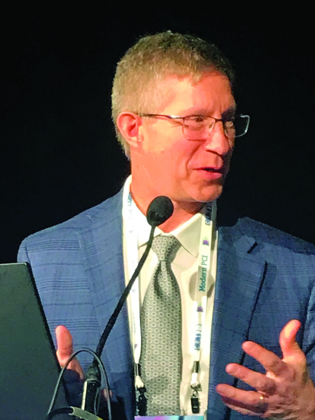
The data were pooled from the CoreValve U.S. Pivotal and SURTAVI randomized trials. Of patients participating in these two trials, 5-year follow-up data were available for 1,128 randomized to the CoreValve/Evolut TAVI and 971 randomized to surgical prosthetic valve replacement.
The major focus of the study was on the cumulative incidence of BVD, but the study also included separate analyses on the relationship between BVD and clinical outcomes. Preprocedural indicators for BVD at 5 years were also analyzed.
SVD was defined as a mean gradient increase of at least 10 mm Hg from discharge to 30 days, along with at least 20 mm Hg at last echo or new-onset aortic regurgitation. Nonstructural valve deterioration (NSVD) was defined as severe PPM at discharge or 30 days or severe paravalvular regurgitation through 5 years. In addition to these two components, the BVD endpoint also included thrombosis and endocarditis.
Surgical valve deterioration high at 5 years
On the basis of these definitions, the rate of BVD at 5 years was 14.2% in the surgery group and 7.8% in the TAVI group, translating into a 50% risk reduction in favor of TAVI (hazard ratio, 0.50; P < .001).
Thrombosis or endocarditis occurred in low rates in both groups, but every other component of BVD favored TAVI significantly, not just numerically. This included SVD (2.2% vs. 4.4%; P = .004), and the two components of NSVD, PPM (3.7% vs. 11.8%; P < .001) and severe paravalvular regurgitation (0.2% vs. 1.2%; P = .02).
When stratified by annular diameter, the relative advantage of TAVI over surgery was greatest in those valves with diameters of up to 23 mm. In this group, the lower relative rate in the TAVI group (8.6% vs. 19.7%) represented a nearly 70% reduction in risk of valve deterioration at 5 years (HR, 0.31; P < .001).
However, the advantage at 5 years also remained substantial and significant in larger valves (8.1% vs. 12.6%), translating into a 40% risk reduction in favor of TAVI (HR, 0.60; P = .002).
Independent of type of valve replacement, BVD at 5 years was associated with worse outcomes, including significantly increased risks for all-cause mortality (HR, 1.46; P = .004), cardiovascular mortality (1.84; P < .001), and hospitalization for valve disease or worsening heart failure (HR, 1.67; P = .001).
The baseline characteristics that were statistically associated with BVD at 5 years on multivariate analysis in pooled data from both the TAVI and surgical groups included age (P = .02), a creatinine clearance less than 30 mL/min per 1.73 m2 (P = .006), and a low relative baseline left ventricular ejection fraction (P < .001).
BVD criteria validated for outcome prediction
The four components of valve performance employed in this analysis (SVD, NSVD, thrombosis, and endocarditis) were drawn from consensus documents issued by the Valve Academic Research Consortium and the European Association of Percutaneous Cardiovascular Interventions, but the relative importance of these components for predicting valve survival was previously unknown, according to Dr. Yakubov.
“This is the first analysis to validate clinical criteria for valve performance and its association with clinical outcomes,” said Dr. Yakubov, medical director of cardiovascular studies, OhioHealth Research Institute at Riverside Methodist Hospital, Columbus.
This is also the first study to employ randomized data to prove an advantage of TAVI over surgery in long-term follow-up.
A 10-year follow-up is planned for the patients who participated in these two trials, but the lower rate of BVD in the TAVI arm at 5 years is already a threat to surgical repairs, acknowledged several surgeons who served as panelists in the session where these results were presented.
“I think that these data are a reflection of the fact that we [surgeons] are not being as aggressive as we should be,” said Gregory P. Fontana, MD, who is national director, cardiothoracic surgery, HCA Healthcare, and is affiliated with Los Robles Health System, Thousand Oaks, Calif. “We need to be employing larger prostheses.”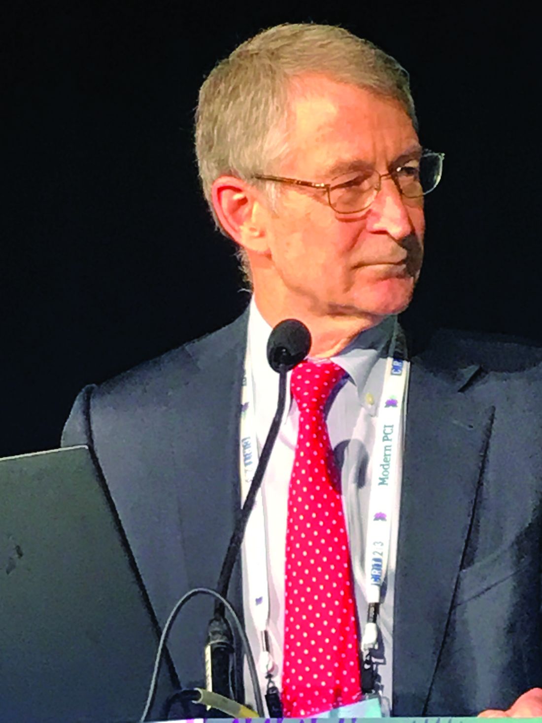
A very similar comment was made by Michael J. Reardon, MD, a professor of cardiothoracic surgery at Houston Methodist Hospital. Pointing to the higher rate of PVL as an example of a common postsurgical complication, he agreed that surgeons should be moving to bigger valve sizes.
While adjustments in valve size might address the steeper rise in NSVD subtypes of BVD observed in the surgical group, but Dr. Reardon and others pointed out that late BVD events also rose at a greater pace in the surgical group. These suggest other improvements in technique might also be needed to keep surgical valve repairs competitive.
Dr. Yakubov reported financial relationships with Medtronic and Boston Scientific, both of which provided funding for this study. Dr. Fontana reported financial relationships with Abbott and Medtronic. Dr. Reardon reported financial relationships with Abbott, Boston Scientific, Medtronic, and Gore Medical.
FROM CRT 2023
NP-PA turf fights: Where the relationship can improve
– The U.S. Bureau of Labor Statistics forecasts a 40% increase in the NP workforce by 2031, coupled with a 28% rise in PAs.
In recent reports on the quality of the relationships involving these health care professions, survey respondents mostly gave positive accounts of collaboration, using words such as like “comradery,” “teamwork,” “congenial,” and “cohesion.” But all was not perfect. Where and how could these important health care provider relationships improve?
PAs: “Competition and collaboration’ with RNs
In a Medscape survey of more than 770 PAs about their working relationships with other health care professionals; 83% of them supported the idea of PAs and NPs practicing more independently from physicians, but sometimes it’s not easy to stay in their individual lanes.
One PA respondent complained that NPs get “more opportunities and preference,” another pointed to PA-NP “turf issues,” and a third griped about NPs’ “strong unions,” which have stoked more fighting about practice abilities and available settings.
Robert Blumm, MA, PA-C, a retired surgical and emergency medicine PA who regards himself as an advocate for both PAs and NPs, describes their interaction as a “mixture of competition and collaboration.”
On one hand, the two groups typically “cooperate and do an excellent job, incurring patient errors similar to or less than physician colleagues or senior residents.” On the other hand, Mr. Blumm conceded, there is some jealousy among PAs over NPs’ advantage in staffing and hiring decisions, “since they don’t need [direct physician] supervision ... and there are limits on how many PAs can be supervised by one physician.”
Most PA-NP interactions are collaborative, although many people emphasize the relatively few conflicts, said Jennifer Orozco, DMSc, PA-C, president and chair of the American Academy of PAs.
“We see that a lot in this country,” she said. “People try to drive a wedge, but it’s often a misnomer that there’s a lot of arguing and infighting.”
NPs: Different backgrounds, same goal
The Medscape survey also included information from 750 NPs on working relationships; 93% of them favored nurses and PAs working more independently from doctors.
April Kapu, DNP, ARPN, has worked closely with PAs for more than 20 years. “In my experience ... they complement one another as health team members, although the education and training are somewhat different,” said Ms. Kapu, , president of the American Association of Nurse Practitioners.
Some respondents noted the different educational trajectories for NPs and PAs. “Doctors and PAs are taught using the same model, but NPs are taught under the nursing model,” wrote a family medicine PA.
In emergency departments where Mr. Blumm has worked, ICU NPs have an edge over PAs in terms of preparation, organization, and the tabulation of formulas. On the other hand, some of Mr. Blumm’s fellow PAs were also emergency medicine technicians or respiratory therapists, who had “2 years of classroom training, on par with that of medical students.”
Must these differences in training and education foment conflict between NPs and PAs? “We all bring something different to the table,” said Ms. Kapu, who also is associate dean for clinical and community partnerships at Vanderbilt University, Nashville, Tenn. “It is important to respect each person’s entry point, education, and training.”
Differing personalities and environments
Numerous PA respondents said that individual personalities and work environments are more likely to trigger issues with NPs than are differences in training.
“It depends on the team and situation and who the people are, not the letters behind their names,” an emergency medicine PA wrote. A surgical PA noted that “group dynamics and work culture differ from place to place,” while a third PA agreed that “it’s personality dependent, not title dependent.”
No single formula will resolve areas of NP-PA conflict, Ms. Orozco said. “What works in Chicago might not work in rural Colorado or Texas or California, but we do have to come together. The overall focus should be on greater flexibility for PAs and NPs. Patients will fare better.”
Joint research, publishing could help
About a decade ago, Mr. Blumm joined with another PA and an NP to form the American College of Clinicians, the first joint PA-NP national professional organization. Although it disbanded after 6 years, owing to low membership, he hopes a similar collaboration will take off in the future.
“I also recommend that PAs and NPs publish articles together, with research as an excellent place to start,” he added. “PAs and NPs should stand together and be a source of healing for all our patients. Regardless of our titles, our responsibility is to bring healing together.”
A version of this article first appeared on Medscape.com.
Docs struggle to keep up with the flood of new medical knowledge. Here’s advice
making it much tougher for physicians to identify innovative findings and newer guidelines for helping patients. Yet not keeping up with the latest information can put doctors at risk.
“Most doctors are feeling lost about keeping up to date,” said John P.A. Ioannidis, MD, professor of medicine at Stanford (Calif.) University School of Medicine. “The vast majority of new studies are either wrong or not useful, but physicians cannot sort out which are those studies.”
The sheer number of new studies may even force some doctors to retreat from areas where they have not kept up, said Stephen A. Martin, MD, professor of family medicine and community health at the University of Massachusetts, Worcester. “When doctors don’t feel they can stay current, they may refer more cases to specialists or narrow their focus,” he said.
Some specialties have a greater challenge than others
Dr. Martin said the deluge of studies heavily impacts generalists because they have a wider field of information to keep up with. However, certain specialties like oncology are particularly flooded with new findings.
Specialties with the greatest number of published studies are reportedly oncology, cardiology, and neurology. A 2021 study found that the number of articles with the word “stroke” in them increased five times from 2000 to 2020. And investigative treatments targeting cancer nearly quadrupled just between 2010 and 2020.
What’s more, physicians spend a great deal of time sifting through studies that are ultimately useless. In a survey of internists by Univadis, which is part of WebMD/Medscape, 82% said that fewer than half of the studies they read actually had an impact on how they practice medicine.
“You often have to dig into an article and learn more about a finding before you now whether it’s useful,” Dr. Martin said. “And in the end, relatively few new findings are truly novel ones that are useful for patient care.”
So what can a physician do? First, find out what you don’t know
Looking for new findings needs to be carried out systematically, according to William B. Cutrer, MD, MEd, a pediatric intensivist who is associate dean for undergraduate medical education at Vanderbilt University School of Medicine, Nashville, Tenn.
“Before you start, you have to know what you don’t know, and that’s often not so easy,” he said. “You may get a spark about what you don’t know in an encounter with a patient or colleague or through patient outcomes data,” he said.
Dr. Martin, on the other hand, advocates a broad approach that involves finding out at least a little about everything in one’s field. “If you have a good base, you’re not starting from zero when you encounter a new clinical situation,” he said.
“The idea is that you don’t need to memorize most things, but you do need to know how to access them,” Dr. Martin said. “I memorize the things I do all the time, such as dosing or indicated testing, but I look up things that I don’t see that often and ones that have some complexity.”
Updating the old ways
For generations, doctors have stayed current by going to meetings, conversing with colleagues, and reading journals, but many physicians have updated these methods through various resources on the internet.
For example, meetings went virtual during the pandemic, and now that face-to-face meetings are back, many of them retain a virtual option, said Kevin Campbell, MD, a cardiologist at Health First Medical Group, Melbourne, Fla. “I typically go to one or two conferences a year, but I also learn a lot digitally,” he said.
As to journal reading, “assessing an article is an essential skill,” Dr. Cutrer said. “It’s important to quickly decide whether a journal article is worth reading or not. One answer to this problem is to consult summaries of important articles. But summaries are sometimes unhelpful, and it is hard to know which articles are significant. Therefore, doctors have been reaching out to others who can research the articles for them.”
For many years, some physicians have pooled their resources in journal clubs. “You get a chance to cross-cultivate your skills with others,” Dr. Ioannidis said. “But you need someone who is well informed and dedicated to run the journal club, using evidence-based principles.”
Dr. Cutrer said physicians like to cast their net wide because they are understandably wary of changing their practice based on one study. “Unless there is one large study that is really well designed, doctors will need two or more findings to be convinced,” he said. This requires having the ability to match studies across many journals.
Using research summaries
In the past two decades, physicians have gained access to countless summaries of journal articles prepared by armies of clinical experts working for review services such as the New England Journal of Medicine’s “Journal Watch,” Annals of Internal Medicine’s “In the Clinic,” and BMJ’s “State of the Arts.”
In addition to summarizing findings from a wide variety of journals in plain language, reviewers may compare them to similar studies and assess the validity of the finding by assigning a level of evidence.
Some commercial ventures provide similar services. Betsy Jones, executive vice president of clinical decisions at EBSCO, said the DynaMed service is now available through an app on the physician’s smartphone or through the electronic health record.
Physicians like this approach. Many specialists have noted that reading full-length articles was not an efficient use of their time, while even more said that reviews are efficient.
Exchanging information online
Physicians are increasingly keeping current by using the internet, especially on social media, Dr. Cutrer said. “Young doctors in particular are more likely to keep up digitally,” he said.
Internet-based information has become so widespread that disparities in health care from region to region have somewhat abated, according to Stuart J. Fischer, MD, an orthopedic surgeon at Summit Orthopaedics and Sports Medicine, New Jersey. “One positive outcome of this plethora of information today is that geographic disparities in clinical practice are not as great as they used to be,” he said.
Rather than chatting up colleagues in the hallway, many physicians have come to rely on internet-based discussion boards.
Blogs, podcasts, and Twitter
Blogs and podcasts, often focused on a specialty, can be a great way for physicians to keep up, said UMass Chan professor Dr. Martin. “Podcasts in particular have enhanced the ability to stay current,” he said. “You want to find someone you trust.”
Internal medicine podcasts include Annals on Call, where doctors discuss articles in the Annals of Internal Medicine, and the Curbsiders, where two internists interview a guest expert.
Orthopedic surgeons can visit podcasts like Nailed it, Orthobullets, the Ortho Show, and Inside Orthopedics. Neurologists can consult Brainwaves, Neurology Podcast, Practical Neurology Podcast, and Clinical Neurology with KD. And pediatricians can drop in on Talking Pediatrics, The Cribsiders, and PedsCases.
Meanwhile, Twitter has become a particularly effective way to broadcast new findings, speeding up the transition from the bench to the bedside, said Dr. Campbell, the Florida cardiologist.
“I visit cardio-specific resources on Twitter,” he said. “They can be real-time video chats or posted messages. They spur discussion like a journal club. Colleagues present cases and drop in and out of the discussion.”
Others are not as enthusiastic. Although Stanford’s Dr. Ioannidis is in the heart of the Silicon Valley, he is leery of some of the new digital methods. “I don’t use Twitter,” he says. “You just add more people to the process, which could only make things more confusing. I want to be able to think a lot about it.”
Cutting-edge knowledge at the point of care
Consulting the literature often takes place at the point of care, when a particular patient requires treatment. This can be done by using clinical decision support (CDS) and by using clinical practice guidelines (CPGs), which are typically developed by panels of doctors at specialty societies.
“It used to be that the doctor was expected to know everything,” said Ms. Jones at DynaMed. “Today there is no way to keep up with it all. Doctors often need a quick memory jog.”
Ms. Jones said the CDS result always requires the doctor’s interpretation. “It is up to the doctor to decide whether a new finding is the best choice for his or her patient,” she said.
Dr. Martin recommends going easy on point-of-care resources. “They can be used for showing a patient a differential diagnosis list or checking the cost of a procedure, but they are harder to use for novel developments that require time and context to evaluate their impact,” he said.
CPGs, meanwhile, have a high profile in the research world. In a 2018 study, Dr. Ioannidis found that 8 of the 15 most-cited articles were CPGs, disease definitions, or disease statistics.
Dr. Fischer said CPGs are typically based on thorough reviews of the literature, but they do involve experts’ interpretation of the science. “It can be difficult to obtain specific answers to some medical questions, especially for problems with complex treatments or variations,” he said.
As a result, Dr. Fischer said doctors have to use their judgment in applying CPGs to a specific patient. “For example, the orthopedic surgeon would normally recommend a total hip replacement for patients with a bad hip, but it might not be appropriate for an overweight patient.”
Stay skeptical
There are many novel ways for physicians to keep current, including summaries of articles, discussion boards, blogs, podcasts, Twitter, clinical decision support, and clinical practice guidelines.
Even with all these new services, though, doctors need to retain a healthy amount of skepticism about new research findings, Dr. Ioannidis said. “Ask yourself questions such as: Does it deal with a real problem? Am I getting the real information? Is it relevant to real patients? Is it offering good value for money?”
A version of this article first appeared on Medscape.com.
Risk of stent infection low, but may be underreported
Infections of coronary stents appear to be uncommon, but it is not clear if they are often missed, underreported, or truly rare, according to a new analysis.
In a search of multiple databases, 79 cases of coronary stent infections (CSI) were found in 65 published reports, according to Venkatakrishnan Ramakumar, MBBS, MD, department of cardiology, All India Institute of Medical Sciences, New Delhi.
Over the period of evaluation, which had no defined starting point but stretched to November 2021, the 79 infections reported worldwide occurred when millions of percutaneous coronary intervention (PCI) procedures were performed. In the United States alone, the current estimated annual number of PCIs is 600,000, according to an article published in the Journal of the American Heart Association.
If the number of reported CSI cases represented even a modest fraction of those that occurred, the risk would still be almost negligible. Yet, Dr. Ramakumar insisted that there has been little attention paid to the potential for CSI, creating a situation in which many or almost all cases are simply being missed.
“We do not know how many infections have gone unrecognized,” Dr. Ramakumar said in presenting his results at the Cardiovascular Research Technologies conference, sponsored by MedStar Heart & Vascular Institute. And even if they are identified and promptly treated, there “is the potential for a publication bias,” he added, referring to the reluctance of investigators to submit and publishers to accept manuscripts with negative results.
Regardless of the frequency with which they occur, CSI is associated with bad outcomes, according to the data evaluated by Dr. Ramakumar. On the basis of in-hospital mortality, the primary endpoint of this analysis, the rate of death in patients developing CSI was 30.3%.
Successful treatment varied by hospital type
This risk was not uniform. Rather, rates of in-hospital mortality and proportion of patients treated successfully varied substantially by type of hospital. At private teaching hospitals for example, successful treatment – whether medical alone or followed by bailout surgery – was 80%. The rates fell to 40% at public teaching hospitals and then to 25% at private nonteaching hospitals.
The full-text articles included in this analysis were evaluated and selected by two reviewers working independently. A CSI diagnosis made clinically or with imaging and treatment outcomes were among criteria for the case studies to be included. Dr. Ramakumar said the study, which he claimed is the largest systematic review of CSI ever conducted, has been registered with PROSPERO, an international prospective registry of systematic reviews.
The presenting symptom was fever in 72% of cases and chest pain in the others, although there was one asymptomatic CSI reported. On angiography, 62% had a concomitant mycotic aneurysm. Intramyocardial abscess (13.9%), rupture (11.3%), and coronary fistula (7.5%) were also common findings, but no angiographic abnormalities could be identified in 53% of patients.
Following PCI, most CSI developed within 8 days (43%) or the first month (23%), but CSI was reported more than 6 months after the procedure in 19%. Complex PCI accounted for 51% of cases. Of stent types, 56% were drug eluting and 13% were bare metal.
When comparing characteristics of those who survived CSI with those who did not, most (89%) of those with a non–ST-segment elevated acute coronary syndrome ultimately survived, while survival from CSI in those with structural heart disease was only 17%.
Microbiological findings were not a criterion for study inclusion, but Staphylococcus species accounted for 65% of the infections for which positive cultures were reported. Pseudomonas accounted for 13%. Less than 4% (3.8%) tested positive for multiple pathogens. A small proportion of patients had unusual infectious organisms.
As part of this analysis, the investigators developed an artificial intelligence model to predict CSI based on patient characteristics and other variables. However, the specificity of only around 70% led Dr. Ramakumar to conclude that it does not yet have practical value.
However, he believes that better methodology to detect CSI is needed, and he proposed a diagnostic algorithm that he believes would both improve detection rates and accelerate the time to diagnosis.
Algorithm proposed for detection of CSI
In this algorithm, the first step in symptomatic patients with a positive blood culture suspected of CSI is imaging, such as transthoracic echocardiography, to identify features of infective endocarditis or endarteritis. If the imaging is positive, further imaging, such as PET, that supports the diagnosis, should be adequate to support a diagnosis and treatment.
If initial imaging is negative, alternative diagnoses should be considered, but Dr. Ramakumar advised repeat imaging after 48 hours if symptoms persist and no other causes are found.
Dr. Ramakumar acknowledged the many limitations of this analysis, including the small sample size and the challenges of assembling coherent data from case reports with variable types of information submitted during different eras of PCI evolution. However, reiterating that CSI might be frequently missed, he emphasized that this problem might be bigger than currently understood.
It is difficult to rule out any possibility that CSI is frequently missed, but Andrew Sharp, MD, PhD, a consultant interventional cardiologist at the University Hospital of Wales, Cardiff, is skeptical.
“One might think this is a potential problem, but I cannot think of one patient in whom this has occurred,” Dr. Sharp said in an interview. He is fairly confident that they are extremely rare.
“When there is infection associated with a foreign body, such as a pacemaker, they do not typically resolve by themselves,” he explained. “Often the device has to be removed. If this was true for CSI, then I think we would be aware of these complications.”
However, he praised the investigators for taking a look at CSI in a systematic approach. An invited panelist during the CRT featured research, which is where these data were presented, Dr. Sharp was more interested in understanding why they do not occur now that data are available to suggest they are rare.
“Is there something in the coronary environment, such as the consistent blood flow, that protects against infection?” he asked. CSI is a valid area of further research, according to Dr. Sharp, but he does not consider infected stents to be a common threat based on his own sizable case series.
Dr. Ramakumar and Dr. Sharp reported no potential conflicts of interest.
FROM CRT 2023
Any level of physical activity tied to better later-life memory
new research suggests.
A prospective study of 1,400 participants showed that those who exercised to any extent in adulthood had significantly better cognitive scores later in life, compared with their peers who were physically inactive.
Maintaining an exercise routine throughout adulthood showed the strongest link to subsequent mental acuity.
Although these associations lessened when investigators controlled for childhood cognitive ability, socioeconomic background, and education, they remained statistically significant.
“Our findings support recommendations for greater participation in physical activity across adulthood,” lead investigator Sarah-Naomi James, PhD, research fellow at the Medical Research Council Unit for Lifelong Health and Ageing at the University College London, told this news organization.
“We provide evidence to encourage inactive adults to be active even to a small extent … at any point during adulthood,” which can improve cognition and memory later in life, Dr. James said.
The findings were published online in the Journal of Neurology, Neurosurgery & Psychiatry.
Exercise timing
Previous studies have established a link between fitness training and cognitive benefit later in life, but the researchers wanted to explore whether the timing or type of exercise influenced cognitive outcomes in later life.
The investigators asked more than 1,400 participants in the 1946 British birth cohort how much they had exercised at ages 36, 43, 60, and 69 years.
The questions changed slightly for each assessment period, but in general, participants were asked whether in the past month they had exercised or participated in such activities as badminton, swimming, fitness exercises, yoga, dancing, football, mountain climbing, jogging, or brisk walks for 30 minutes or more; and if so, how many times they participated per month.
Prior research showed that when the participants were aged 60 years, the most commonly reported activities were walking (71%), swimming (33%), floor exercises (24%), and cycling (15%).
When they turned 69, researchers tested participants’ cognitive performance using the Addenbrooke’s Cognitive Examination–III, which measures attention and orientation, verbal fluency, memory, language, and visuospatial function. In this study sample, 53% were women, and all were White.
Physical activity levels were classified as inactive, moderately active (one to four times per month), and most active (five or more times per month). In addition, they were summed across all five assessments to create a total score ranging from 0 (inactive at all ages) to 5 (active at all ages).
Overall, 11% of participants were physically inactive at all five time points; 17% were active at one time point; 20% were active at two and three time points; 17% were active at four time points; and 15% were active at all five time points.
‘Cradle to grave’ study?
Results showed that being physically active at all study time points was significantly associated with higher cognitive performance, verbal memory, and processing speed when participants were aged 69 (P < .01).
Those who exercised to any extent in adulthood – even just once a month during one of the time periods, fared better cognitively in later life, compared with physically inactive participants. (P < .01).
Study limitations cited include a lack of diversity among participants and a disproportionately high attrition rate among those who were socially disadvantaged.
“Our findings show that being active during every decade from their 30s on was associated with better cognition at around 70. Indeed, those who were active for longer had the highest cognitive function,” Dr. James said.
“However, it is also never too late to start. People in our study who only started being active in their 50s or 60s still had higher cognitive scores at age 70, compared to people of the same age who had never been active,” she added.
Dr. James intends to continue following the study sample to determine whether physical activity is linked to preserved cognitive aging “and buffers the effects of cognitive deterioration in the presence of disease markers that cause dementia, ultimately delaying dementia onset.
“We hope the cohort we study will be the first ‘cradle to grave’ study in the world, where we have followed people for their entire lives,” she said.
Encouraging finding
In a comment, Joel Hughes, PhD, professor of psychology and director of clinical training at Kent (Ohio) State University, said the study contributes to the idea that “accumulation of physical activity over one’s lifetime fits the data better than a ‘sensitive period’ – which suggests that it’s never too late to start exercising.”
Dr. Hughes, who was not involved in the research, noted that “exercise can improve cerebral blood flow and hemodynamic function, as well as greater activation of relevant brain regions such as the frontal lobes.”
While observing that the effects of exercise on cognition are likely complex from a mechanistic point of view, the finding that “exercise preserves or improves cognition later in life is encouraging,” he said.
The study received funding from the UK Medical Research Council and Alzheimer’s Research UK. The investigators and Dr. Hughes report no relevant financial relationships.
A version of this article first appeared on Medscape.com.
FROM THE JOURNAL OF NEUROLOGY, NEUROSURGERY & PSYCHIATRY