User login
If you’ve never had COVID, should you relax or worry?
If you’re among those people in the United States who never had COVID-19, how should you think about your risk?
According to the Centers for Disease Control and Prevention (CDC), more than half of people in the United States are in the never-got-COVID category.
The CDC estimated that by the end of January, 43.4% in the United States had developed antibodies to SARS-CoV-2 triggered by infection, not by vaccination — suggesting nearly 60% of people have never been infected.
Now mask mandates are lifting, and daily case and death numbers are plunging. According to the New York Times tracker, new cases are down 51% for the past 2 weeks, and deaths have fallen 30% in that period.
So as those who have so far escaped the virus venture further out into reopened environments, should they worry more or less about risk than their previously infected counterparts?
No “suit of armor”
William Schaffner, MD, an infectious disease expert at Vanderbilt University School of Medicine in Nashville, Tenn., said in an interview that science has not been able to determine why some people have been able to be stay COVID-free when the virus was raging and exposure was ubiquitous.
He said it’s important to remember that though some people think they have never had COVID, they may have been asymptomatic or attributed mild symptoms to something else.
“People may have conceivably — but we can’t define them yet — different capacities to ward off viruses or bacteria,” Dr. Schaffner said.
Could it be that some people have a better immune system or genetic component or environmental reason that they are less susceptible to infectious disease? “We can’t define that in 2022 medicine, but it could be,” he said.
More is known about why people with the same COVID exposure may have different levels of illness severity.
“They’re more likely to get seriously ill if they have a list of predisposing conditions — if they’re older, if they’re frail, if they have underlying illness or are obese. All of those things clearly impair the body’s response to virus,” Dr. Schaffner said.
He warns those who have never been infected, though, not to assume they have “a suit of armor.”
All should continue to follow guidance on getting vaccinated, and those vaccinated should continue to get boosted, Dr. Schaffner said.
“Clearly, the data show that if you are vaccinated and boosted, you’re protected much more securely against severe disease,” he said.
If the never-COVIDs develop a respiratory infection, they should still get tested for COVID, Dr. Schaffner said.
He said while both vaccines and previous natural infection offer protection, the duration of that protection is not yet known.
“We have to stay tuned,” Dr. Schaffner said. “There may be a recommendation in the future to get a booster annually or something like that. We need to be open to those down the road.”
Amesh Adalja, MD, senior scholar at the Johns Hopkins Center for Health Security in Baltimore, says it’s unclear why some people have been able to avoid COVID.
“The explanation is likely multifactorial and involves behaviors as well as possible idiosyncrasies with their immune systems that are genetically based,” he said. “It also may be the case that inapparent infections occurred and went undiagnosed.”
Dr. Adalja agrees, though, that this isn’t the time to get overconfident with risk-taking where COVID is concerned.
“People who have not knowingly been infected with COVID should be vaccinated, and after that, be assured that they are protected against serious disease from this virus,” he said.
Genetic protection?
A new study in Nature Genetics explains a potential genetic relationship. Study authors found evidence that levels of expression of angiotensin-converting enzyme 2 (ACE2) – which helps regulate blood pressure, wound healing, and inflammation, but has also been shown to serve as an entry point into cells for some coronaviruses like SARS-CoV-2 — influence COVID-19 risk.
Manuel A. Ferreira, PhD, an executive director for analytic genetics at Regeneron Pharmaceuticals, said in an interview that ACE2 receptors — what he calls the “gateways” for SARS-CoV-2 to enter the body — are different in people who have inherited a particular allele.
The researchers have found that that allele is associated with lower risk of SARS-CoV-2 infection.
“It’s quite substantial -- a 40% risk reduction if you carry the allele that reduces ACE2 expression,” he said. They were not able to discern from this study, however, whether that could predict severity of disease.
The team also looked at a series of six genetic variants elsewhere in the genome and developed a risk score to see if it was possible to predict who might be more susceptible to severe COVID.
Dr. Ferreira said the score only slightly improved predictive abilities beyond factors such as age, sex, weight, and comorbidities. Further information will help hone the ability to predict the likelihood of developing severe disease on the basis of genetics, Ferreira said.
“As we identify more genetic risk factors for COVID — variants like the ACE2 variant that will affect your risk of having COVID — the more informative the risk score will be,” he said.
Several authors of the Nature Genetics article are current and/or former employees of AncestryDNA and may hold equity in AncestryDNA. Several are Regeneron employees and/or hold stock in the company. Dr. Ferreira is an employee of Regeneron and holds stock in the company. Dr. Schaffner and Dr. Adalja report no relevant financial relationships.
A version of this article first appeared on Medscape.com.
Height an ‘overlooked risk factor’ for colorectal cancer?
A new meta-analysis provides more evidence that
“There are well-known modifiable dietary associations for colorectal cancer, such as processed red meats and smoking, but guidelines currently are fixated on family history, and height is clinically neglected when it comes to risk screening,” study investigator Gerard Mullin, MD, with Johns Hopkins University, Baltimore, said in a news release. This large study “builds on evidence that taller height is an overlooked risk factor and should be considered when evaluating and recommending patients for colorectal cancer screenings.”
The study was published online March 1 in Cancer Epidemiology, Biomarkers & Prevention.
The evidence: Height and cancer risk
Height has been actively studied as a potential nonmodifiable risk factor for a range of cancers, including CRC.
In one large prospective study of postmenopausal women, researchers found a modest but statistically significant positive association between height and risk for any cancer and for melanoma, multiple myeloma, and cancers of the thyroid, ovary, colorectum, and endometrium.
A separate study found that tall men, especially those who are long-legged, may be at increased risk for prostate cancer, including high-grade tumors, relative to men of more modest stature.
However, the study authors point out, past studies have also produced mixed results, used inconsistent measures of height, and failed to include the risk of adenomas.
In the current meta-analysis, the investigators included 47 international, observational studies involving 280,644 adults with CRC and 14,139 cases of colorectal adenoma.
Because the definition of tallness differs around the world, the researchers compared the highest versus the lowest height percentile of various study groups. The findings were adjusted for demographic, socioeconomic, behavioral, and other known risk factors for CRC.
Overall, the investigators found that the tallest individuals within the highest percentile of height had a 24% higher risk of developing CRC compared to the shortest individuals within the lowest percentile (hazard ratio [HR], 1.24; P < .001).
In addition, they found that every 10-cm increase (about 4 inches) in height was associated with a 14% increased risk of developing CRC (HR, 1.14; P < .001) and a 6% increased likelihood of adenomas (odds ratio [OR], 1.06; P = .03).
In the United States, the average height for men is 5 feet, 9 inches, and for women it is 5 feet, 4 inches, which means men who are 6 feet, 1 inch and women who are 5 feet, 8 inches or taller have a 14% increased risk of CRC and a 6% increased risk of adenomas, the researchers explained.
According to co–first author Elinor Zhou, MD, also with Johns Hopkins University, a potential explanation for this link “is that adult height correlates with body organ size. More active proliferation in organs of taller people could increase the possibility of mutations leading to malignant transformation.”
The study authors said more research is needed to identify particular subgroups of tall people at risk for CRC.
“For instance, tall athletes and individuals with inherited tallness, such as those with Marfan syndrome, could be screened earlier and the impact of height further explored,” Dr. Zhou said.
Plus, Dr. Zhou added, more studies are needed to “definitively say at what height you would need earlier colorectal cancer screening.”
The current study was supported by grants from Bloomberg Philanthropies, intramural funds, and the Johns Hopkins Cancer Center Support Grant. The authors declared no conflicts of interest.
A version of this article first appeared on Medscape.com.
FROM CANCER EPIDEMIOLOGY, BIOMARKERS & PREVENTION
Wake Forest Cancer Center director fired, advisory board resigns
and withdrew their endorsement for renewal of the center’s National Cancer Institute comprehensive cancer center support grant.
The move was prompted by the abrupt firing of center director Boris Pasche, MD, PhD, on February 10, one day after NCI renewed a multimillion dollar grant.
The Cancer Letter broke the story and published the resignation letter from the EAB. It was signed by board chair Gerold Bepler, MD, PhD, CEO and director of the Karmanos Cancer Institute, Detroit, on behalf of the board.
The mass resignation of an EAB, a panel of outside experts that help shepherd cancer centers through the NCI grant process, is “highly unusual,” according to The Cancer Letter. It also raises concerns about the “immediate future” of Wake Forest’s cancer center, the publication added.
Numerous people involved with the situation did not respond or declined to comment when this news organization requested additional information and updates, including questions about the reason for Dr. Pasche’s termination; whether or not withdrawal of the endorsement puts Wake’s NCI designation in jeopardy; and if the EAB is being reconstituted.
A written statement from Wake Forest simply said that “the situation involving Dr. Pasche is an administrative decision. Various administrative changes occur regularly in organizations across the country. Dr. Pasche remains employed by Atrium Health Wake Forest Baptist. We are very grateful to Dr. Pasche for his years of service and many contributions to the mission and vision of our NCI-designated Comprehensive Cancer Center in Winston-Salem.”
Wake’s cancer center is in the process of combining with the Atrium Health Levine Cancer Center, which is not NCI-designated, following Atrium Health system’s recent acquisition of the Wake Forest Baptist Medical Center.
The NCI renewal notice, dated February 9, states that Dr. Pasche “and his leadership team have built a robust, transdisciplinary center that includes 140 scientists.”
Dr. Pasche was fired a day later.
The EAB resignation letter states that during Wake Forest’s recent NCI review process, “leadership gave their glowing endorsement of Dr. Pasche...This endorsement included unequivocal statements of support for Dr. Pasche’s oversight of the combined Atrium-Wake Forest cancer program.”
“What followed was his rapid dismissal after the...notice of award was issued, following a period during which the approach to integration was apparently being revisited,” Dr. Bepler said on behalf of the board.
“It is with sadness and dismay that we witnessed the change in approach by the institutional leadership towards” the merger, he wrote.
The Cancer Letter quotes an unnamed board member as saying, “EABs for cancer centers can only provide value to the center when there is openness and transparency in the process. In the absence of such, I believe the members felt that there was no further utility in providing guidance to the organization.”
The resignation letter was sent to the interim director of Wake’s cancer center, radiation oncologist William Blackstock, Jr, MD, and also copied to Atrium-Wake and NCI leadership.
The resignation letter endorsed Dr. Blackstock’s qualifications to run the center, and noted that as the board is reconstituted, “some of us would be honored to discuss participation...if there is unequivocal evidence from the health system’s senior management for support of a single, academically driven, comprehensive, and integrated cancer center.”
A version of this article first appeared on Medscape.com.
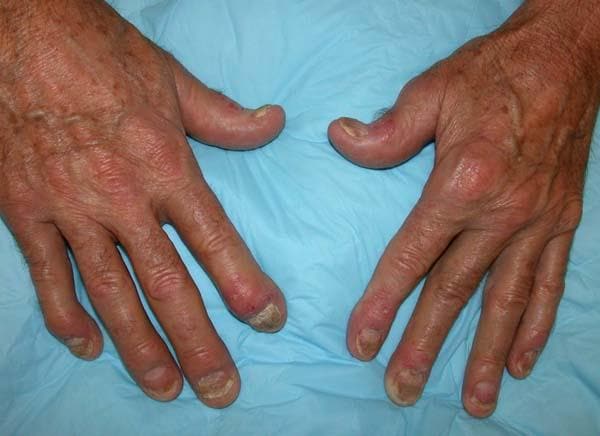
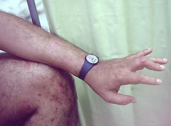
Painful swelling of fingers and toe
Although psoriatic arthritis is not the only disease associated with dactylitis — other culprits are sarcoidosis, septic arthritis, tuberculosis, and gout — dactylitis is one of the characteristic symptoms of psoriatic arthritis. Dactylitis is seen in as many as 35% of patients with psoriatic disease. Dactylitis clinically presents — as in this patient — with sausage-like swelling of the digits. It is included in the Classification Criteria for Psoriatic Arthritis (CASPAR) as one of the hallmarks of psoriatic arthritis.
Dactylitis has been thought to be a result of the concomitant swelling and inflammation of the flexor tendon sheaths of the metacarpophalangeal, metatarsophalangeal, or interphalangeal joints. Flexor tenosynovitis can be detected by examination with MRI and ultrasound. Dactylitis is associated with radiologically evident erosive damage to the joints.
Patients with psoriatic arthritis are typically seronegative for rheumatoid factor and antinuclear antibody; antinuclear antibody titers in persons with psoriatic arthritis do not differ from those of age- and sex-matched controls. C-reactive protein may be elevated but is often normal. Lack of C-reactive protein elevation, however, does not mean that systemic inflammation is absent, but rather indicates that different markers are needed that allow better quantification of systemic inflammation in psoriasis and psoriatic arthritis.
Herbert S. Diamond, MD, Professor of Medicine (retired), Temple University School of Medicine, University of Pittsburgh; Chairman, Department of Medicine Emeritus, Western Pennsylvania Hospital, Pittsburgh, PA.
Herbert S. Diamond, MD, has disclosed no relevant financial relationships.

A 35-year-old man presents with painful swelling of his right index and ring fingers as well as the fourth toe on his right foot, which has persisted for 5 days. He cannot perform his daily activities owing to severe pain in the affected fingers and toes. His medical history is unremarkable. His paternal uncle had psoriasis, which was successfully treated with adalimumab.
Physical assessment reveals tender, fusiform, swollen soft tissues in the affected fingertips, the fourth toe, and swollen palms. Nails are pitted. Hand radiography reveals mild edema of the soft tissue of the index and ring fingers but no significant joint abnormalities. Enthesitis is not present. Laboratory tests reveal a negative human leukocyte antigen B27 (HLA-B27) test, negative rheumatoid factor, negative antinuclear antibody, and normal C-reactive protein.
Dactylitis was diagnosed on the basis of clinical symptoms, radiographic results, and laboratory findings.
ESC suspension of Russia, Belarus societies sparks controversy
, provoking a heated discussion on whether medical organizations should become involved in politics.
“In the light of the continued aggression against Ukraine by the leaderships of the Russian Federation and Belarus, the European Society of Cardiology (ESC) has temporarily suspended the memberships of the Russian Society of Cardiology and the Belarussian Society of Cardiologists in the ESC,” the ESC statement reads.
“Individuals based in the Russian Federation or in Belarus are excluded from active participation in any ESC event or activity,” it states.
“The ESC very much regrets the effect this may have on individual Russian and Belarussian cardiologists and scientists, but the message to Russian and Belarussian leadership must be distinct and unequivocal,” it adds.
This action from the ESC has provoked a storm of heated discussions on the issue.
In a Twitter thread on the subject, Italian cardiologist Giuseppe Galati, MD, writes: “An astonishing decision by ESC that’s excluding all the Russian and Belarussian scientists from ESC congresses and activities. Treating doctors and scientists as [if] they are Putin and are responsible for the war.”
Dr. Galati adds: “A strong message that brings us to 70 years ago. ESC is promoting exclusion and not inclusion and diversity.”
Another commentator on the thread says: “It is a very unfortunate decision. Science, medicine should not be involved in politics. We are colleagues gathering together during congresses to exchange information for the sake of our patients. Politics should not overshadow this.”
And another added: “I think most cardiologists from Russia will not be able to participate in the events anyway, since international payments will soon be impossible from Russia. But it is wrong to limit the rights of doctors because of their nationality.”
But others support the ESC’s stance. Polish cardiologist Blazej Michalski, MD, says: “I think it is [a] good decision. Russians if they do not actively support dictatorship of Putin, the silence is also an agreement.” He adds: “I am proud of ESC. They did what they were supposed to do.”
A Twitter poll started by Ali Elzieny, MD, a cardiologist from Boston, titled “Do you agree that ESC suspend membership of Russian Society of Cardiology?” as of March 8 had 1,300 votes, with 61% of respondents disagreeing with the ESC decision and 39% in favor.
Medical societies respond
Several other medical societies have issued communications appearing to disagree with the action by the ESC.
The American College of Cardiology issued a statement saying medicine should be above politics.
“The American College of Cardiology believes that patients come first, and now, more than ever, there is a need to rally around our members across the globe to ensure that they have the support and resources they need to care for their patients,” said ACC President Dipti Itchhaporia, MD.
“Medicine is above politics and ACC will not exclude any of our colleagues who are working toward a shared mission of improving heart health. The College has a long history of working across borders to improve heart health and remains committed to that now and in the future. The ACC continues to express its support and concern for our members in the Ukraine and the patients they are working to treat on the frontlines,” the ACC statement added.
The Tele-Cardiology Working Group of the International Society for Telemedicine & eHealth (ISFTEH) also issued a statement disagreeing with the action from the ESC.
“In light of recent events, the cardiology working group of the ISFTEH will not restrict access to its events to cardiologists with regards to their nationality, religious beliefs or other characteristics that may seem discriminatory. We believe medical information should be widely available for all, especially for those doctors that find themselves in difficulty,” it said.
The European Academy of Neurology (EAN) said: “EAN is looking at ways to give practical support to Ukrainian neurologists and healthcare professionals there. EAN is not considering suspension of any individual member based on country of residence or nationality or any National Society member.”
But one oncology professional group has also cut ties with Russia.
The international cancer specialist network, OncoAlert, issued a statement saying it has severed all cooperation with doctors in Russia as part of the Western sanctions.
“The OncoAlert Network is non-political, but we cannot stand idle and not take a stand against this aggression towards our Ukrainian friends & colleagues,” OncoAlert said, adding that it will be pulling out of all collaborations and congresses in Russia. That statement was also greeted with a barrage of criticism on Twitter, mainly from Russian users.
A version of this article first appeared on Medscape.com.
Intermittent joint aches
Fundamental changes in the initial pharmacologic approach to psoriatic arthritis were made in the 2018 American College Rheumatology/National Psoriasis (ACR/NPF) guidelines for the treatment of psoriatic arthritis. Previously, methotrexate had been widely used as the first-line agent. The 2018 guidelines recommend a tumor necrosis factor (TNF) inhibitor over methotrexate and other oral small molecules (leflunomide, cyclosporine, and apremilast).
Herein is a broad summary of the guidelines:
· Treat with TNF inhibitor over oral small molecule; may consider oral small molecule with mild psoriatic arthritis and psoriasis, patient preference, and/or contraindication to TNF inhibitor
· Treat with TNF inhibitor over interleukin (IL)-17 inhibitor; may consider IL-17 inhibitor with severe psoriasis or contraindication to TNF inhibitor
· Treat with TNF inhibitor over interleukin (IL)-12/23 inhibitor; may consider IL-12/23 inhibitor with severe psoriasis or contraindication to TNF inhibitor
· Treat with oral small molecule over IL-17 inhibitor; may consider IL-17 inhibitor with severe psoriasis and/or psoriatic arthritis
· Treat with oral small molecule over IL-12/23 inhibitor; may consider IL-12/23 inhibitor with severe psoriasis or psoriatic arthritis, or concomitant inflammatory bowel disease
· Treat with methotrexate over nonsteroidal anti-inflammatory drugs; may consider nonsteroidals for mild psoriatic arthritis and psoriasis
· Treat with IL-17 inhibitor over IL-12/23 inhibitor; may consider IL-12/23 inhibitor in a patient with concomitant inflammatory bowel disease
Note that these recommendations are based on conditional evidence (ie, low to very low quality). In fact, in the entire guideline document, only 6% of the recommendations are strong, whereas 96% are conditional. This emphasizes the importance of evaluating each patient individually and engaging in a discussion to choose optimal therapy.
Another set of guidelines, from the Group for Research and Assessment of Psoriasis and Psoriatic Arthritis (GRAPPA), was last updated in 2015. Since then, many of the agents above have been introduced. Updated GRAPPA guidelines are expected to be released later this year.
Herbert S. Diamond, MD, Professor of Medicine (retired), Temple University School of Medicine, University of Pittsburgh; Chairman, Department of Medicine Emeritus, Western Pennsylvania Hospital, Pittsburgh, PA.
Herbert S. Diamond, MD, has disclosed no relevant financial relationships.
A 56-year-old man presents because of intermittent joint aches and difficulty picking up his grandchild and cleaning his home. He has a 6-year history of scalp psoriasis that he has controlled with a salicylic acid shampoo. On physical examination, he has tenderness over both elbows and in his metacarpophalangeal and proximal interphalangeal (PIP) joints on both hands. Swollen joints are noted in the proximal and distal joints of the right hand. His fingernails show uniform pitting.
Neurologic examination shows no sensory deficits or hyperesthesia. Motor examination is unremarkable, and chest and abdominal findings are unremarkable. Blood pressure is 138/90 mm Hg. Radiographic imaging shows asymmetric erosive changes with very small areas of bony proliferation in the PIP joints.There is asymmetric narrowing of the joint space in the interphalangeal joints. Laboratory findings reveal an erythrocyte sedimentation rate of 35 mm/h, negative rheumatoid factor, negative antinuclear antibody, and C-reactive protein of 7 mg/dL.
These findings are consistent with a diagnosis of psoriatic arthritis. This patient has severe psoriatic arthritis based on radiographic evidence of erosive disease.

Home blood pressure testing better than at clinics: Study
Everyone’s been there. Which one’s right?
The answer: Perhaps neither. Individual measures of blood pressure are not as accurate as taking multiple readings over a day and averaging them.
Blood pressure varies throughout the day – by about 30 points for systolic pressure, or the pressure when the heart beats – and one or two measurements in a doctor’s office may not accurately reflect the average figure, said Beverly B. Green, MD, a senior investigator at Kaiser Permanente Washington Health Research Institute in Seattle.
Average blood pressure reading is the only measurement on which a doctor can accurately diagnose and treat high blood pressure, she said. A new study by Dr. Green and other researchers at Kaiser Permanente showed that giving patients the chance to monitor their blood pressure at home could help get more reliable measurements.
Nearly one in four adults in the United States with high blood pressure are unaware they have the condition and are not getting treatment to control it. Without treatment, the condition can cause heart attacks, strokes, kidney damage, and other potentially life-threatening health problems.
Current guidelines for diagnosing high blood pressure recommend that patients whose pressure is high in the clinic get tested again to confirm the results. While the guidelines recommend home monitoring before diagnosing high blood pressure, research shows that doctors continue to measure blood pressure in their clinics for the second reading.
In their study, Dr. Green and colleagues found that home readings were more accurate than measurements taken in clinics or at pharmacy kiosks.
“Home blood pressure monitoring was a better option, because it was more accurate” than clinic blood pressure readings, Green said. A companion study found that patients preferred taking their blood pressure at home.
For their study, Dr. Green’s group used Kaiser’s electronic health record system to identify people at high risk for high blood pressure based on a recent clinic visit. They then randomly assigned the participants to get their follow-up blood pressure readings in the clinic, at home, or at kiosks in clinics or pharmacies.
Each participant also received a 24-hour ambulatory blood pressure monitor (ABPM). These devices, which people must wear continuously for 24 hours, have cuffs that inflate every 20-0 minutes during the day and every 30-60 minutes at night. Although ABPMs are the preferred test for accurately diagnosing high blood pressure, they aren’t available for widespread use.
The Kaiser researchers found that people’s systolic BP readings at clinics were generally lower than their ABPM measurements, leading to undiagnosed high BP in more than 50% of cases. Kiosk readings were much higher than the ABPM measurements and tended to overdiagnose high BP.
The value of home monitoring
Branden Villavaso, a 48-year-old attorney in New Orleans who was diagnosed with high BP at age 32, attributes his condition to genetics. He says an at-home monitor plus the occasional use of an ABPM finally provided his doctor with an accurate assessment of his condition.
Thanks to this aggressive approach, over the past 3 years, Mr. Villavaso’s diastolic reading has dropped from a previous range of between 90 and 100 to a healthier but not quite ideal value of about 80. Meanwhile, his systolic pressure has dropped to about 120, well below the goal of 130.
Mr. Villavaso said his doctor has relied on the averages of the BP readings to tailor his medication, and he also credited his wife, Chloe, a clinical nurse specialist, for monitoring his progress.
While previous studies have found similar benefits for measuring BP at home, Dr. Green said the latest study may offer the most powerful evidence to date because of the large number of people who took part, the involvement of primary care clinics, and the use of real-world health care professionals to take measurements instead of people who usually do health research. She said this study is the first to compare kiosk and ABPM results.
“The study indicates that assisting patients with getting access to valid blood pressure readings so they can measure their blood pressure at home will give a better picture of the true burden of [high BP],” said Keith C. Ferdinand, MD, a cardiologist at Tulane University, New Orleans.
He recommended patients select a home monitoring device from www.validatebp.org, a noncommercial website that lists home BP systems that have proven to be accurate.
“We know that [high blood pressure] is the most common and powerful cause of heart disease and death,” Dr. Ferdinand said. “Patients are pleased to participate in shared decision-making and actively assist in the control of a potentially deadly disease.”
A version of this article first appeared on WebMD.com.
FROM THE JOURNAL OF GENERAL INTERNAL MEDICINE
Discoid lupus
THE COMPARISON
A Multicolored (pink, brown, and white) indurated plaques in a butterfly distribution on the face of a 30-year-old woman with a darker skin tone.
B Pink, elevated, indurated plaques with hypopigmentation in a butterfly distribution on the face of a 19-year-old woman with a lighter skin tone.
Cutaneous lupus erythematosus may occur with or without systemic lupus erythematosus. Discoid lupus erythematosus (DLE), a form of chronic cutaneous lupus, is most commonly found on the scalp, face, and ears.1

Epidemiology
DLE is most common in adult women (age range, 20–40 years).2 It occurs more frequently in women of African descent.3,4
Key clinical features in people with darker skin tones
Clinical features of DLE lesions include erythema, induration, follicular plugging, dyspigmentation, and scarring alopecia.1 In patients of African descent, lesions may be annular and hypopigmented to depigmented centrally with a border of hyperpigmentation. Active lesions may be painful and/or pruritic.2
DLE lesions occur in photodistributed areas, although not exclusively. Photoprotective clothing and sunscreen are an important part of the treatment plan.1 Although sunscreen is recommended for patients with DLE, those with darker skin tones may find some sunscreens cosmetically unappealing due to a mismatch with their normal skin color.5 Tinted sunscreens may be beneficial additions.
Worth noting
Approximately 5% to 25% of patients with cutaneous lupus go on to develop systemic lupus erythematosus.6
Health disparity highlight
Discoid lesions may cause cutaneous scars that are quite disfiguring and may negatively impact quality of life. Some patients may have a few scattered lesions, whereas others have extensive disease covering most of the scalp. DLE lesions of the scalp have classic clinical features including hair loss, erythema, hypopigmentation, and hyperpigmentation. The clinician’s comfort with performing a scalp examination with cultural humility is an important acquired skill and is especially important when the examination is performed on patients with more tightly coiled hair.7 For example, physicians may adopt the “compliment, discuss, and suggest” method when counseling patients.8
1. Bolognia JL, Jorizzo JJ, Schaffer JV, et al. Dermatology. 3rd ed. Elsevier; 2012.
2. Otberg N, Wu W-Y, McElwee KJ, et al. Diagnosis and management of primary cicatricial alopecia: part I. Skinmed. 2008;7:19-26. doi:10.1111/j.1540-9740.2007.07163.x
3. Callen JP. Chronic cutaneous lupus erythematosus. clinical, laboratory, therapeutic, and prognostic examination of 62 patients. Arch Dermatol. 1982;118:412-416. doi:10.1001/archderm.118.6.412
4. McCarty DJ, Manzi S, Medsger TA Jr, et al. Incidence of systemic lupus erythematosus. race and gender differences. Arthritis Rheum. 1995;38:1260-1270. doi:10.1002/art.1780380914
5. Morquette AJ, Waples ER, Heath CR. The importance of cosmetically elegant sunscreen in skin of color populations. J Cosmet Dermatol. In press.
6. Zhou W, Wu H, Zhao M, et al. New insights into the progression from cutaneous lupus to systemic lupus erythematosus. Expert Rev Clin Immunol. 2020;16:829-837. doi:10.1080/17446 66X.2020.1805316
7. Grayson C, Heath C. An approach to examining tightly coiled hair among patients with hair loss in race-discordant patient-physician interactions. JAMA Dermatol. 2021;157:505-506. doi:10.1001/jamadermatol.2021.0338
8. Grayson C, Heath CR. Counseling about traction alopecia: a “compliment, discuss, and suggest” method. Cutis. 2021;108:20-22.
THE COMPARISON
A Multicolored (pink, brown, and white) indurated plaques in a butterfly distribution on the face of a 30-year-old woman with a darker skin tone.
B Pink, elevated, indurated plaques with hypopigmentation in a butterfly distribution on the face of a 19-year-old woman with a lighter skin tone.
Cutaneous lupus erythematosus may occur with or without systemic lupus erythematosus. Discoid lupus erythematosus (DLE), a form of chronic cutaneous lupus, is most commonly found on the scalp, face, and ears.1
Epidemiology
DLE is most common in adult women (age range, 20–40 years).2 It occurs more frequently in women of African descent.3,4
Key clinical features in people with darker skin tones
Clinical features of DLE lesions include erythema, induration, follicular plugging, dyspigmentation, and scarring alopecia.1 In patients of African descent, lesions may be annular and hypopigmented to depigmented centrally with a border of hyperpigmentation. Active lesions may be painful and/or pruritic.2
DLE lesions occur in photodistributed areas, although not exclusively. Photoprotective clothing and sunscreen are an important part of the treatment plan.1 Although sunscreen is recommended for patients with DLE, those with darker skin tones may find some sunscreens cosmetically unappealing due to a mismatch with their normal skin color.5 Tinted sunscreens may be beneficial additions.
Worth noting
Approximately 5% to 25% of patients with cutaneous lupus go on to develop systemic lupus erythematosus.6
Health disparity highlight
Discoid lesions may cause cutaneous scars that are quite disfiguring and may negatively impact quality of life. Some patients may have a few scattered lesions, whereas others have extensive disease covering most of the scalp. DLE lesions of the scalp have classic clinical features including hair loss, erythema, hypopigmentation, and hyperpigmentation. The clinician’s comfort with performing a scalp examination with cultural humility is an important acquired skill and is especially important when the examination is performed on patients with more tightly coiled hair.7 For example, physicians may adopt the “compliment, discuss, and suggest” method when counseling patients.8
1. Bolognia JL, Jorizzo JJ, Schaffer JV, et al. Dermatology. 3rd ed. Elsevier; 2012.
2. Otberg N, Wu W-Y, McElwee KJ, et al. Diagnosis and management of primary cicatricial alopecia: part I. Skinmed. 2008;7:19-26. doi:10.1111/j.1540-9740.2007.07163.x
3. Callen JP. Chronic cutaneous lupus erythematosus. clinical, laboratory, therapeutic, and prognostic examination of 62 patients. Arch Dermatol. 1982;118:412-416. doi:10.1001/archderm.118.6.412
4. McCarty DJ, Manzi S, Medsger TA Jr, et al. Incidence of systemic lupus erythematosus. race and gender differences. Arthritis Rheum. 1995;38:1260-1270. doi:10.1002/art.1780380914
5. Morquette AJ, Waples ER, Heath CR. The importance of cosmetically elegant sunscreen in skin of color populations. J Cosmet Dermatol. In press.
6. Zhou W, Wu H, Zhao M, et al. New insights into the progression from cutaneous lupus to systemic lupus erythematosus. Expert Rev Clin Immunol. 2020;16:829-837. doi:10.1080/17446 66X.2020.1805316
7. Grayson C, Heath C. An approach to examining tightly coiled hair among patients with hair loss in race-discordant patient-physician interactions. JAMA Dermatol. 2021;157:505-506. doi:10.1001/jamadermatol.2021.0338
8. Grayson C, Heath CR. Counseling about traction alopecia: a “compliment, discuss, and suggest” method. Cutis. 2021;108:20-22.
 indurated plaques in a butterfly distribution on the face of a 30-year-old woman with a darker skin tone")
Mutation drives persistent Pseudomonas in COPD
Pseudomonas aeruginosa persisted in the airways of patients with chronic obstructive pulmonary disease (COPD), based on data from 23 patients over a 1-year period.
P. aeruginosa is cultured in as many as 20% of bacterial exacerbations and has been linked to increased morbidity and mortality in patients with COPD, wrote Josefin Eklöf, MD, of the University of Copenhagen and colleagues. However, its patterns and characteristics have not been well studied, and researchers proposed that P. aerunginosa persists in COPD patients in part because of genetic adaptations in the genes related to antibiotic resistance.
In a study published in Clinical Microbiology and Infection, the researchers identified 23 consecutive patients enrolled in an ongoing randomized clinical trial at four sites in Denmark between Jan. 2018 and Jan. 2020. Participants were randomized 1:1 to targeted antipseudomonal antibiotic treatment for 14 days (between visit day 1 and visit day 14) or no antipseudomonal treatment. Sputum samples were collected at baseline on day 1 and on days 14, 30, 60, 90, and 365.
The researchers sequenced isolates from 23 adult patients over 365 days of follow-up. The recurrence of P. aeruginosa occurred in 19 patients (83%) during this period. Ultimately, a total of 153 isolates were analyzed. The researchers found that each patient carried their own unique lineage, with the except of one patient in whom two distinct lineages were identified.
“Independent mutation of the same gene across multiple lineages may be the result of positive selection of adaptive mutations,” Dr. Eklöf and colleagues wrote. They found 38 genes for P. aeruginosa that were mutated in at least two lineages, which suggested adaptive mutations. Some of the more frequently mutated genes were those important to antibiotic resistance and chronic infections, the researchers said. Specifically, mutations occurred in 40 of 140 pathoadaptive genes, compared with 265 of 5,572 other genes (P < .001). In addition, the 24 total lineages carried 4-6 antibiotic resistance genes, and no evidence suggested that lineages acquired or lost these genes during carriage.
Overall, the results indicate that the recurrence of P. aeruginosa was caused by persistence of the same clonal lineage in each patient. “This pattern of persistence was associated with genetic adaptation related to phenotypes considered important for P. aeruginosa infections,” the researchers said.
The study findings were limited by the relatively small number of samples and isolates per sample, the follow-up of only 1 year, and the inability to account for mutations in the early stage because few patients were naive to P. aeruginosa at the start of the study, the researchers noted. However, the results were strengthened by the relatively large and well-defined study population and high rate of sampling compliance, they said.
Overall, “the findings warrant research to improve therapy, including trial data on possible clinical benefits of attempting antibiotic eradication of P. aeruginosa in this vulnerable group of patients,” they concluded.
The study was supported by the Independent Research Fund Denmark and the Research committee at Copenhagen University Hospital-Herlev and Gentofte Hospital. The researchers had no financial conflicts to disclose.
FROM CLINICAL MICROBIOLOGY AND INFECTION
First ‘before-and-after’ COVID-19 brain imaging study shows structural changes
, a new imaging study shows.
In the first study to use magnetic resonance brain imaging, before and after COVID-19, investigators found “greater reduction in grey matter thickness and tissue-contrast in the orbitofrontal cortex and parahippocampal gyrus, greater changes in markers of tissue damage in regions functionally connected to the primary olfactory cortex and greater reduction in global brain size.” However, the researchers urge caution when interpreting the findings.
Gwenaëlle Douaud, PhD, Wellcome Center for Integrative Neuroimaging, Nuffield Department of Clinical Neurosciences, University of Oxford, England, and colleagues describe these brain changes as “modest.”
“Whether these abnormal changes are the hallmark of the spread of the pathogenic effects in the brain, or of the virus itself, and whether these may prefigure a future vulnerability of the limbic system in particular, including memory, for these participants, remains to be investigated,” the researchers wrote.
The findings were published online March 7 in the journal Nature.
Gray matter loss
The investigators analyzed data from the UK Biobank, a large-scale biomedical database with genetic and health information for about 500,000 individuals living in the UK. They identified 785 adults aged 51-81 years who had undergone two brain MRIs about 3 years apart. Of these, 401 tested positive for SARS-CoV-2 before the second scan.
Participants also completed cognitive tests at the time of both scans.
Biobank centers use identical MRI scans and scanning methods, including six types of MRI scans, to image distinct regions of the brain and brain function. Results showed that although some loss of gray matter over time is normal, individuals who were infected with SARS-CoV-2 showed a 0.2% to 2% brain tissue loss in the parahippocampal gyrus, the orbitofrontal cortex, and the insula – all of which are largely involved in the sense of smell.
Participants who had contracted COVID-19 also showed a greater reduction in overall brain volume and a decrease in cognitive function.
Most of those with COVID-19 had only mild or moderate symptoms. However, the findings held even after the researchers excluded patients who had been hospitalized.
More research needed
“These findings might help explain why some people experience brain symptoms long after the acute infection,” Max Taquet, PhD, National Institute for Health Research Oxford Health BRC senior research fellow, University of Oxford, said in a press release.
Dr. Taquet, who was not a part of the study, noted the causes of these brain changes remain to be determined. Questions remain as to “whether they can be prevented or even reverted, as well as whether similar changes are observed in hospitalized patients,” children, younger adults, and minority groups.
“It is possible that these brain changes are not caused by COVID-19 but represent the natural progression of a disease that itself increased the risk of COVID-19,” Dr. Taquet said.
Other experts expressed concern over the findings and emphasized the need for more research.
“I am very concerned by the alarming use of language in the report with terms such as ‘neurodegenerative,’ “ Alan Carson, MD, professor of neuropsychiatry at the Center for Clinical Brain Sciences at the University of Edinburgh, Scotland, said in a press release. “The size and magnitude of brain changes found is very modest and such changes can be caused by a simple change in mental experience,” Dr. Carson said.
“What this study almost certainly shows is the impact, in terms of neural changes, of being disconnected from one’s sense of smell,” he added.
The study was funded by the Wellcome Trust Collaborative. Full financial conflict information for the study authors is included in the original article. Dr. Taquet has collaborated previously with some of the investigators.
A version of this article first appeared on Medscape.com.
From Nature