User login
How often do neurologists escalate MS therapy after detecting MRI activity?
WEST PALM BEACH, FLA. – About a third of patients with multiple sclerosis (MS) switch to a more potent disease-modifying therapy (DMT) within 1 year of disease activity being detected on MRI, according to a study of prescribing practices. The number of T2 lesions on MRI may be associated with the likelihood of switching DMTs, said Ryan Canissario, MD, a neurology resident at University of Rochester (New York) Medical Center, and colleagues.
The researchers had hypothesized that “the majority of patients would undergo a change in DMT in response to MRI activity,” they said. Delays in follow-up or therapy start times may partly explain the relatively low rates of switching during the first few months. “We speculate that other reasons ... include clinician or patient risk tolerance, patient age, prior longstanding stability on existing therapy, recent therapy change prior to MRI, or high baseline DMT potency,” the researchers said. Future studies will try to clarify the findings and assess outcomes related to prescribing practices.
Preventing new lesions on MRI is a primary treatment target in MS. “Following this principle, change in [DMT] should be considered in the setting of MRI evidence of disease activity,” but prescribing practices have not been well characterized, Dr. Canissario and colleagues said.
To identify and characterize patients who underwent a DMT change after the detection of brain MRI disease activity, Dr. Canissario and colleagues analyzed data from more than 1,300 patients in MS PATHS (MS Partners Advancing Technology and Health Solutions), a research network of 10 health care institutions. The investigators presented their results at ACTRIMS Forum 2020, the meeting held by the Americas Committee for Treatment and Research in Multiple Sclerosis.
By consensus, the investigators classified DMTs as low potency (for example, interferons, immunoglobulin G, glatiramer acetate, and teriflunomide), medium potency (azathioprine, cladribine, daclizumab, dimethyl fumarate, fingolimod, methotrexate, and mycophenolate mofetil), or high potency (alemtuzumab, cyclophosphamide, mitoxantrone, natalizumab, ocrelizumab, ofatumumab, and rituximab).
The researchers reviewed available imaging data from Apr. 2015 to Aug. 2019 to identify patients with new T2 or gadolinium-enhancing lesions. They determined whether these patients had an escalation in DMT potency or a lateral switch at 3, 6, 9, and 12 months after a radiologist reviewed the MRI.
The number of patients with MRI evidence of disease activity and complete DMT data ranged from 1,364 at 3 months to 952 at 12 months. The proportion of patients who had an escalation in therapy was 17.4% at 3 months, 25.5% at 6 months, 30.4% at 9 months, and 34.3% at 12 months. The proportion with a lateral change was 2% at 3 months, 3.4% at 6 months, 4.3% at 9 months, and 6% at 12 months.
The percentage of patients with DMT escalation or lateral change at 9 months increased with an increasing number of new T2 lesions. About 27% of patients with one new lesion switched therapy, compared with 43.5% of those with more than three new lesions.
Dr. Canissario had no disclosures. Coauthors disclosed research support from and consulting for pharmaceutical companies. MS PATHS is funded by Biogen.
SOURCE: Canissario R et al. ACTRIMS Forum 2020. Abstract P112.
WEST PALM BEACH, FLA. – About a third of patients with multiple sclerosis (MS) switch to a more potent disease-modifying therapy (DMT) within 1 year of disease activity being detected on MRI, according to a study of prescribing practices. The number of T2 lesions on MRI may be associated with the likelihood of switching DMTs, said Ryan Canissario, MD, a neurology resident at University of Rochester (New York) Medical Center, and colleagues.
The researchers had hypothesized that “the majority of patients would undergo a change in DMT in response to MRI activity,” they said. Delays in follow-up or therapy start times may partly explain the relatively low rates of switching during the first few months. “We speculate that other reasons ... include clinician or patient risk tolerance, patient age, prior longstanding stability on existing therapy, recent therapy change prior to MRI, or high baseline DMT potency,” the researchers said. Future studies will try to clarify the findings and assess outcomes related to prescribing practices.
Preventing new lesions on MRI is a primary treatment target in MS. “Following this principle, change in [DMT] should be considered in the setting of MRI evidence of disease activity,” but prescribing practices have not been well characterized, Dr. Canissario and colleagues said.
To identify and characterize patients who underwent a DMT change after the detection of brain MRI disease activity, Dr. Canissario and colleagues analyzed data from more than 1,300 patients in MS PATHS (MS Partners Advancing Technology and Health Solutions), a research network of 10 health care institutions. The investigators presented their results at ACTRIMS Forum 2020, the meeting held by the Americas Committee for Treatment and Research in Multiple Sclerosis.
By consensus, the investigators classified DMTs as low potency (for example, interferons, immunoglobulin G, glatiramer acetate, and teriflunomide), medium potency (azathioprine, cladribine, daclizumab, dimethyl fumarate, fingolimod, methotrexate, and mycophenolate mofetil), or high potency (alemtuzumab, cyclophosphamide, mitoxantrone, natalizumab, ocrelizumab, ofatumumab, and rituximab).
The researchers reviewed available imaging data from Apr. 2015 to Aug. 2019 to identify patients with new T2 or gadolinium-enhancing lesions. They determined whether these patients had an escalation in DMT potency or a lateral switch at 3, 6, 9, and 12 months after a radiologist reviewed the MRI.
The number of patients with MRI evidence of disease activity and complete DMT data ranged from 1,364 at 3 months to 952 at 12 months. The proportion of patients who had an escalation in therapy was 17.4% at 3 months, 25.5% at 6 months, 30.4% at 9 months, and 34.3% at 12 months. The proportion with a lateral change was 2% at 3 months, 3.4% at 6 months, 4.3% at 9 months, and 6% at 12 months.
The percentage of patients with DMT escalation or lateral change at 9 months increased with an increasing number of new T2 lesions. About 27% of patients with one new lesion switched therapy, compared with 43.5% of those with more than three new lesions.
Dr. Canissario had no disclosures. Coauthors disclosed research support from and consulting for pharmaceutical companies. MS PATHS is funded by Biogen.
SOURCE: Canissario R et al. ACTRIMS Forum 2020. Abstract P112.
WEST PALM BEACH, FLA. – About a third of patients with multiple sclerosis (MS) switch to a more potent disease-modifying therapy (DMT) within 1 year of disease activity being detected on MRI, according to a study of prescribing practices. The number of T2 lesions on MRI may be associated with the likelihood of switching DMTs, said Ryan Canissario, MD, a neurology resident at University of Rochester (New York) Medical Center, and colleagues.
The researchers had hypothesized that “the majority of patients would undergo a change in DMT in response to MRI activity,” they said. Delays in follow-up or therapy start times may partly explain the relatively low rates of switching during the first few months. “We speculate that other reasons ... include clinician or patient risk tolerance, patient age, prior longstanding stability on existing therapy, recent therapy change prior to MRI, or high baseline DMT potency,” the researchers said. Future studies will try to clarify the findings and assess outcomes related to prescribing practices.
Preventing new lesions on MRI is a primary treatment target in MS. “Following this principle, change in [DMT] should be considered in the setting of MRI evidence of disease activity,” but prescribing practices have not been well characterized, Dr. Canissario and colleagues said.
To identify and characterize patients who underwent a DMT change after the detection of brain MRI disease activity, Dr. Canissario and colleagues analyzed data from more than 1,300 patients in MS PATHS (MS Partners Advancing Technology and Health Solutions), a research network of 10 health care institutions. The investigators presented their results at ACTRIMS Forum 2020, the meeting held by the Americas Committee for Treatment and Research in Multiple Sclerosis.
By consensus, the investigators classified DMTs as low potency (for example, interferons, immunoglobulin G, glatiramer acetate, and teriflunomide), medium potency (azathioprine, cladribine, daclizumab, dimethyl fumarate, fingolimod, methotrexate, and mycophenolate mofetil), or high potency (alemtuzumab, cyclophosphamide, mitoxantrone, natalizumab, ocrelizumab, ofatumumab, and rituximab).
The researchers reviewed available imaging data from Apr. 2015 to Aug. 2019 to identify patients with new T2 or gadolinium-enhancing lesions. They determined whether these patients had an escalation in DMT potency or a lateral switch at 3, 6, 9, and 12 months after a radiologist reviewed the MRI.
The number of patients with MRI evidence of disease activity and complete DMT data ranged from 1,364 at 3 months to 952 at 12 months. The proportion of patients who had an escalation in therapy was 17.4% at 3 months, 25.5% at 6 months, 30.4% at 9 months, and 34.3% at 12 months. The proportion with a lateral change was 2% at 3 months, 3.4% at 6 months, 4.3% at 9 months, and 6% at 12 months.
The percentage of patients with DMT escalation or lateral change at 9 months increased with an increasing number of new T2 lesions. About 27% of patients with one new lesion switched therapy, compared with 43.5% of those with more than three new lesions.
Dr. Canissario had no disclosures. Coauthors disclosed research support from and consulting for pharmaceutical companies. MS PATHS is funded by Biogen.
SOURCE: Canissario R et al. ACTRIMS Forum 2020. Abstract P112.
REPORTING FROM ACTRIMS FORUM 2020
Incidence of cardiovascular events is doubled in patients with MS
WEST PALM BEACH, FLA. – The incidence rate of many cardiovascular events is more than doubled in patients with multiple sclerosis (MS), compared with matched controls without MS, according to a study presented at ACTRIMS Forum 2020. The risk of a major adverse cardiac event (MACE) – that is, a first myocardial infarction, stroke, or cardiac arrest – is approximately twofold higher. Venous thromboembolism and peripheral vascular disease also occur at notably increased rates, reported Rebecca Persson, MPH, and colleagues at the meeting held by the Americas Committee for Treatment and Research in Multiple Sclerosis. Ms. Persson is an epidemiologist at the Boston Collaborative Drug Surveillance Program in Lexington, Mass.
Vascular comorbidities are more prevalent in patients with MS than in the general population, but few studies have reported on the incidence of cardiovascular disease after MS diagnosis. To describe rates of incident cardiovascular disease after MS diagnosis and compare them with rates in a matched population without MS, the researchers analyzed data from a U.S. Department of Defense database.
The study included a cohort of 6,406 patients with MS diagnosed and treated during Jan. 2004–Aug. 2017 who had at least one prescription for an MS disease-modifying treatment.
A cohort of 66,281 patients without MS were matched to the patients with MS 10:1 based on age, sex, geographic region, and cohort entry date. The researchers excluded patients with a history of cardiovascular disease or select comorbidities such as dyslipidemia, atrial fibrillation, or a disorder related to peripheral vascular disease. They also excluded patients with a history of treated hypertension or treated type 2 diabetes, defined as diagnosis and treatment within 90 days of each other.
Researchers considered a patient to have a cardiovascular disease outcome – including MI, stroke, cardiac arrest, heart failure, angina or unspecified ischemic heart disease, transient ischemic attack or unspecified cerebrovascular disease, venous thromboembolism, peripheral vascular disease, pericardial disease, bradycardia or heart block, or arrhythmia other than atrial fibrillation or atrial flutter – if the disease was recorded five or more times.
The researchers followed patients from cohort entry until study outcome (separate for each outcome), loss of eligibility, death, or end of data collection. Ms. Persson and colleagues calculated incidence rates (IRs) using the Byar method and incidence rate ratios (IRRs) using Poisson regression for each outcome.
The median age at MS diagnosis or at the matched date was 38 years, and 71% were female. The median duration of record after patients entered the cohort was 7.2 years for patients with MS and 5.3 years for patients without MS.
The IRs of all cardiovascular disease types, with the exception of bradycardia or heart block, were higher for patients with MS, compared with non-MS patients, the researchers reported. Many cardiovascular disease outcomes had IRRs greater than 2. “The incidence of MI was higher among MS patients than among non-MS patients,” the researchers said (IR, 12.4 vs. 5.9 per 10,000 person-years; IRR, 2.11).
“Risk of MACE and risk of stroke were higher among MS patients than among non-MS patients,” the researchers said. Relative risks also were higher among women than among men (2.47 vs. 1.55 for MACE, and 2.19 vs. 1.71 for stroke). When the investigators performed a sensitivity analysis to address the possibility that physicians might misdiagnosis MS symptoms as stroke, the rate of stroke was attenuated among patients with MS, but remained elevated relative to the rate among patients without MS (IRR, 1.63).
The IR of venous thromboembolism was more than 2 times higher among patients with MS than among non-MS patients (38.4 vs. 15.1 per 10,000 person-years; IRR, 2.54), as was the risk of peripheral vascular disease (14.9 vs. 6.0 per 10,000 person-years; IRR, 2.49). The relative risk of peripheral vascular disease was higher in women than men, and the risk in patients with MS increased after age 40 years.
The study was funded by a grant from Celgene, a subsidiary of Bristol-Myers Squibb. Four of Ms. Persson’s coauthors are employees of BMS, and one works for a company that has a business relationship with Celgene.
SOURCE: Persson R et al. ACTRIMS Forum 2020. Abstract P082.
WEST PALM BEACH, FLA. – The incidence rate of many cardiovascular events is more than doubled in patients with multiple sclerosis (MS), compared with matched controls without MS, according to a study presented at ACTRIMS Forum 2020. The risk of a major adverse cardiac event (MACE) – that is, a first myocardial infarction, stroke, or cardiac arrest – is approximately twofold higher. Venous thromboembolism and peripheral vascular disease also occur at notably increased rates, reported Rebecca Persson, MPH, and colleagues at the meeting held by the Americas Committee for Treatment and Research in Multiple Sclerosis. Ms. Persson is an epidemiologist at the Boston Collaborative Drug Surveillance Program in Lexington, Mass.
Vascular comorbidities are more prevalent in patients with MS than in the general population, but few studies have reported on the incidence of cardiovascular disease after MS diagnosis. To describe rates of incident cardiovascular disease after MS diagnosis and compare them with rates in a matched population without MS, the researchers analyzed data from a U.S. Department of Defense database.
The study included a cohort of 6,406 patients with MS diagnosed and treated during Jan. 2004–Aug. 2017 who had at least one prescription for an MS disease-modifying treatment.
A cohort of 66,281 patients without MS were matched to the patients with MS 10:1 based on age, sex, geographic region, and cohort entry date. The researchers excluded patients with a history of cardiovascular disease or select comorbidities such as dyslipidemia, atrial fibrillation, or a disorder related to peripheral vascular disease. They also excluded patients with a history of treated hypertension or treated type 2 diabetes, defined as diagnosis and treatment within 90 days of each other.
Researchers considered a patient to have a cardiovascular disease outcome – including MI, stroke, cardiac arrest, heart failure, angina or unspecified ischemic heart disease, transient ischemic attack or unspecified cerebrovascular disease, venous thromboembolism, peripheral vascular disease, pericardial disease, bradycardia or heart block, or arrhythmia other than atrial fibrillation or atrial flutter – if the disease was recorded five or more times.
The researchers followed patients from cohort entry until study outcome (separate for each outcome), loss of eligibility, death, or end of data collection. Ms. Persson and colleagues calculated incidence rates (IRs) using the Byar method and incidence rate ratios (IRRs) using Poisson regression for each outcome.
The median age at MS diagnosis or at the matched date was 38 years, and 71% were female. The median duration of record after patients entered the cohort was 7.2 years for patients with MS and 5.3 years for patients without MS.
The IRs of all cardiovascular disease types, with the exception of bradycardia or heart block, were higher for patients with MS, compared with non-MS patients, the researchers reported. Many cardiovascular disease outcomes had IRRs greater than 2. “The incidence of MI was higher among MS patients than among non-MS patients,” the researchers said (IR, 12.4 vs. 5.9 per 10,000 person-years; IRR, 2.11).
“Risk of MACE and risk of stroke were higher among MS patients than among non-MS patients,” the researchers said. Relative risks also were higher among women than among men (2.47 vs. 1.55 for MACE, and 2.19 vs. 1.71 for stroke). When the investigators performed a sensitivity analysis to address the possibility that physicians might misdiagnosis MS symptoms as stroke, the rate of stroke was attenuated among patients with MS, but remained elevated relative to the rate among patients without MS (IRR, 1.63).
The IR of venous thromboembolism was more than 2 times higher among patients with MS than among non-MS patients (38.4 vs. 15.1 per 10,000 person-years; IRR, 2.54), as was the risk of peripheral vascular disease (14.9 vs. 6.0 per 10,000 person-years; IRR, 2.49). The relative risk of peripheral vascular disease was higher in women than men, and the risk in patients with MS increased after age 40 years.
The study was funded by a grant from Celgene, a subsidiary of Bristol-Myers Squibb. Four of Ms. Persson’s coauthors are employees of BMS, and one works for a company that has a business relationship with Celgene.
SOURCE: Persson R et al. ACTRIMS Forum 2020. Abstract P082.
WEST PALM BEACH, FLA. – The incidence rate of many cardiovascular events is more than doubled in patients with multiple sclerosis (MS), compared with matched controls without MS, according to a study presented at ACTRIMS Forum 2020. The risk of a major adverse cardiac event (MACE) – that is, a first myocardial infarction, stroke, or cardiac arrest – is approximately twofold higher. Venous thromboembolism and peripheral vascular disease also occur at notably increased rates, reported Rebecca Persson, MPH, and colleagues at the meeting held by the Americas Committee for Treatment and Research in Multiple Sclerosis. Ms. Persson is an epidemiologist at the Boston Collaborative Drug Surveillance Program in Lexington, Mass.
Vascular comorbidities are more prevalent in patients with MS than in the general population, but few studies have reported on the incidence of cardiovascular disease after MS diagnosis. To describe rates of incident cardiovascular disease after MS diagnosis and compare them with rates in a matched population without MS, the researchers analyzed data from a U.S. Department of Defense database.
The study included a cohort of 6,406 patients with MS diagnosed and treated during Jan. 2004–Aug. 2017 who had at least one prescription for an MS disease-modifying treatment.
A cohort of 66,281 patients without MS were matched to the patients with MS 10:1 based on age, sex, geographic region, and cohort entry date. The researchers excluded patients with a history of cardiovascular disease or select comorbidities such as dyslipidemia, atrial fibrillation, or a disorder related to peripheral vascular disease. They also excluded patients with a history of treated hypertension or treated type 2 diabetes, defined as diagnosis and treatment within 90 days of each other.
Researchers considered a patient to have a cardiovascular disease outcome – including MI, stroke, cardiac arrest, heart failure, angina or unspecified ischemic heart disease, transient ischemic attack or unspecified cerebrovascular disease, venous thromboembolism, peripheral vascular disease, pericardial disease, bradycardia or heart block, or arrhythmia other than atrial fibrillation or atrial flutter – if the disease was recorded five or more times.
The researchers followed patients from cohort entry until study outcome (separate for each outcome), loss of eligibility, death, or end of data collection. Ms. Persson and colleagues calculated incidence rates (IRs) using the Byar method and incidence rate ratios (IRRs) using Poisson regression for each outcome.
The median age at MS diagnosis or at the matched date was 38 years, and 71% were female. The median duration of record after patients entered the cohort was 7.2 years for patients with MS and 5.3 years for patients without MS.
The IRs of all cardiovascular disease types, with the exception of bradycardia or heart block, were higher for patients with MS, compared with non-MS patients, the researchers reported. Many cardiovascular disease outcomes had IRRs greater than 2. “The incidence of MI was higher among MS patients than among non-MS patients,” the researchers said (IR, 12.4 vs. 5.9 per 10,000 person-years; IRR, 2.11).
“Risk of MACE and risk of stroke were higher among MS patients than among non-MS patients,” the researchers said. Relative risks also were higher among women than among men (2.47 vs. 1.55 for MACE, and 2.19 vs. 1.71 for stroke). When the investigators performed a sensitivity analysis to address the possibility that physicians might misdiagnosis MS symptoms as stroke, the rate of stroke was attenuated among patients with MS, but remained elevated relative to the rate among patients without MS (IRR, 1.63).
The IR of venous thromboembolism was more than 2 times higher among patients with MS than among non-MS patients (38.4 vs. 15.1 per 10,000 person-years; IRR, 2.54), as was the risk of peripheral vascular disease (14.9 vs. 6.0 per 10,000 person-years; IRR, 2.49). The relative risk of peripheral vascular disease was higher in women than men, and the risk in patients with MS increased after age 40 years.
The study was funded by a grant from Celgene, a subsidiary of Bristol-Myers Squibb. Four of Ms. Persson’s coauthors are employees of BMS, and one works for a company that has a business relationship with Celgene.
SOURCE: Persson R et al. ACTRIMS Forum 2020. Abstract P082.
REPORTING FROM ACTRIMS FORUM 2020
Functional connectivity model identifies MS impairment
WEST PALM BEACH, FLA. – A machine learning model that combines data on the brain’s functional connectivity with clinical information such as age, sex and disease duration shows the potential to provide an accurate assessment of clinical impairment in patients with multiple sclerosis (MS).
“This is the first study to show that dynamic functional connectivity is useful to identify the impairment level in MS, and can be used for personalized treatment by clinicians,” first author Ceren Tozlu, PhD, of Weill Cornell Medicine, New York, said in an interview.
“We found out that structural connectivity is the most important feature that distinguishes MS patients from healthy controls, while dynamic functional connectivity was more discriminative compared to the static functional connectivity in MS patient classification regarding their impairment level.”
The findings were presented at the meeting held by the Americas Committee for Treatment and Research in Multiple Sclerosis.
Statistical assessment of the clinical impairment of MS using MRI is hindered by a relatively weak correlation between the impairment and disease burden, such as lesion load.
However, the brain’s functional connectivity network, which is indicative of the disruption of the transmission of signals of gray matter regions, could provide a deeper understanding of connectome-level mechanisms that underlie variability in MS-related impairments, Dr. Tozlu and colleagues say.
With no previous study pulling together multimodal imaging data including static and dynamic functional connectivity to classify MS patients with a clinically significant impairment versus non–clinically significant impairment, Dr. Tozlu and the team sought to build a machine-learning–based model to do so.
For the study, they enrolled 79 patients with MS, including 42 with Expanded Disability Status scores of 2 or higher, representing clinically significant impairment at baseline.
The patients, who had a mean age of 45 years, were 66% female and had a mean disease duration of 12.48 years. The ensemble model that was used incorporated functional connectivity and a clinical dataset of age, sex, and disease duration. Functional connectivity was measured by evaluating blood oxygen level dependent (BOLD) signal activity between 86 FreeSurfer-based gray matter regions.
“Functional connectivity is a statistical correlation (Pearson’s correlation coefficient) between two time series of BOLD signals measured on two distinct region of interest of the brain during MRI scan,” Dr. Tozlu explained. “In our study, BOLD time series were measured using resting-state functional MRI technique that last 7 minutes.”
The ensemble model was able to classify low-adapting MS patients with an area under ROC curve (AUC) of 0.638 and a balanced accuracy of 0.659. The model performed well in accurately classifying the MS patients with clinically significant impairment with a sensitivity of 0.719.
“The models in which we applied functional and structural connectivity showed a high performance in classifying MS patients regarding their impairment level,” Dr. Tozlu said.
She noted that “these models may be extended to predict change in impairment level in a longitudinal study, for instance, identifying MS patients who may have a clinically significant impairment.”
In further evaluating which particular functional connections were most related to MS disease activity, Dr. Tozlu and colleagues found the most discriminative areas were between the right superior parietal and right inferior temporal, between right lateral occipital and left pericalcarine, and between right pericalcarine and right side of frontal pole.
If further validated, the approach could have important, broader clinical implications, Dr. Tozlu said.
“If the validation of these models on a larger dataset is successful, this model may be used to decide for personalized treatment,” Dr. Tozlu added. “The model could offer guidance in providing more powerful treatment for MS patients who may have a clinically significant impairment and less powerful treatment for MS patients who may not have a clinically significant impairment in order to avoid the side effects of treatments.
“Therefore, we believe that dynamics in functional connectivity should be taken into account in the next studies in MS.”
In commenting on the research, Eric Klawiter, MD, associate professor of neurology, Harvard Medical School and associate neurologist at Massachusetts General Hospital, both in Boston, said the findings offer valuable insights in the use of machine learning and MS imaging.
“This research shows very nicely the power of machine learning and connectivity techniques to differentiate MS phenotypes based on disability level,” he said in an interview.
“The future direction of this work is to develop predictive markers for disability progression and this would have significant impact in how we evaluate newly diagnosed patients and counsel their treatment decisions.”
Dr. Tozlu and Dr. Klawiter had no disclosures to report.
SOURCE: Tozlu C et al. ACTRIMS Forum 2020. Abstract P025.
WEST PALM BEACH, FLA. – A machine learning model that combines data on the brain’s functional connectivity with clinical information such as age, sex and disease duration shows the potential to provide an accurate assessment of clinical impairment in patients with multiple sclerosis (MS).
“This is the first study to show that dynamic functional connectivity is useful to identify the impairment level in MS, and can be used for personalized treatment by clinicians,” first author Ceren Tozlu, PhD, of Weill Cornell Medicine, New York, said in an interview.
“We found out that structural connectivity is the most important feature that distinguishes MS patients from healthy controls, while dynamic functional connectivity was more discriminative compared to the static functional connectivity in MS patient classification regarding their impairment level.”
The findings were presented at the meeting held by the Americas Committee for Treatment and Research in Multiple Sclerosis.
Statistical assessment of the clinical impairment of MS using MRI is hindered by a relatively weak correlation between the impairment and disease burden, such as lesion load.
However, the brain’s functional connectivity network, which is indicative of the disruption of the transmission of signals of gray matter regions, could provide a deeper understanding of connectome-level mechanisms that underlie variability in MS-related impairments, Dr. Tozlu and colleagues say.
With no previous study pulling together multimodal imaging data including static and dynamic functional connectivity to classify MS patients with a clinically significant impairment versus non–clinically significant impairment, Dr. Tozlu and the team sought to build a machine-learning–based model to do so.
For the study, they enrolled 79 patients with MS, including 42 with Expanded Disability Status scores of 2 or higher, representing clinically significant impairment at baseline.
The patients, who had a mean age of 45 years, were 66% female and had a mean disease duration of 12.48 years. The ensemble model that was used incorporated functional connectivity and a clinical dataset of age, sex, and disease duration. Functional connectivity was measured by evaluating blood oxygen level dependent (BOLD) signal activity between 86 FreeSurfer-based gray matter regions.
“Functional connectivity is a statistical correlation (Pearson’s correlation coefficient) between two time series of BOLD signals measured on two distinct region of interest of the brain during MRI scan,” Dr. Tozlu explained. “In our study, BOLD time series were measured using resting-state functional MRI technique that last 7 minutes.”
The ensemble model was able to classify low-adapting MS patients with an area under ROC curve (AUC) of 0.638 and a balanced accuracy of 0.659. The model performed well in accurately classifying the MS patients with clinically significant impairment with a sensitivity of 0.719.
“The models in which we applied functional and structural connectivity showed a high performance in classifying MS patients regarding their impairment level,” Dr. Tozlu said.
She noted that “these models may be extended to predict change in impairment level in a longitudinal study, for instance, identifying MS patients who may have a clinically significant impairment.”
In further evaluating which particular functional connections were most related to MS disease activity, Dr. Tozlu and colleagues found the most discriminative areas were between the right superior parietal and right inferior temporal, between right lateral occipital and left pericalcarine, and between right pericalcarine and right side of frontal pole.
If further validated, the approach could have important, broader clinical implications, Dr. Tozlu said.
“If the validation of these models on a larger dataset is successful, this model may be used to decide for personalized treatment,” Dr. Tozlu added. “The model could offer guidance in providing more powerful treatment for MS patients who may have a clinically significant impairment and less powerful treatment for MS patients who may not have a clinically significant impairment in order to avoid the side effects of treatments.
“Therefore, we believe that dynamics in functional connectivity should be taken into account in the next studies in MS.”
In commenting on the research, Eric Klawiter, MD, associate professor of neurology, Harvard Medical School and associate neurologist at Massachusetts General Hospital, both in Boston, said the findings offer valuable insights in the use of machine learning and MS imaging.
“This research shows very nicely the power of machine learning and connectivity techniques to differentiate MS phenotypes based on disability level,” he said in an interview.
“The future direction of this work is to develop predictive markers for disability progression and this would have significant impact in how we evaluate newly diagnosed patients and counsel their treatment decisions.”
Dr. Tozlu and Dr. Klawiter had no disclosures to report.
SOURCE: Tozlu C et al. ACTRIMS Forum 2020. Abstract P025.
WEST PALM BEACH, FLA. – A machine learning model that combines data on the brain’s functional connectivity with clinical information such as age, sex and disease duration shows the potential to provide an accurate assessment of clinical impairment in patients with multiple sclerosis (MS).
“This is the first study to show that dynamic functional connectivity is useful to identify the impairment level in MS, and can be used for personalized treatment by clinicians,” first author Ceren Tozlu, PhD, of Weill Cornell Medicine, New York, said in an interview.
“We found out that structural connectivity is the most important feature that distinguishes MS patients from healthy controls, while dynamic functional connectivity was more discriminative compared to the static functional connectivity in MS patient classification regarding their impairment level.”
The findings were presented at the meeting held by the Americas Committee for Treatment and Research in Multiple Sclerosis.
Statistical assessment of the clinical impairment of MS using MRI is hindered by a relatively weak correlation between the impairment and disease burden, such as lesion load.
However, the brain’s functional connectivity network, which is indicative of the disruption of the transmission of signals of gray matter regions, could provide a deeper understanding of connectome-level mechanisms that underlie variability in MS-related impairments, Dr. Tozlu and colleagues say.
With no previous study pulling together multimodal imaging data including static and dynamic functional connectivity to classify MS patients with a clinically significant impairment versus non–clinically significant impairment, Dr. Tozlu and the team sought to build a machine-learning–based model to do so.
For the study, they enrolled 79 patients with MS, including 42 with Expanded Disability Status scores of 2 or higher, representing clinically significant impairment at baseline.
The patients, who had a mean age of 45 years, were 66% female and had a mean disease duration of 12.48 years. The ensemble model that was used incorporated functional connectivity and a clinical dataset of age, sex, and disease duration. Functional connectivity was measured by evaluating blood oxygen level dependent (BOLD) signal activity between 86 FreeSurfer-based gray matter regions.
“Functional connectivity is a statistical correlation (Pearson’s correlation coefficient) between two time series of BOLD signals measured on two distinct region of interest of the brain during MRI scan,” Dr. Tozlu explained. “In our study, BOLD time series were measured using resting-state functional MRI technique that last 7 minutes.”
The ensemble model was able to classify low-adapting MS patients with an area under ROC curve (AUC) of 0.638 and a balanced accuracy of 0.659. The model performed well in accurately classifying the MS patients with clinically significant impairment with a sensitivity of 0.719.
“The models in which we applied functional and structural connectivity showed a high performance in classifying MS patients regarding their impairment level,” Dr. Tozlu said.
She noted that “these models may be extended to predict change in impairment level in a longitudinal study, for instance, identifying MS patients who may have a clinically significant impairment.”
In further evaluating which particular functional connections were most related to MS disease activity, Dr. Tozlu and colleagues found the most discriminative areas were between the right superior parietal and right inferior temporal, between right lateral occipital and left pericalcarine, and between right pericalcarine and right side of frontal pole.
If further validated, the approach could have important, broader clinical implications, Dr. Tozlu said.
“If the validation of these models on a larger dataset is successful, this model may be used to decide for personalized treatment,” Dr. Tozlu added. “The model could offer guidance in providing more powerful treatment for MS patients who may have a clinically significant impairment and less powerful treatment for MS patients who may not have a clinically significant impairment in order to avoid the side effects of treatments.
“Therefore, we believe that dynamics in functional connectivity should be taken into account in the next studies in MS.”
In commenting on the research, Eric Klawiter, MD, associate professor of neurology, Harvard Medical School and associate neurologist at Massachusetts General Hospital, both in Boston, said the findings offer valuable insights in the use of machine learning and MS imaging.
“This research shows very nicely the power of machine learning and connectivity techniques to differentiate MS phenotypes based on disability level,” he said in an interview.
“The future direction of this work is to develop predictive markers for disability progression and this would have significant impact in how we evaluate newly diagnosed patients and counsel their treatment decisions.”
Dr. Tozlu and Dr. Klawiter had no disclosures to report.
SOURCE: Tozlu C et al. ACTRIMS Forum 2020. Abstract P025.
REPORTING FROM ACTRIMS FORUM 2020
Key clinical point:
Major finding: The model classified low-adapting MS patients with an ROC curve (AUC) of 0.638 and a balanced accuracy of 0.659.
Study details: Modeling study based on 79 patients with MS, including low adapters.
Disclosures: Dr. Tozlu and Dr. Klawiter had no disclosures to report.
Source: Tozlu C et al. ACTRIMS Forum 2020. Abstract P025.
Inebilizumab benefits patients with NMOSD
WEST PALM BEACH, FLA. – Compared with placebo, inebilizumab reduces the risk of attacks, the risk of disability worsening, the number of hospitalizations, and the number of new MRI lesions in patients with neuromyelitis optica spectrum disorder (NMOSD), according to a study presented at the meeting held by the Americas Committee for Treatment and Research in Multiple Sclerosis. The drug’s efficacy was sustained for one year in the study, and the treatment had an acceptable safety profile.
“Multiple lines of evidence suggest that NMO is predominantly a B-cell–mediated disorder,” said Bruce Cree, MD, PhD, professor of neurology at the University of California, San Francisco, Weill Institute for Neurosciences. Inebilizumab depletes B cells and reduces inflammatory disease activity in NMO potentially by altering immune networks that are dependent on B cells for cytokine production or antigen presentation.” Inebilizumab is an anti-CD19 monoclonal antibody.
Dr. Cree and colleagues conducted a randomized, controlled trial called N-MOmentum to characterize the long-term efficacy and safety of inebilizumab in patients with NMOSD. The investigators randomized patients with NMOSD to inebilizumab or placebo monotherapy in a 3:1 ratio for 6.5 months. The study’s primary outcome was the time to the first adjudicated attack. Patients who had an adjudicated attack or completed the trial could receive inebilizumab in an ongoing open-label extension.
The study was conducted at 99 sites in 25 countries. In all, 230 patients were randomized and dosed (174 received inebilizumab, and 56 received placebo). About 91% of the population was aquaporin-4-IgG–positive (AQP4-IgG+), and 91% was female. The population’s mean age at baseline was 43 years. The population’s median baseline Expanded Disability Status Scale score was approximately 3.5, and the range was from 0 to 8.0. Approximately 50% of participants were white, 20% were Asian, and 9% were of African descent. “The demographic profile is similar to that of many published studies,” said Dr. Cree.
Because of clear evidence of efficacy, the independent data monitoring committee recommended that the study be stopped early. In the randomized, controlled trial, inebilizumab reduced the risk of attack by 77.3% among AQP4-IgG+ patients and by 72.8% in the total population. The number needed to treat for 6.5 months to prevent one attack was 3.2 for the AQP4-IgG+ group and 3.7 for the total population.
Furthermore, inebilizumab significantly reduced the risk of worsening disability, the number of new MRI lesions, and NMOSD-related hospitalizations. After 1 year on inebilizumab, 85% of patients were free of an NMOSD attack. In safety analyses that combined data from the randomized, controlled trial and interim data from the open-label extension, the mean duration of inebilizumab treatment was 1.5 years.
“The rapid effect of inebilizumab on attack prevention is not mediated by decreasing AQP4-IgG, although it is possible that long-term inebilizumab treatment might eventually reduce AQP4-IgG production,” said Dr. Cree.
The most common adverse events (AEs) were urinary tract infection (UTI, 19.6%), nasopharyngitis (12.9%), and infusion-related reactions (IRRs, 11.6%). IRRs were most common with the first infusion. The proportion of inebilizumab-treated patients with IgG levels below the lower limit of normal was 7.5% at 1 year and 13.4% at 2 years. Serious AEs occurred in 12% of patients, and UTI was the most common (2.2%). Two patients died in the open-label extension; one death resulted from NMOSD, and one from new presumed inflammatory brain lesions of undetermined etiology.
“The open-label results show a striking durability of treatment effect,” said Dr. Cree. “Most of the attacks that occurred during the open-label extension occurred early on, suggesting that the risk of attack decreases with duration of B-cell depletion. The open-label study also is important for assessing the longer-term AE profile of inebilizumab treatment. One potentially important observation from the open-label extension is that the extent of B-cell depletion correlates with reduced attack risk. Approximately 95% of participants with deep B-cell depletion were free of attacks. Participants who either incompletely depleted B cells or who began to reconstitute B cells more rapidly were at increased risk of attack. Therefore, by monitoring B-cell counts in inebilizumab-treated patients, it may be possible to further reduce the risk of attack in patients who partially deplete, or replete B cells early, with an extra inebilizumab treatment.”
Viela Bio, which is developing inebilizumab, and MedImmune sponsored the study. Dr. Cree has received compensation for consulting services that he provided to Alexion, Atara, Biogen, EMD Serono, Novartis, and TG Therapeutics.
SOURCE: Cree BA et al. ACTRIMS 2020. Abstract P207.
WEST PALM BEACH, FLA. – Compared with placebo, inebilizumab reduces the risk of attacks, the risk of disability worsening, the number of hospitalizations, and the number of new MRI lesions in patients with neuromyelitis optica spectrum disorder (NMOSD), according to a study presented at the meeting held by the Americas Committee for Treatment and Research in Multiple Sclerosis. The drug’s efficacy was sustained for one year in the study, and the treatment had an acceptable safety profile.
“Multiple lines of evidence suggest that NMO is predominantly a B-cell–mediated disorder,” said Bruce Cree, MD, PhD, professor of neurology at the University of California, San Francisco, Weill Institute for Neurosciences. Inebilizumab depletes B cells and reduces inflammatory disease activity in NMO potentially by altering immune networks that are dependent on B cells for cytokine production or antigen presentation.” Inebilizumab is an anti-CD19 monoclonal antibody.
Dr. Cree and colleagues conducted a randomized, controlled trial called N-MOmentum to characterize the long-term efficacy and safety of inebilizumab in patients with NMOSD. The investigators randomized patients with NMOSD to inebilizumab or placebo monotherapy in a 3:1 ratio for 6.5 months. The study’s primary outcome was the time to the first adjudicated attack. Patients who had an adjudicated attack or completed the trial could receive inebilizumab in an ongoing open-label extension.
The study was conducted at 99 sites in 25 countries. In all, 230 patients were randomized and dosed (174 received inebilizumab, and 56 received placebo). About 91% of the population was aquaporin-4-IgG–positive (AQP4-IgG+), and 91% was female. The population’s mean age at baseline was 43 years. The population’s median baseline Expanded Disability Status Scale score was approximately 3.5, and the range was from 0 to 8.0. Approximately 50% of participants were white, 20% were Asian, and 9% were of African descent. “The demographic profile is similar to that of many published studies,” said Dr. Cree.
Because of clear evidence of efficacy, the independent data monitoring committee recommended that the study be stopped early. In the randomized, controlled trial, inebilizumab reduced the risk of attack by 77.3% among AQP4-IgG+ patients and by 72.8% in the total population. The number needed to treat for 6.5 months to prevent one attack was 3.2 for the AQP4-IgG+ group and 3.7 for the total population.
Furthermore, inebilizumab significantly reduced the risk of worsening disability, the number of new MRI lesions, and NMOSD-related hospitalizations. After 1 year on inebilizumab, 85% of patients were free of an NMOSD attack. In safety analyses that combined data from the randomized, controlled trial and interim data from the open-label extension, the mean duration of inebilizumab treatment was 1.5 years.
“The rapid effect of inebilizumab on attack prevention is not mediated by decreasing AQP4-IgG, although it is possible that long-term inebilizumab treatment might eventually reduce AQP4-IgG production,” said Dr. Cree.
The most common adverse events (AEs) were urinary tract infection (UTI, 19.6%), nasopharyngitis (12.9%), and infusion-related reactions (IRRs, 11.6%). IRRs were most common with the first infusion. The proportion of inebilizumab-treated patients with IgG levels below the lower limit of normal was 7.5% at 1 year and 13.4% at 2 years. Serious AEs occurred in 12% of patients, and UTI was the most common (2.2%). Two patients died in the open-label extension; one death resulted from NMOSD, and one from new presumed inflammatory brain lesions of undetermined etiology.
“The open-label results show a striking durability of treatment effect,” said Dr. Cree. “Most of the attacks that occurred during the open-label extension occurred early on, suggesting that the risk of attack decreases with duration of B-cell depletion. The open-label study also is important for assessing the longer-term AE profile of inebilizumab treatment. One potentially important observation from the open-label extension is that the extent of B-cell depletion correlates with reduced attack risk. Approximately 95% of participants with deep B-cell depletion were free of attacks. Participants who either incompletely depleted B cells or who began to reconstitute B cells more rapidly were at increased risk of attack. Therefore, by monitoring B-cell counts in inebilizumab-treated patients, it may be possible to further reduce the risk of attack in patients who partially deplete, or replete B cells early, with an extra inebilizumab treatment.”
Viela Bio, which is developing inebilizumab, and MedImmune sponsored the study. Dr. Cree has received compensation for consulting services that he provided to Alexion, Atara, Biogen, EMD Serono, Novartis, and TG Therapeutics.
SOURCE: Cree BA et al. ACTRIMS 2020. Abstract P207.
WEST PALM BEACH, FLA. – Compared with placebo, inebilizumab reduces the risk of attacks, the risk of disability worsening, the number of hospitalizations, and the number of new MRI lesions in patients with neuromyelitis optica spectrum disorder (NMOSD), according to a study presented at the meeting held by the Americas Committee for Treatment and Research in Multiple Sclerosis. The drug’s efficacy was sustained for one year in the study, and the treatment had an acceptable safety profile.
“Multiple lines of evidence suggest that NMO is predominantly a B-cell–mediated disorder,” said Bruce Cree, MD, PhD, professor of neurology at the University of California, San Francisco, Weill Institute for Neurosciences. Inebilizumab depletes B cells and reduces inflammatory disease activity in NMO potentially by altering immune networks that are dependent on B cells for cytokine production or antigen presentation.” Inebilizumab is an anti-CD19 monoclonal antibody.
Dr. Cree and colleagues conducted a randomized, controlled trial called N-MOmentum to characterize the long-term efficacy and safety of inebilizumab in patients with NMOSD. The investigators randomized patients with NMOSD to inebilizumab or placebo monotherapy in a 3:1 ratio for 6.5 months. The study’s primary outcome was the time to the first adjudicated attack. Patients who had an adjudicated attack or completed the trial could receive inebilizumab in an ongoing open-label extension.
The study was conducted at 99 sites in 25 countries. In all, 230 patients were randomized and dosed (174 received inebilizumab, and 56 received placebo). About 91% of the population was aquaporin-4-IgG–positive (AQP4-IgG+), and 91% was female. The population’s mean age at baseline was 43 years. The population’s median baseline Expanded Disability Status Scale score was approximately 3.5, and the range was from 0 to 8.0. Approximately 50% of participants were white, 20% were Asian, and 9% were of African descent. “The demographic profile is similar to that of many published studies,” said Dr. Cree.
Because of clear evidence of efficacy, the independent data monitoring committee recommended that the study be stopped early. In the randomized, controlled trial, inebilizumab reduced the risk of attack by 77.3% among AQP4-IgG+ patients and by 72.8% in the total population. The number needed to treat for 6.5 months to prevent one attack was 3.2 for the AQP4-IgG+ group and 3.7 for the total population.
Furthermore, inebilizumab significantly reduced the risk of worsening disability, the number of new MRI lesions, and NMOSD-related hospitalizations. After 1 year on inebilizumab, 85% of patients were free of an NMOSD attack. In safety analyses that combined data from the randomized, controlled trial and interim data from the open-label extension, the mean duration of inebilizumab treatment was 1.5 years.
“The rapid effect of inebilizumab on attack prevention is not mediated by decreasing AQP4-IgG, although it is possible that long-term inebilizumab treatment might eventually reduce AQP4-IgG production,” said Dr. Cree.
The most common adverse events (AEs) were urinary tract infection (UTI, 19.6%), nasopharyngitis (12.9%), and infusion-related reactions (IRRs, 11.6%). IRRs were most common with the first infusion. The proportion of inebilizumab-treated patients with IgG levels below the lower limit of normal was 7.5% at 1 year and 13.4% at 2 years. Serious AEs occurred in 12% of patients, and UTI was the most common (2.2%). Two patients died in the open-label extension; one death resulted from NMOSD, and one from new presumed inflammatory brain lesions of undetermined etiology.
“The open-label results show a striking durability of treatment effect,” said Dr. Cree. “Most of the attacks that occurred during the open-label extension occurred early on, suggesting that the risk of attack decreases with duration of B-cell depletion. The open-label study also is important for assessing the longer-term AE profile of inebilizumab treatment. One potentially important observation from the open-label extension is that the extent of B-cell depletion correlates with reduced attack risk. Approximately 95% of participants with deep B-cell depletion were free of attacks. Participants who either incompletely depleted B cells or who began to reconstitute B cells more rapidly were at increased risk of attack. Therefore, by monitoring B-cell counts in inebilizumab-treated patients, it may be possible to further reduce the risk of attack in patients who partially deplete, or replete B cells early, with an extra inebilizumab treatment.”
Viela Bio, which is developing inebilizumab, and MedImmune sponsored the study. Dr. Cree has received compensation for consulting services that he provided to Alexion, Atara, Biogen, EMD Serono, Novartis, and TG Therapeutics.
SOURCE: Cree BA et al. ACTRIMS 2020. Abstract P207.
REPORTING FROM ACTRIMS FORUM 2020
Eculizumab reduces relapse-related hospitalizations in patients with NMOSD
WEST PALM BEACH, FLA. – , according to research presented at the meeting held by the Americas Committee for Treatment and Research in Multiple Sclerosis. The results suggest that eculizumab may have a favorable effect on health-resource utilization.
Many patients with NMOSD, a rare autoimmune inflammatory disease, have relapses that result in hospitalization. Eculizumab (Soliris)is a humanized monoclonal antibody that inhibits the terminal complement protein C5. In the randomized, double-blind, placebo-controlled PREVENT study, Dean M. Wingerchuk, MD, chair of neurology at Mayo Clinic in Phoenix, , and colleagues found that eculizumab was associated with a 94% reduction in the risk of relapse, compared with placebo, in AQP4-IgG-positive patients with NMOSD.
In a new analysis of the PREVENT data, the investigators sought to evaluate rates of relapse-related hospitalization and associated treatment among study participants. The researchers chose time to first adjudicated relapse as their primary endpoint.
In the PREVENT study, Dr. Wingerchuk and colleagues randomized patients with AQP4-IgG-positive NMOSD to eculizumab (1,200 mg/2 weeks) or placebo. The annualized relapse-related hospitalization and treatment rates were calculated as the number of relapses requiring hospitalization or treatment divided by the total number of patient-years during the study.
Approximately 91% of participants were female. Mean age at initial clinical presentation was about 37 years. Participants’ median Expanded Disability Status Scale score at baseline was 4, and their mean annualized relapse rate in the 24 months before screening was 2. In all, 96 patients received eculizumab, and 47 received placebo. The median length of exposure to treatment was 89.4 weeks in the eculizumab group and 41.3 weeks among controls.
The rate of adverse events requiring hospitalization was 29% in the eculizumab group and 53% in the placebo group. The most common events requiring hospitalization were physician-determined relapses. Infections were the next most common events requiring hospitalization.
The overall annualized hospitalization rate was 0.26 in the eculizumab group and 0.78 in the placebo group. The difference between groups was statistically significant. In addition, the annualized relapse-related hospitalization rate was lower in the eculizumab group than in the placebo group (0.04 vs. 0.31, respectively).
The annualized relapse-related use of intravenous methylprednisolone for the eculizumab and placebo groups were 0.07 and 0.42, respectively; for use of plasma exchange, 0.02 and 0.19; and for use of high-dose oral corticosteroids, 0.04 and 0.11. The differences between groups in use of intravenous methylprednisolone and plasma exchange were statistically significant.
Alexion Pharmaceuticals, which markets eculizumab, sponsored the study. Dr. Wingerchuk received grants from Alexion during the study. He also received personal fees from Biogen, BrainStorm Therapeutics, Celgene, MedImmune, Novartis, and ONO Pharmaceutical.
SOURCE: Kim H et al. ACTRIMS 2020. Abstract P197.
WEST PALM BEACH, FLA. – , according to research presented at the meeting held by the Americas Committee for Treatment and Research in Multiple Sclerosis. The results suggest that eculizumab may have a favorable effect on health-resource utilization.
Many patients with NMOSD, a rare autoimmune inflammatory disease, have relapses that result in hospitalization. Eculizumab (Soliris)is a humanized monoclonal antibody that inhibits the terminal complement protein C5. In the randomized, double-blind, placebo-controlled PREVENT study, Dean M. Wingerchuk, MD, chair of neurology at Mayo Clinic in Phoenix, , and colleagues found that eculizumab was associated with a 94% reduction in the risk of relapse, compared with placebo, in AQP4-IgG-positive patients with NMOSD.
In a new analysis of the PREVENT data, the investigators sought to evaluate rates of relapse-related hospitalization and associated treatment among study participants. The researchers chose time to first adjudicated relapse as their primary endpoint.
In the PREVENT study, Dr. Wingerchuk and colleagues randomized patients with AQP4-IgG-positive NMOSD to eculizumab (1,200 mg/2 weeks) or placebo. The annualized relapse-related hospitalization and treatment rates were calculated as the number of relapses requiring hospitalization or treatment divided by the total number of patient-years during the study.
Approximately 91% of participants were female. Mean age at initial clinical presentation was about 37 years. Participants’ median Expanded Disability Status Scale score at baseline was 4, and their mean annualized relapse rate in the 24 months before screening was 2. In all, 96 patients received eculizumab, and 47 received placebo. The median length of exposure to treatment was 89.4 weeks in the eculizumab group and 41.3 weeks among controls.
The rate of adverse events requiring hospitalization was 29% in the eculizumab group and 53% in the placebo group. The most common events requiring hospitalization were physician-determined relapses. Infections were the next most common events requiring hospitalization.
The overall annualized hospitalization rate was 0.26 in the eculizumab group and 0.78 in the placebo group. The difference between groups was statistically significant. In addition, the annualized relapse-related hospitalization rate was lower in the eculizumab group than in the placebo group (0.04 vs. 0.31, respectively).
The annualized relapse-related use of intravenous methylprednisolone for the eculizumab and placebo groups were 0.07 and 0.42, respectively; for use of plasma exchange, 0.02 and 0.19; and for use of high-dose oral corticosteroids, 0.04 and 0.11. The differences between groups in use of intravenous methylprednisolone and plasma exchange were statistically significant.
Alexion Pharmaceuticals, which markets eculizumab, sponsored the study. Dr. Wingerchuk received grants from Alexion during the study. He also received personal fees from Biogen, BrainStorm Therapeutics, Celgene, MedImmune, Novartis, and ONO Pharmaceutical.
SOURCE: Kim H et al. ACTRIMS 2020. Abstract P197.
WEST PALM BEACH, FLA. – , according to research presented at the meeting held by the Americas Committee for Treatment and Research in Multiple Sclerosis. The results suggest that eculizumab may have a favorable effect on health-resource utilization.
Many patients with NMOSD, a rare autoimmune inflammatory disease, have relapses that result in hospitalization. Eculizumab (Soliris)is a humanized monoclonal antibody that inhibits the terminal complement protein C5. In the randomized, double-blind, placebo-controlled PREVENT study, Dean M. Wingerchuk, MD, chair of neurology at Mayo Clinic in Phoenix, , and colleagues found that eculizumab was associated with a 94% reduction in the risk of relapse, compared with placebo, in AQP4-IgG-positive patients with NMOSD.
In a new analysis of the PREVENT data, the investigators sought to evaluate rates of relapse-related hospitalization and associated treatment among study participants. The researchers chose time to first adjudicated relapse as their primary endpoint.
In the PREVENT study, Dr. Wingerchuk and colleagues randomized patients with AQP4-IgG-positive NMOSD to eculizumab (1,200 mg/2 weeks) or placebo. The annualized relapse-related hospitalization and treatment rates were calculated as the number of relapses requiring hospitalization or treatment divided by the total number of patient-years during the study.
Approximately 91% of participants were female. Mean age at initial clinical presentation was about 37 years. Participants’ median Expanded Disability Status Scale score at baseline was 4, and their mean annualized relapse rate in the 24 months before screening was 2. In all, 96 patients received eculizumab, and 47 received placebo. The median length of exposure to treatment was 89.4 weeks in the eculizumab group and 41.3 weeks among controls.
The rate of adverse events requiring hospitalization was 29% in the eculizumab group and 53% in the placebo group. The most common events requiring hospitalization were physician-determined relapses. Infections were the next most common events requiring hospitalization.
The overall annualized hospitalization rate was 0.26 in the eculizumab group and 0.78 in the placebo group. The difference between groups was statistically significant. In addition, the annualized relapse-related hospitalization rate was lower in the eculizumab group than in the placebo group (0.04 vs. 0.31, respectively).
The annualized relapse-related use of intravenous methylprednisolone for the eculizumab and placebo groups were 0.07 and 0.42, respectively; for use of plasma exchange, 0.02 and 0.19; and for use of high-dose oral corticosteroids, 0.04 and 0.11. The differences between groups in use of intravenous methylprednisolone and plasma exchange were statistically significant.
Alexion Pharmaceuticals, which markets eculizumab, sponsored the study. Dr. Wingerchuk received grants from Alexion during the study. He also received personal fees from Biogen, BrainStorm Therapeutics, Celgene, MedImmune, Novartis, and ONO Pharmaceutical.
SOURCE: Kim H et al. ACTRIMS 2020. Abstract P197.
REPORTING FROM ACTRIMS FORUM 2020
Isotretinoin data provide postmeal absorption guidance
LAHAINA, HAWAII – Recent , Hilary E. Baldwin, MD, said at the Hawaii Dermatology Seminar provided by Global Academy for Medical Education/Skin Disease Education Foundation.
It is recommended that isotretinoin, which is fat-soluble, be taken with food, preferably high-fat foods. So it has been unclear what the effect would be when taken with lower-fat food, such as low-fat cereal and raspberries, for example, Dr. Baldwin, medical director of the Acne Treatment and Research Center in New York, pointed out.
“We’ve been trying for years to figure out how we’re going to get around this,” and there have not been any relevant data available until recently, other than in the setting of taking isotretinoin on an empty stomach or with a high-fat meal, she commented.
She referred to a open-label, single-dose, randomized crossover study that compared the bioavailability of the lidose formulation of isotretinoin (Absorica) and brand name Accutane, at a dose of 40 mg either on top of a fatty meal (the Food and Drug Administration-stipulated high-fat, high-calorie diet) or after a 10-hour fast; 60 patients did all four arms, with a 21-day washout period between them (J Am Acad Dermatol. 2013 Nov;69[5]:762-7).
In the fed state, both isotretinoin formulations were absorbed to the same extent, “but in the fasting state, there was a considerable difference,” Dr. Baldwin said. Absorption of both dropped in the fasting state, but the drop was more extreme with Accutane, “about a 50% difference between the two, in terms of how much drug was getting into the system,” she noted.
That is important because weight-based dosing is considered with isotretinoin, so at the end of treatment, a patient who has been taking it on an empty stomach may be getting a 60% lower dose than prescribed, “which could lead to a lessening of the effectiveness of the drug and also an increase in relapse over time.”
But how would a low-fat meal, like low-fat cereal and raspberries, affect the absorption, and ultimate efficacy?
This question was addressed in an open-label, single-arm study of 163 patients with acne, who were taking the lidose isotretinoin formulation without food, at the standard dose, for no longer than 20 weeks. Whether they relapsed was evaluated in a 2-year observational phase of the study, Dr. Baldwin said.
At the end of the trial, the drug was considered effective, with improvements in IGA (the 5-point Investigator’s Global Assessment scale). But the change from baseline was maintained at the 2-year posttreatment period, so the benefits of treatment lasted, which indicates that patients can take it “on top of absolutely no food whatsoever ... so if they eat anything, we are headed in the right direction,” including a low-fat meal. During the 2-year period, most patients did not need to be retreated. Of those people who needed treatment, only 4.2% needed treatment with isotretinoin, which is better than the historical relapse rates with isotretinoin, she noted.
Dr. Baldwin’s disclosures included being on the speakers’ bureau, serving as an advisor, and/or an investigator for companies that include Almirall, BioPharmx, Foamix, Galderma, Ortho Dermatologics, Sun Pharmaceuticals, Johnson & Johnson, and La Roche–Posay.
SDEF/Global Academy for Medical Education and this news organization are owned by the same parent company.
LAHAINA, HAWAII – Recent , Hilary E. Baldwin, MD, said at the Hawaii Dermatology Seminar provided by Global Academy for Medical Education/Skin Disease Education Foundation.
It is recommended that isotretinoin, which is fat-soluble, be taken with food, preferably high-fat foods. So it has been unclear what the effect would be when taken with lower-fat food, such as low-fat cereal and raspberries, for example, Dr. Baldwin, medical director of the Acne Treatment and Research Center in New York, pointed out.
“We’ve been trying for years to figure out how we’re going to get around this,” and there have not been any relevant data available until recently, other than in the setting of taking isotretinoin on an empty stomach or with a high-fat meal, she commented.
She referred to a open-label, single-dose, randomized crossover study that compared the bioavailability of the lidose formulation of isotretinoin (Absorica) and brand name Accutane, at a dose of 40 mg either on top of a fatty meal (the Food and Drug Administration-stipulated high-fat, high-calorie diet) or after a 10-hour fast; 60 patients did all four arms, with a 21-day washout period between them (J Am Acad Dermatol. 2013 Nov;69[5]:762-7).
In the fed state, both isotretinoin formulations were absorbed to the same extent, “but in the fasting state, there was a considerable difference,” Dr. Baldwin said. Absorption of both dropped in the fasting state, but the drop was more extreme with Accutane, “about a 50% difference between the two, in terms of how much drug was getting into the system,” she noted.
That is important because weight-based dosing is considered with isotretinoin, so at the end of treatment, a patient who has been taking it on an empty stomach may be getting a 60% lower dose than prescribed, “which could lead to a lessening of the effectiveness of the drug and also an increase in relapse over time.”
But how would a low-fat meal, like low-fat cereal and raspberries, affect the absorption, and ultimate efficacy?
This question was addressed in an open-label, single-arm study of 163 patients with acne, who were taking the lidose isotretinoin formulation without food, at the standard dose, for no longer than 20 weeks. Whether they relapsed was evaluated in a 2-year observational phase of the study, Dr. Baldwin said.
At the end of the trial, the drug was considered effective, with improvements in IGA (the 5-point Investigator’s Global Assessment scale). But the change from baseline was maintained at the 2-year posttreatment period, so the benefits of treatment lasted, which indicates that patients can take it “on top of absolutely no food whatsoever ... so if they eat anything, we are headed in the right direction,” including a low-fat meal. During the 2-year period, most patients did not need to be retreated. Of those people who needed treatment, only 4.2% needed treatment with isotretinoin, which is better than the historical relapse rates with isotretinoin, she noted.
Dr. Baldwin’s disclosures included being on the speakers’ bureau, serving as an advisor, and/or an investigator for companies that include Almirall, BioPharmx, Foamix, Galderma, Ortho Dermatologics, Sun Pharmaceuticals, Johnson & Johnson, and La Roche–Posay.
SDEF/Global Academy for Medical Education and this news organization are owned by the same parent company.
LAHAINA, HAWAII – Recent , Hilary E. Baldwin, MD, said at the Hawaii Dermatology Seminar provided by Global Academy for Medical Education/Skin Disease Education Foundation.
It is recommended that isotretinoin, which is fat-soluble, be taken with food, preferably high-fat foods. So it has been unclear what the effect would be when taken with lower-fat food, such as low-fat cereal and raspberries, for example, Dr. Baldwin, medical director of the Acne Treatment and Research Center in New York, pointed out.
“We’ve been trying for years to figure out how we’re going to get around this,” and there have not been any relevant data available until recently, other than in the setting of taking isotretinoin on an empty stomach or with a high-fat meal, she commented.
She referred to a open-label, single-dose, randomized crossover study that compared the bioavailability of the lidose formulation of isotretinoin (Absorica) and brand name Accutane, at a dose of 40 mg either on top of a fatty meal (the Food and Drug Administration-stipulated high-fat, high-calorie diet) or after a 10-hour fast; 60 patients did all four arms, with a 21-day washout period between them (J Am Acad Dermatol. 2013 Nov;69[5]:762-7).
In the fed state, both isotretinoin formulations were absorbed to the same extent, “but in the fasting state, there was a considerable difference,” Dr. Baldwin said. Absorption of both dropped in the fasting state, but the drop was more extreme with Accutane, “about a 50% difference between the two, in terms of how much drug was getting into the system,” she noted.
That is important because weight-based dosing is considered with isotretinoin, so at the end of treatment, a patient who has been taking it on an empty stomach may be getting a 60% lower dose than prescribed, “which could lead to a lessening of the effectiveness of the drug and also an increase in relapse over time.”
But how would a low-fat meal, like low-fat cereal and raspberries, affect the absorption, and ultimate efficacy?
This question was addressed in an open-label, single-arm study of 163 patients with acne, who were taking the lidose isotretinoin formulation without food, at the standard dose, for no longer than 20 weeks. Whether they relapsed was evaluated in a 2-year observational phase of the study, Dr. Baldwin said.
At the end of the trial, the drug was considered effective, with improvements in IGA (the 5-point Investigator’s Global Assessment scale). But the change from baseline was maintained at the 2-year posttreatment period, so the benefits of treatment lasted, which indicates that patients can take it “on top of absolutely no food whatsoever ... so if they eat anything, we are headed in the right direction,” including a low-fat meal. During the 2-year period, most patients did not need to be retreated. Of those people who needed treatment, only 4.2% needed treatment with isotretinoin, which is better than the historical relapse rates with isotretinoin, she noted.
Dr. Baldwin’s disclosures included being on the speakers’ bureau, serving as an advisor, and/or an investigator for companies that include Almirall, BioPharmx, Foamix, Galderma, Ortho Dermatologics, Sun Pharmaceuticals, Johnson & Johnson, and La Roche–Posay.
SDEF/Global Academy for Medical Education and this news organization are owned by the same parent company.
EXPERT ANALYSIS FROM SDEF HAWAII DERMATOLOGY SEMINAR
CDC revises COVID-19 test kits, broadens ‘person under investigation’ definition
In a telebriefing on the COVID-19 outbreak, Nancy Messonnier, MD, director of the National Center for Immunization and Respiratory Diseases at .
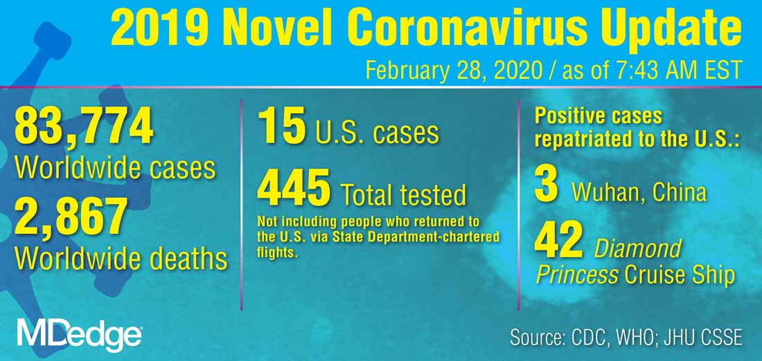
The definition has been revised “to meet the needs of this rapidly evolving situation,” she said. The new PUI definition includes travel to more geographic areas to reflect this past week’s marked uptick in coronavirus activity in Italy and Iran. In addition to these countries and China, recent travel to Japan or South Korea also constitutes an epidemiologic risk factor which, in conjunction with clinical features, warrant an individual being classified as a PUI. These five countries each now have widespread person-to-person transmission of the virus.
Dr. Messonnier left open the possibility that the PUI definition would continue to evolve if such transmission within communities becomes more common. Asked whether the small number of U.S. cases thus might be an artifact of low test volumes, she said, “We aggressively controlled our borders to slow the spread. This was an intentional U.S. strategy. The CDC has always had the capacity to test rapidly from the time the sequence was available. ...We have been testing aggressively.”
The original PUI definition, she explained, emphasized individuals with fever, cough, or trouble breathing who had traveled recently from areas with COVID-19 activity, in particular China’s Hubei province. “We have been most focused on symptomatic people who are closely linked to, or who had, travel history, but our criteria also allow for clinical discretion,” she said. “There is no substitute for an astute clinician on the front lines of patient care.”
The first COVID-19 case from person-to-person spread was reported on Feb. 27. “At this time, we don’t know how or where this person became infected,” said Dr. Messonnier, although investigations are still underway. She responded to a question about whether the CDC delayed allowing COVID-19 testing for the patient for several days, as was reported in some media accounts. “According to CDC records, the first call we got was Feb. 23,” when public health officials in California reported a severely ill person with no travel abroad and no known contacts with individuals that would trigger suspicions for coronavirus. The CDC recommended COVID-19 testing on that day, she said.
Dr. Messonnier declined to answer questions about a whistleblower report alleging improper training and inadequate protective measures for Department of Health & Human Services workers at the quarantine center at Travis Air Force Base, Calif.
Dr. Messonnier said that the CDC has been working closely with the Food and Drug Administration to address problems with the COVID-19 test kits that were unusable because of a large number of indeterminate results. The two agencies together have determined that of the three reactions that were initially deemed necessary for a definitive COVID-19 diagnosis, just two are sufficient, so new kits that omit the problematic chemical are being manufactured and distributed.
These new kits are rapidly being made available; the goal, said Dr. Messonnier, is to have to state and local public health departments equipped with test kits by about March 7.
As local tests become available, the most updated information will be coming from state and local public health departments, she stressed, adding that the CDC would continue to update case counts on Monday, Wednesday, and Friday of each week. Procedures are being developed for the management of patients presumed to have COVID-19, where local health departments see positive tests but the mandatory CDC confirmatory test hasn’t been completed.
While new cases emerge across Europe and Asia, China’s earlier COVID-19 explosion seems to be slowing. “It’s really good news that the case counts in China are decreasing,” both for the well-being of that country’s citizens, and as a sign of the disease’s potential global effects, said Dr. Messonnier. She added that epidemiologists and mathematical modelers are parsing case fatality rates as well.
She advised health care providers and public health officials to keep abreast of changes in CDC guidance by checking frequently at https://www.cdc.gov/coronavirus/2019-ncov/index.html.
In a telebriefing on the COVID-19 outbreak, Nancy Messonnier, MD, director of the National Center for Immunization and Respiratory Diseases at .
The definition has been revised “to meet the needs of this rapidly evolving situation,” she said. The new PUI definition includes travel to more geographic areas to reflect this past week’s marked uptick in coronavirus activity in Italy and Iran. In addition to these countries and China, recent travel to Japan or South Korea also constitutes an epidemiologic risk factor which, in conjunction with clinical features, warrant an individual being classified as a PUI. These five countries each now have widespread person-to-person transmission of the virus.
Dr. Messonnier left open the possibility that the PUI definition would continue to evolve if such transmission within communities becomes more common. Asked whether the small number of U.S. cases thus might be an artifact of low test volumes, she said, “We aggressively controlled our borders to slow the spread. This was an intentional U.S. strategy. The CDC has always had the capacity to test rapidly from the time the sequence was available. ...We have been testing aggressively.”
The original PUI definition, she explained, emphasized individuals with fever, cough, or trouble breathing who had traveled recently from areas with COVID-19 activity, in particular China’s Hubei province. “We have been most focused on symptomatic people who are closely linked to, or who had, travel history, but our criteria also allow for clinical discretion,” she said. “There is no substitute for an astute clinician on the front lines of patient care.”
The first COVID-19 case from person-to-person spread was reported on Feb. 27. “At this time, we don’t know how or where this person became infected,” said Dr. Messonnier, although investigations are still underway. She responded to a question about whether the CDC delayed allowing COVID-19 testing for the patient for several days, as was reported in some media accounts. “According to CDC records, the first call we got was Feb. 23,” when public health officials in California reported a severely ill person with no travel abroad and no known contacts with individuals that would trigger suspicions for coronavirus. The CDC recommended COVID-19 testing on that day, she said.
Dr. Messonnier declined to answer questions about a whistleblower report alleging improper training and inadequate protective measures for Department of Health & Human Services workers at the quarantine center at Travis Air Force Base, Calif.
Dr. Messonnier said that the CDC has been working closely with the Food and Drug Administration to address problems with the COVID-19 test kits that were unusable because of a large number of indeterminate results. The two agencies together have determined that of the three reactions that were initially deemed necessary for a definitive COVID-19 diagnosis, just two are sufficient, so new kits that omit the problematic chemical are being manufactured and distributed.
These new kits are rapidly being made available; the goal, said Dr. Messonnier, is to have to state and local public health departments equipped with test kits by about March 7.
As local tests become available, the most updated information will be coming from state and local public health departments, she stressed, adding that the CDC would continue to update case counts on Monday, Wednesday, and Friday of each week. Procedures are being developed for the management of patients presumed to have COVID-19, where local health departments see positive tests but the mandatory CDC confirmatory test hasn’t been completed.
While new cases emerge across Europe and Asia, China’s earlier COVID-19 explosion seems to be slowing. “It’s really good news that the case counts in China are decreasing,” both for the well-being of that country’s citizens, and as a sign of the disease’s potential global effects, said Dr. Messonnier. She added that epidemiologists and mathematical modelers are parsing case fatality rates as well.
She advised health care providers and public health officials to keep abreast of changes in CDC guidance by checking frequently at https://www.cdc.gov/coronavirus/2019-ncov/index.html.
In a telebriefing on the COVID-19 outbreak, Nancy Messonnier, MD, director of the National Center for Immunization and Respiratory Diseases at .
The definition has been revised “to meet the needs of this rapidly evolving situation,” she said. The new PUI definition includes travel to more geographic areas to reflect this past week’s marked uptick in coronavirus activity in Italy and Iran. In addition to these countries and China, recent travel to Japan or South Korea also constitutes an epidemiologic risk factor which, in conjunction with clinical features, warrant an individual being classified as a PUI. These five countries each now have widespread person-to-person transmission of the virus.
Dr. Messonnier left open the possibility that the PUI definition would continue to evolve if such transmission within communities becomes more common. Asked whether the small number of U.S. cases thus might be an artifact of low test volumes, she said, “We aggressively controlled our borders to slow the spread. This was an intentional U.S. strategy. The CDC has always had the capacity to test rapidly from the time the sequence was available. ...We have been testing aggressively.”
The original PUI definition, she explained, emphasized individuals with fever, cough, or trouble breathing who had traveled recently from areas with COVID-19 activity, in particular China’s Hubei province. “We have been most focused on symptomatic people who are closely linked to, or who had, travel history, but our criteria also allow for clinical discretion,” she said. “There is no substitute for an astute clinician on the front lines of patient care.”
The first COVID-19 case from person-to-person spread was reported on Feb. 27. “At this time, we don’t know how or where this person became infected,” said Dr. Messonnier, although investigations are still underway. She responded to a question about whether the CDC delayed allowing COVID-19 testing for the patient for several days, as was reported in some media accounts. “According to CDC records, the first call we got was Feb. 23,” when public health officials in California reported a severely ill person with no travel abroad and no known contacts with individuals that would trigger suspicions for coronavirus. The CDC recommended COVID-19 testing on that day, she said.
Dr. Messonnier declined to answer questions about a whistleblower report alleging improper training and inadequate protective measures for Department of Health & Human Services workers at the quarantine center at Travis Air Force Base, Calif.
Dr. Messonnier said that the CDC has been working closely with the Food and Drug Administration to address problems with the COVID-19 test kits that were unusable because of a large number of indeterminate results. The two agencies together have determined that of the three reactions that were initially deemed necessary for a definitive COVID-19 diagnosis, just two are sufficient, so new kits that omit the problematic chemical are being manufactured and distributed.
These new kits are rapidly being made available; the goal, said Dr. Messonnier, is to have to state and local public health departments equipped with test kits by about March 7.
As local tests become available, the most updated information will be coming from state and local public health departments, she stressed, adding that the CDC would continue to update case counts on Monday, Wednesday, and Friday of each week. Procedures are being developed for the management of patients presumed to have COVID-19, where local health departments see positive tests but the mandatory CDC confirmatory test hasn’t been completed.
While new cases emerge across Europe and Asia, China’s earlier COVID-19 explosion seems to be slowing. “It’s really good news that the case counts in China are decreasing,” both for the well-being of that country’s citizens, and as a sign of the disease’s potential global effects, said Dr. Messonnier. She added that epidemiologists and mathematical modelers are parsing case fatality rates as well.
She advised health care providers and public health officials to keep abreast of changes in CDC guidance by checking frequently at https://www.cdc.gov/coronavirus/2019-ncov/index.html.
REPORTING FROM A CDC BRIEFING
Serum NfL levels may predict 10-year deep gray matter volumes
WEST PALM BEACH, FLA. – , according to research presented at the meeting held by the Americas Committee for Treatment and Research in Multiple Sclerosis.

Researchers have begun to study longitudinal changes in serum NfL levels as a way to monitor axonal damage in patients with MS and see how they relate to other measures of neuronal loss, such as brain atrophy, and clinical outcomes over the long-term. “Deep gray matter volumes have been shown to correlate with neurological outcomes in MS patients. In particular, thalamic volume has been shown to correlate with measures of cognitive processing speed, such as the Symbol Digit Modalities Test,” said senior author Tanuja Chitnis, MD, professor of neurology at Harvard Medical School in Boston.
She and her colleagues sought to determine whether annual serum NfL measures could predict 10-year deep gray matter atrophy measured by volumetric MRI in patients with MS. They examined patients who were enrolled in the Comprehensive Longitudinal Investigations in MS at Brigham and Women’s Hospital (CLIMB) study. Eligible participants were enrolled within 5 years of disease onset and had had annual blood samples drawn for as long as 10 years. In all, 122 patients met these criteria. The investigators measured serum NfL and compared it against deep gray matter volume in the thalamus, caudate, putamen, and globus pallidus from high-resolution 3-T MRI scans taken at year 10. Dr. Chitnis and colleagues assessed correlations between averaged annual NfL and 10-year MRI outcomes using univariate and multivariate linear regression models.
About 96% of participants were white, and about 2% were black. Approximately 73% of participants were female. Average age at the first symptom was 36 years, and average age at the first sample collection was 38 years.
The investigators found several negative associations between averaged NfL values and various MRI volumetric outcomes. Averaged annual serum NfL levels for the first 5 years were significantly and negatively associated with 10-year thalamic volumes in the unadjusted analysis. A 1-pg/mL increase in the average sNfL value was associated with a decrease of 0.0000272 cm3 in thalamic volume. The association remained significant in an analysis adjusted for age, sex, and disease duration. In this analysis, a 1-pg/mL increase in the average sNfL value was associated with a decrease of 0.0000259 cm3 in thalamic volume. Analyzing serum NfL levels beyond year 5 did not reveal a stronger association. Serum NfL levels during the first 5 years accounted for about 24% of the variance in 10-year thalamic volumes. Dr. Chitnis and colleagues found similar statistically significant associations between serum NfL levels and caudate, putamen, and globus pallidus volumes. “Therefore, early serum NfL levels contribute to the identification of patients who may require highly effective therapies,” she said.
“We will continue to validate these results. As well, we are exploring other early biomarkers that increase predictive power of long-term outcomes in MS, with the goal of identifying patients most appropriate for high-efficacy treatments.”
The study was supported by funds from the U.S. Department of Defense, Novartis, and the Swiss National Research Foundation. Dr. Chitnis has received personal compensation for consulting and advisory board membership from Biogen, Merck Serono, Novartis, and Sanofi.
SOURCE: Lokhande H et al. ACTRIMS Forum 2020, Abstract P018.
WEST PALM BEACH, FLA. – , according to research presented at the meeting held by the Americas Committee for Treatment and Research in Multiple Sclerosis.
Researchers have begun to study longitudinal changes in serum NfL levels as a way to monitor axonal damage in patients with MS and see how they relate to other measures of neuronal loss, such as brain atrophy, and clinical outcomes over the long-term. “Deep gray matter volumes have been shown to correlate with neurological outcomes in MS patients. In particular, thalamic volume has been shown to correlate with measures of cognitive processing speed, such as the Symbol Digit Modalities Test,” said senior author Tanuja Chitnis, MD, professor of neurology at Harvard Medical School in Boston.
She and her colleagues sought to determine whether annual serum NfL measures could predict 10-year deep gray matter atrophy measured by volumetric MRI in patients with MS. They examined patients who were enrolled in the Comprehensive Longitudinal Investigations in MS at Brigham and Women’s Hospital (CLIMB) study. Eligible participants were enrolled within 5 years of disease onset and had had annual blood samples drawn for as long as 10 years. In all, 122 patients met these criteria. The investigators measured serum NfL and compared it against deep gray matter volume in the thalamus, caudate, putamen, and globus pallidus from high-resolution 3-T MRI scans taken at year 10. Dr. Chitnis and colleagues assessed correlations between averaged annual NfL and 10-year MRI outcomes using univariate and multivariate linear regression models.
About 96% of participants were white, and about 2% were black. Approximately 73% of participants were female. Average age at the first symptom was 36 years, and average age at the first sample collection was 38 years.
The investigators found several negative associations between averaged NfL values and various MRI volumetric outcomes. Averaged annual serum NfL levels for the first 5 years were significantly and negatively associated with 10-year thalamic volumes in the unadjusted analysis. A 1-pg/mL increase in the average sNfL value was associated with a decrease of 0.0000272 cm3 in thalamic volume. The association remained significant in an analysis adjusted for age, sex, and disease duration. In this analysis, a 1-pg/mL increase in the average sNfL value was associated with a decrease of 0.0000259 cm3 in thalamic volume. Analyzing serum NfL levels beyond year 5 did not reveal a stronger association. Serum NfL levels during the first 5 years accounted for about 24% of the variance in 10-year thalamic volumes. Dr. Chitnis and colleagues found similar statistically significant associations between serum NfL levels and caudate, putamen, and globus pallidus volumes. “Therefore, early serum NfL levels contribute to the identification of patients who may require highly effective therapies,” she said.
“We will continue to validate these results. As well, we are exploring other early biomarkers that increase predictive power of long-term outcomes in MS, with the goal of identifying patients most appropriate for high-efficacy treatments.”
The study was supported by funds from the U.S. Department of Defense, Novartis, and the Swiss National Research Foundation. Dr. Chitnis has received personal compensation for consulting and advisory board membership from Biogen, Merck Serono, Novartis, and Sanofi.
SOURCE: Lokhande H et al. ACTRIMS Forum 2020, Abstract P018.
WEST PALM BEACH, FLA. – , according to research presented at the meeting held by the Americas Committee for Treatment and Research in Multiple Sclerosis.
Researchers have begun to study longitudinal changes in serum NfL levels as a way to monitor axonal damage in patients with MS and see how they relate to other measures of neuronal loss, such as brain atrophy, and clinical outcomes over the long-term. “Deep gray matter volumes have been shown to correlate with neurological outcomes in MS patients. In particular, thalamic volume has been shown to correlate with measures of cognitive processing speed, such as the Symbol Digit Modalities Test,” said senior author Tanuja Chitnis, MD, professor of neurology at Harvard Medical School in Boston.
She and her colleagues sought to determine whether annual serum NfL measures could predict 10-year deep gray matter atrophy measured by volumetric MRI in patients with MS. They examined patients who were enrolled in the Comprehensive Longitudinal Investigations in MS at Brigham and Women’s Hospital (CLIMB) study. Eligible participants were enrolled within 5 years of disease onset and had had annual blood samples drawn for as long as 10 years. In all, 122 patients met these criteria. The investigators measured serum NfL and compared it against deep gray matter volume in the thalamus, caudate, putamen, and globus pallidus from high-resolution 3-T MRI scans taken at year 10. Dr. Chitnis and colleagues assessed correlations between averaged annual NfL and 10-year MRI outcomes using univariate and multivariate linear regression models.
About 96% of participants were white, and about 2% were black. Approximately 73% of participants were female. Average age at the first symptom was 36 years, and average age at the first sample collection was 38 years.
The investigators found several negative associations between averaged NfL values and various MRI volumetric outcomes. Averaged annual serum NfL levels for the first 5 years were significantly and negatively associated with 10-year thalamic volumes in the unadjusted analysis. A 1-pg/mL increase in the average sNfL value was associated with a decrease of 0.0000272 cm3 in thalamic volume. The association remained significant in an analysis adjusted for age, sex, and disease duration. In this analysis, a 1-pg/mL increase in the average sNfL value was associated with a decrease of 0.0000259 cm3 in thalamic volume. Analyzing serum NfL levels beyond year 5 did not reveal a stronger association. Serum NfL levels during the first 5 years accounted for about 24% of the variance in 10-year thalamic volumes. Dr. Chitnis and colleagues found similar statistically significant associations between serum NfL levels and caudate, putamen, and globus pallidus volumes. “Therefore, early serum NfL levels contribute to the identification of patients who may require highly effective therapies,” she said.
“We will continue to validate these results. As well, we are exploring other early biomarkers that increase predictive power of long-term outcomes in MS, with the goal of identifying patients most appropriate for high-efficacy treatments.”
The study was supported by funds from the U.S. Department of Defense, Novartis, and the Swiss National Research Foundation. Dr. Chitnis has received personal compensation for consulting and advisory board membership from Biogen, Merck Serono, Novartis, and Sanofi.
SOURCE: Lokhande H et al. ACTRIMS Forum 2020, Abstract P018.
REPORTING FROM ACTRIMS FORUM 2020

DDSEP® 9 Quick Quiz Question 2
Q2. Correct answer: D
Rationale
This patient has tropical sprue based on her travel to an endemic country, negative celiac serologies, labs revealing a macrocytic anemia and low albumin and characteristic histology (villous blunting, increased intraepithelial lymphocytes). Treatment is with tetracycline and folate. Diagnosis of tropical sprue is ultimately confirmed by a response to treatment. A gluten-free diet is not appropriate, as the patient does not have celiac disease, confirmed by normal celiac serologies. Ceftriaxone IV followed by Bactrim PO is the correct treatment for Whipple's disease. A diet low in fermentable oligo-, di-, and monosaccharides and polyols (FODMAPs) is beneficial treatment in some patients with IBS with abdominal bloating or pain. Rifaximin is the correct treatment for small intestine bacterial overgrowth or IBS-D.
References
1. Brown IS, Bettington A, Bettington M, Rosty C. Tropical sprue: revisiting an underrecognized disease. Am J Surg Pathol. 2014;38:666.
2. Shah VH, Rotterdam H, Kotler DP, et al. All that scallops is not celiac disease. Gastrointest Endosc. 2000;51:717.
ginews@gastro.org
Q2. Correct answer: D
Rationale
This patient has tropical sprue based on her travel to an endemic country, negative celiac serologies, labs revealing a macrocytic anemia and low albumin and characteristic histology (villous blunting, increased intraepithelial lymphocytes). Treatment is with tetracycline and folate. Diagnosis of tropical sprue is ultimately confirmed by a response to treatment. A gluten-free diet is not appropriate, as the patient does not have celiac disease, confirmed by normal celiac serologies. Ceftriaxone IV followed by Bactrim PO is the correct treatment for Whipple's disease. A diet low in fermentable oligo-, di-, and monosaccharides and polyols (FODMAPs) is beneficial treatment in some patients with IBS with abdominal bloating or pain. Rifaximin is the correct treatment for small intestine bacterial overgrowth or IBS-D.
References
1. Brown IS, Bettington A, Bettington M, Rosty C. Tropical sprue: revisiting an underrecognized disease. Am J Surg Pathol. 2014;38:666.
2. Shah VH, Rotterdam H, Kotler DP, et al. All that scallops is not celiac disease. Gastrointest Endosc. 2000;51:717.
ginews@gastro.org
Q2. Correct answer: D
Rationale
This patient has tropical sprue based on her travel to an endemic country, negative celiac serologies, labs revealing a macrocytic anemia and low albumin and characteristic histology (villous blunting, increased intraepithelial lymphocytes). Treatment is with tetracycline and folate. Diagnosis of tropical sprue is ultimately confirmed by a response to treatment. A gluten-free diet is not appropriate, as the patient does not have celiac disease, confirmed by normal celiac serologies. Ceftriaxone IV followed by Bactrim PO is the correct treatment for Whipple's disease. A diet low in fermentable oligo-, di-, and monosaccharides and polyols (FODMAPs) is beneficial treatment in some patients with IBS with abdominal bloating or pain. Rifaximin is the correct treatment for small intestine bacterial overgrowth or IBS-D.
References
1. Brown IS, Bettington A, Bettington M, Rosty C. Tropical sprue: revisiting an underrecognized disease. Am J Surg Pathol. 2014;38:666.
2. Shah VH, Rotterdam H, Kotler DP, et al. All that scallops is not celiac disease. Gastrointest Endosc. 2000;51:717.
ginews@gastro.org
An 18-year-old woman presents for evaluation of chronic diarrhea, fatigue, and abdominal cramping. She was recently in Puerto Rico for 6 months visiting family and returned a few weeks ago. Her labs are significant for a hemoglobin of 11 g/L with an MCV of 109 fL. Her albumin is 3.6 g/dL. She had stool studies which ruled out infection, including parasites. TtG IgA and total IgA were within normal limits. EGD with multiple duodenal biopsies showed villous blunting with increased intraepithelial lymphocytes.
March 2020 - Question 1
Correct answer: C
Rationale
This patient has been exposed to HBV in the past and has cleared the virus. The HBVcore total Ab is indicative of prior exposure while the HBV surface Ab is detectable and gives immunity against reinfection under most routine clinical scenarios. Patients who have been exposed to HBV still have HBV ccc DNA within their hepatocytes that is dormant, but under extreme combined B and T cell immunosuppression, the patients are at risk for reverse seroconversion where they can lose HBV surface Ab and manifest HBV surface antigen and present as an acute HBV infection. Prophylaxis is required during therapy and for at least 12-18 months after therapy due to the long-lasting effects of anti-B cell monoclonal antibodies like rituximab. Reactivation of HCV in HCV Ab–positive, RNA-negative patients has not been reported.
Reference
1. Pauly MP, Tucker LY, Szpakowski JL, et al. Incidence of hepatitis B virus reactivation and hepatotoxicity in patients receiving long-term treatment with tumor necrosis factor antagonists. Clin Gastroenterol Hepatol. 2018 Apr 24. doi: 10.1016/j. cgh.2018.04.033.
ginews@gastro.org
Correct answer: C
Rationale
This patient has been exposed to HBV in the past and has cleared the virus. The HBVcore total Ab is indicative of prior exposure while the HBV surface Ab is detectable and gives immunity against reinfection under most routine clinical scenarios. Patients who have been exposed to HBV still have HBV ccc DNA within their hepatocytes that is dormant, but under extreme combined B and T cell immunosuppression, the patients are at risk for reverse seroconversion where they can lose HBV surface Ab and manifest HBV surface antigen and present as an acute HBV infection. Prophylaxis is required during therapy and for at least 12-18 months after therapy due to the long-lasting effects of anti-B cell monoclonal antibodies like rituximab. Reactivation of HCV in HCV Ab–positive, RNA-negative patients has not been reported.
Reference
1. Pauly MP, Tucker LY, Szpakowski JL, et al. Incidence of hepatitis B virus reactivation and hepatotoxicity in patients receiving long-term treatment with tumor necrosis factor antagonists. Clin Gastroenterol Hepatol. 2018 Apr 24. doi: 10.1016/j. cgh.2018.04.033.
ginews@gastro.org
Correct answer: C
Rationale
This patient has been exposed to HBV in the past and has cleared the virus. The HBVcore total Ab is indicative of prior exposure while the HBV surface Ab is detectable and gives immunity against reinfection under most routine clinical scenarios. Patients who have been exposed to HBV still have HBV ccc DNA within their hepatocytes that is dormant, but under extreme combined B and T cell immunosuppression, the patients are at risk for reverse seroconversion where they can lose HBV surface Ab and manifest HBV surface antigen and present as an acute HBV infection. Prophylaxis is required during therapy and for at least 12-18 months after therapy due to the long-lasting effects of anti-B cell monoclonal antibodies like rituximab. Reactivation of HCV in HCV Ab–positive, RNA-negative patients has not been reported.
Reference
1. Pauly MP, Tucker LY, Szpakowski JL, et al. Incidence of hepatitis B virus reactivation and hepatotoxicity in patients receiving long-term treatment with tumor necrosis factor antagonists. Clin Gastroenterol Hepatol. 2018 Apr 24. doi: 10.1016/j. cgh.2018.04.033.
ginews@gastro.org
Q1. A 45-year-old man has recently been diagnosed with leukemia. The chemotherapeutic regimen will include rituximab and high-dose steroids. He is a former IV drug user but has been sober for 20 years. His lab work is as follows: ALT 25 U/L, HAV total antibody positive, HBs antibody positive, HBs antigen negative, HBc total positive, HCV antibody positive, HCV RNA undetected.