User login
Trial of epicutaneous immunotherapy in eosinophilic esophagitis
For children with milk-induced eosinophilic esophagitis (EoE), 9 months of epicutaneous immunotherapy (EPIT) with Viaskin Milk did not significantly improve eosinophil counts or symptoms, compared with placebo, according to the results of an intention-to-treat analysis of a randomized, double-blinded pilot study.
Average maximum eosinophil counts were 50.1 per high-power field in the Viaskin Milk group versus 48.2 in the placebo group, said Jonathan M. Spergel, MD, of the Children’s Hospital of Philadelphia and associates. However, in the per-protocol analysis, the seven patients who received Viaskin Milk had mean eosinophil counts of 25.6 per high-power field, compared with 95.0 for the two children who received placebo (P = .038). Moreover, 47% of patients had fewer than 15 eosinophils per high-power field after an additional 11 months of open-label treatment with Viaskin Milk. Taken together, the findings justify larger, multicenter studies to evaluate EPIT for treating EoE and other non-IgE mediated food diseases, Dr. Spergel and associates wrote in Clinical Gastroenterology and Hepatology.
EoE results from an immune response to specific food allergens, including milk. Classic symptoms include difficulty feeding and failure to thrive in infants, abdominal pain in young children, and dysphagia in older children and adults. Definitive diagnosis requires an esophageal biopsy with an eosinophil count of 15 or more cells per high-power field. “There are no approved therapies [for eosinophilic esophagitis] beyond avoidance of the allergen(s) or treatment of inflammation,” the investigators wrote.
In prior studies, exposure to EPIT was found to mitigate eosinophilic gastrointestinal disease in mice and pigs. In humans, milk is the most common dietary cause of eosinophilic esophagitis. Accordingly, Viaskin Milk is an EPIT containing an allergen extract of milk that is administered epicutaneously using a specialized delivery system. To evaluate its use for the treatment of pediatric milk-induced EoE (at least 15 eosinophils per high-power frame despite at least 2 months of high-dose proton pump–inhibitor therapy at 1-2 mg/kg twice daily), the researchers randomly assigned 20 children on a 3:1 basis to receive either Viaskin Milk or placebo for 9 months. Patients and investigators were double-blinded for this phase of the study, during most of which patients abstained from milk. Toward the end of the 9 months, patients resumed consuming milk and continued doing so if their upper endoscopy biopsy showed resolution of EoE (eosinophil count less than 15 per high-power field).
In the intention-to-treat analysis, Viaskin Milk did not meet the primary endpoint of the difference in least squares mean compared with placebo (8.6; 95% confidence interval, –35.36 to 52.56). Symptom scores also were similar between groups. In contrast, at the end of the 11-month, open-label period, 9 of 19 evaluable patients had eosinophil biopsy counts of fewer than 15 per high-power field, for a response rate of 47%. “The number of adverse events did not differ significantly between the Viaskin Milk and placebo groups,” the researchers added.
Protocol violations might explain why EPIT failed to meet the primary endpoint in the intention-to-treat analysis, they wrote. “For example, the patients on the active therapy wanted to ingest more milk, while the patients in the placebo group wanted less milk,” they reported. “Three patients in the active therapy went on binge milk diets drinking 4 to 8 times the amount of milk compared with baseline.” The use of proton pump inhibitors also was inconsistent between groups, they added. “The major limitation in the [per-protocol] population was the small sample size of this pilot study, raising the possibility of false-positive results.”
The study was funded by DBV Technologies and by the Children’s Hospital of Philadelphia Eosinophilic Esophagitis Family Fund. Dr. Spergel disclosed consulting agreements, grants funding, and stock equity with DBV Technologies. Three coinvestigators also disclosed ties to DBV. The remaining five coinvestigators reported having no conflicts of interest.
SOURCE: Spergel JM et al. Clin Gastroenterol Hepatol. 2019 May 14. doi: 10.1016/j.cgh.2019.05.014.

Eosinophilic esophagitis (EoE) is a chronic immune-mediated disease that is primarily triggered by food antigens. Though many patients can be treated with dietary elimination or pharmacologic therapies, when foods are added back, elimination diets are not followed, or medications stopped, the disease will flare. Further, unlike some other atopic conditions, patients with EoE do not “grow out of it.” A true cure for EoE has been elusive. In this study by Spergel and colleagues, they build on intriguing data from animal models showing induction of immune tolerance to food antigens with epicutaneous immunotherapy (EPIT).
The investigators conducted a proof-of-concept, double-blind, placebo-controlled randomized trial of epicutaneous desensitization with a milk patch in children with EoE who had milk as a confirmed dietary trigger. The primary intention-to-treat results showed that there was no difference between placebo and active patches for decreasing esophageal eosinophil counts. However, in the small set of patients who were able to adhere fully to the protocol, the per-protocol analysis suggested that there was a lower eosinophil count with active treatment. Additionally, in an 11-month, open-label extension, there were patients who maintained histologic response (less than 15 eosinophils/hpf) after reintroducing milk.
These data suggest that EPIT potentially can desensitize milk-triggered EoE patients and that this treatment method should be pursued in future studies, with protocol alterations based on lessons learned regarding adherence in this study. Should this line of investigation be successful, then EoE patients who have milk as their EoE trigger, and who undergo successful desensitization with mild reintroduction while maintaining disease remission, may be able to be deemed cured.
Evan S. Dellon, MD, MPH, professor of medicine and epidemiology, division of gastroenterology and hepatology, University of North Carolina at Chapel Hill. He has received research funding from and consulted for Adare, Allakos, GSK, Celgene/Receptos, and Shire/Takeda among other pharmaceutical companies.
Eosinophilic esophagitis (EoE) is a chronic immune-mediated disease that is primarily triggered by food antigens. Though many patients can be treated with dietary elimination or pharmacologic therapies, when foods are added back, elimination diets are not followed, or medications stopped, the disease will flare. Further, unlike some other atopic conditions, patients with EoE do not “grow out of it.” A true cure for EoE has been elusive. In this study by Spergel and colleagues, they build on intriguing data from animal models showing induction of immune tolerance to food antigens with epicutaneous immunotherapy (EPIT).
The investigators conducted a proof-of-concept, double-blind, placebo-controlled randomized trial of epicutaneous desensitization with a milk patch in children with EoE who had milk as a confirmed dietary trigger. The primary intention-to-treat results showed that there was no difference between placebo and active patches for decreasing esophageal eosinophil counts. However, in the small set of patients who were able to adhere fully to the protocol, the per-protocol analysis suggested that there was a lower eosinophil count with active treatment. Additionally, in an 11-month, open-label extension, there were patients who maintained histologic response (less than 15 eosinophils/hpf) after reintroducing milk.
These data suggest that EPIT potentially can desensitize milk-triggered EoE patients and that this treatment method should be pursued in future studies, with protocol alterations based on lessons learned regarding adherence in this study. Should this line of investigation be successful, then EoE patients who have milk as their EoE trigger, and who undergo successful desensitization with mild reintroduction while maintaining disease remission, may be able to be deemed cured.
Evan S. Dellon, MD, MPH, professor of medicine and epidemiology, division of gastroenterology and hepatology, University of North Carolina at Chapel Hill. He has received research funding from and consulted for Adare, Allakos, GSK, Celgene/Receptos, and Shire/Takeda among other pharmaceutical companies.
Eosinophilic esophagitis (EoE) is a chronic immune-mediated disease that is primarily triggered by food antigens. Though many patients can be treated with dietary elimination or pharmacologic therapies, when foods are added back, elimination diets are not followed, or medications stopped, the disease will flare. Further, unlike some other atopic conditions, patients with EoE do not “grow out of it.” A true cure for EoE has been elusive. In this study by Spergel and colleagues, they build on intriguing data from animal models showing induction of immune tolerance to food antigens with epicutaneous immunotherapy (EPIT).
The investigators conducted a proof-of-concept, double-blind, placebo-controlled randomized trial of epicutaneous desensitization with a milk patch in children with EoE who had milk as a confirmed dietary trigger. The primary intention-to-treat results showed that there was no difference between placebo and active patches for decreasing esophageal eosinophil counts. However, in the small set of patients who were able to adhere fully to the protocol, the per-protocol analysis suggested that there was a lower eosinophil count with active treatment. Additionally, in an 11-month, open-label extension, there were patients who maintained histologic response (less than 15 eosinophils/hpf) after reintroducing milk.
These data suggest that EPIT potentially can desensitize milk-triggered EoE patients and that this treatment method should be pursued in future studies, with protocol alterations based on lessons learned regarding adherence in this study. Should this line of investigation be successful, then EoE patients who have milk as their EoE trigger, and who undergo successful desensitization with mild reintroduction while maintaining disease remission, may be able to be deemed cured.
Evan S. Dellon, MD, MPH, professor of medicine and epidemiology, division of gastroenterology and hepatology, University of North Carolina at Chapel Hill. He has received research funding from and consulted for Adare, Allakos, GSK, Celgene/Receptos, and Shire/Takeda among other pharmaceutical companies.
For children with milk-induced eosinophilic esophagitis (EoE), 9 months of epicutaneous immunotherapy (EPIT) with Viaskin Milk did not significantly improve eosinophil counts or symptoms, compared with placebo, according to the results of an intention-to-treat analysis of a randomized, double-blinded pilot study.
Average maximum eosinophil counts were 50.1 per high-power field in the Viaskin Milk group versus 48.2 in the placebo group, said Jonathan M. Spergel, MD, of the Children’s Hospital of Philadelphia and associates. However, in the per-protocol analysis, the seven patients who received Viaskin Milk had mean eosinophil counts of 25.6 per high-power field, compared with 95.0 for the two children who received placebo (P = .038). Moreover, 47% of patients had fewer than 15 eosinophils per high-power field after an additional 11 months of open-label treatment with Viaskin Milk. Taken together, the findings justify larger, multicenter studies to evaluate EPIT for treating EoE and other non-IgE mediated food diseases, Dr. Spergel and associates wrote in Clinical Gastroenterology and Hepatology.
EoE results from an immune response to specific food allergens, including milk. Classic symptoms include difficulty feeding and failure to thrive in infants, abdominal pain in young children, and dysphagia in older children and adults. Definitive diagnosis requires an esophageal biopsy with an eosinophil count of 15 or more cells per high-power field. “There are no approved therapies [for eosinophilic esophagitis] beyond avoidance of the allergen(s) or treatment of inflammation,” the investigators wrote.
In prior studies, exposure to EPIT was found to mitigate eosinophilic gastrointestinal disease in mice and pigs. In humans, milk is the most common dietary cause of eosinophilic esophagitis. Accordingly, Viaskin Milk is an EPIT containing an allergen extract of milk that is administered epicutaneously using a specialized delivery system. To evaluate its use for the treatment of pediatric milk-induced EoE (at least 15 eosinophils per high-power frame despite at least 2 months of high-dose proton pump–inhibitor therapy at 1-2 mg/kg twice daily), the researchers randomly assigned 20 children on a 3:1 basis to receive either Viaskin Milk or placebo for 9 months. Patients and investigators were double-blinded for this phase of the study, during most of which patients abstained from milk. Toward the end of the 9 months, patients resumed consuming milk and continued doing so if their upper endoscopy biopsy showed resolution of EoE (eosinophil count less than 15 per high-power field).
In the intention-to-treat analysis, Viaskin Milk did not meet the primary endpoint of the difference in least squares mean compared with placebo (8.6; 95% confidence interval, –35.36 to 52.56). Symptom scores also were similar between groups. In contrast, at the end of the 11-month, open-label period, 9 of 19 evaluable patients had eosinophil biopsy counts of fewer than 15 per high-power field, for a response rate of 47%. “The number of adverse events did not differ significantly between the Viaskin Milk and placebo groups,” the researchers added.
Protocol violations might explain why EPIT failed to meet the primary endpoint in the intention-to-treat analysis, they wrote. “For example, the patients on the active therapy wanted to ingest more milk, while the patients in the placebo group wanted less milk,” they reported. “Three patients in the active therapy went on binge milk diets drinking 4 to 8 times the amount of milk compared with baseline.” The use of proton pump inhibitors also was inconsistent between groups, they added. “The major limitation in the [per-protocol] population was the small sample size of this pilot study, raising the possibility of false-positive results.”
The study was funded by DBV Technologies and by the Children’s Hospital of Philadelphia Eosinophilic Esophagitis Family Fund. Dr. Spergel disclosed consulting agreements, grants funding, and stock equity with DBV Technologies. Three coinvestigators also disclosed ties to DBV. The remaining five coinvestigators reported having no conflicts of interest.
SOURCE: Spergel JM et al. Clin Gastroenterol Hepatol. 2019 May 14. doi: 10.1016/j.cgh.2019.05.014.
For children with milk-induced eosinophilic esophagitis (EoE), 9 months of epicutaneous immunotherapy (EPIT) with Viaskin Milk did not significantly improve eosinophil counts or symptoms, compared with placebo, according to the results of an intention-to-treat analysis of a randomized, double-blinded pilot study.
Average maximum eosinophil counts were 50.1 per high-power field in the Viaskin Milk group versus 48.2 in the placebo group, said Jonathan M. Spergel, MD, of the Children’s Hospital of Philadelphia and associates. However, in the per-protocol analysis, the seven patients who received Viaskin Milk had mean eosinophil counts of 25.6 per high-power field, compared with 95.0 for the two children who received placebo (P = .038). Moreover, 47% of patients had fewer than 15 eosinophils per high-power field after an additional 11 months of open-label treatment with Viaskin Milk. Taken together, the findings justify larger, multicenter studies to evaluate EPIT for treating EoE and other non-IgE mediated food diseases, Dr. Spergel and associates wrote in Clinical Gastroenterology and Hepatology.
EoE results from an immune response to specific food allergens, including milk. Classic symptoms include difficulty feeding and failure to thrive in infants, abdominal pain in young children, and dysphagia in older children and adults. Definitive diagnosis requires an esophageal biopsy with an eosinophil count of 15 or more cells per high-power field. “There are no approved therapies [for eosinophilic esophagitis] beyond avoidance of the allergen(s) or treatment of inflammation,” the investigators wrote.
In prior studies, exposure to EPIT was found to mitigate eosinophilic gastrointestinal disease in mice and pigs. In humans, milk is the most common dietary cause of eosinophilic esophagitis. Accordingly, Viaskin Milk is an EPIT containing an allergen extract of milk that is administered epicutaneously using a specialized delivery system. To evaluate its use for the treatment of pediatric milk-induced EoE (at least 15 eosinophils per high-power frame despite at least 2 months of high-dose proton pump–inhibitor therapy at 1-2 mg/kg twice daily), the researchers randomly assigned 20 children on a 3:1 basis to receive either Viaskin Milk or placebo for 9 months. Patients and investigators were double-blinded for this phase of the study, during most of which patients abstained from milk. Toward the end of the 9 months, patients resumed consuming milk and continued doing so if their upper endoscopy biopsy showed resolution of EoE (eosinophil count less than 15 per high-power field).
In the intention-to-treat analysis, Viaskin Milk did not meet the primary endpoint of the difference in least squares mean compared with placebo (8.6; 95% confidence interval, –35.36 to 52.56). Symptom scores also were similar between groups. In contrast, at the end of the 11-month, open-label period, 9 of 19 evaluable patients had eosinophil biopsy counts of fewer than 15 per high-power field, for a response rate of 47%. “The number of adverse events did not differ significantly between the Viaskin Milk and placebo groups,” the researchers added.
Protocol violations might explain why EPIT failed to meet the primary endpoint in the intention-to-treat analysis, they wrote. “For example, the patients on the active therapy wanted to ingest more milk, while the patients in the placebo group wanted less milk,” they reported. “Three patients in the active therapy went on binge milk diets drinking 4 to 8 times the amount of milk compared with baseline.” The use of proton pump inhibitors also was inconsistent between groups, they added. “The major limitation in the [per-protocol] population was the small sample size of this pilot study, raising the possibility of false-positive results.”
The study was funded by DBV Technologies and by the Children’s Hospital of Philadelphia Eosinophilic Esophagitis Family Fund. Dr. Spergel disclosed consulting agreements, grants funding, and stock equity with DBV Technologies. Three coinvestigators also disclosed ties to DBV. The remaining five coinvestigators reported having no conflicts of interest.
SOURCE: Spergel JM et al. Clin Gastroenterol Hepatol. 2019 May 14. doi: 10.1016/j.cgh.2019.05.014.
FROM CLINICAL GASTROENTEROLOGY AND HEPATOLOGY

Eradicating H. pylori may cut risk of gastric cancer
Eradication of Helicobacter pylori infection was associated with a more than 75% decrease in hazard of subsequent stomach cancer in a large retrospective cohort study.
Simply being treated for H. pylori infection did not mitigate the risk of gastric adenocarcinoma, and patients whose H. pylori was not eradicated were at increased risk, said Shria Kumar, MD, of the Perelman Center for Advanced Medicine in Philadelphia, and with her associates. “This speaks to the ability of H. pylori eradication to modify future risks of gastric adenocarcinoma, and the need to not only treat those diagnosed with H. pylori, but to confirm eradication, and re-treat those who fail eradication,” they wrote in Gastroenterology.
Gastric adenocarcinoma remains a grave diagnosis, with a 5-year survival rate of less than 30%. Although H. pylori infection is an established risk factor for gastric cancer (particularly nonproximal disease), most studies have used national cancer databases that do not track H. pylori infection. Accordingly, Dr. Kumar and her associates analyzed data for 371,813 patients diagnosed with H. pylori infection at U.S. Veterans Health Administration facilities between 1994 and 2018. A total of 92% of patients were men, 58% were white, 24% were black, and approximately 1% each were Native American, Asian, or Native Hawaiian/Pacific Islander. Median age was 62 years.
Patients with H. pylori infection who subsequently were diagnosed with nonproximal gastric cancer were significantly (P less than .001) more likely to be older (median age, 65.1 vs. 62.0 years), current or historical smokers, or racial or ethnic minorities (black or African American, Asian, or Hispanic/Latino), compared with patients with H. pylori who did not develop cancer. In the multivariable analysis, standardized hazard ratios for these variables remained statistically significant, with point estimates ranging from 1.13 (for each 5-year increase in age at diagnosis of infection) to 2.00 (for black or African American race). Cumulative incidence rates of distal gastric adenocarcinoma following H. pylori infection were 0.37% at 5 years, 0.5% at 10 years, and 0.65% at 20 year.
Patients whose infections were confirmed to have been eradicated were at markedly lower risk for subsequent gastric cancer than were patients whose infections were not eradicated (SHR, 0.24; 95% confidence interval, 0.15-0.41; P less than .001). Importantly, simply being treated for H. pylori did not significantly affect cancer risk (SHR, 1.16; 95% CI, 0.74-1.83).
Rates in Japan are approximately five times higher, while in sub-Saharan Africa, H. pylori infection is prevalent but gastric cancer is uncommon, the researchers noted. These discrepancies support the idea that carcinogenesis depends on additional genetic or environmental variables in addition to H. pylori infection alone, they said. They called for future studies of protective factors.
Dr. Kumar is supported by a training grant from the National Institutes of Health. She disclosed travel support from Boston Scientific and Olympus. Her coinvestigators disclosed ties to Takeda, Novartis, Janssen, Gilead, Bayer, and several other companies.
SOURCE: Kumar S et al. Gastroenterology. 2019 Jul 31. doi: 10.1053/j.gastro.2019.10.019.
Eradication of Helicobacter pylori infection was associated with a more than 75% decrease in hazard of subsequent stomach cancer in a large retrospective cohort study.
Simply being treated for H. pylori infection did not mitigate the risk of gastric adenocarcinoma, and patients whose H. pylori was not eradicated were at increased risk, said Shria Kumar, MD, of the Perelman Center for Advanced Medicine in Philadelphia, and with her associates. “This speaks to the ability of H. pylori eradication to modify future risks of gastric adenocarcinoma, and the need to not only treat those diagnosed with H. pylori, but to confirm eradication, and re-treat those who fail eradication,” they wrote in Gastroenterology.
Gastric adenocarcinoma remains a grave diagnosis, with a 5-year survival rate of less than 30%. Although H. pylori infection is an established risk factor for gastric cancer (particularly nonproximal disease), most studies have used national cancer databases that do not track H. pylori infection. Accordingly, Dr. Kumar and her associates analyzed data for 371,813 patients diagnosed with H. pylori infection at U.S. Veterans Health Administration facilities between 1994 and 2018. A total of 92% of patients were men, 58% were white, 24% were black, and approximately 1% each were Native American, Asian, or Native Hawaiian/Pacific Islander. Median age was 62 years.
Patients with H. pylori infection who subsequently were diagnosed with nonproximal gastric cancer were significantly (P less than .001) more likely to be older (median age, 65.1 vs. 62.0 years), current or historical smokers, or racial or ethnic minorities (black or African American, Asian, or Hispanic/Latino), compared with patients with H. pylori who did not develop cancer. In the multivariable analysis, standardized hazard ratios for these variables remained statistically significant, with point estimates ranging from 1.13 (for each 5-year increase in age at diagnosis of infection) to 2.00 (for black or African American race). Cumulative incidence rates of distal gastric adenocarcinoma following H. pylori infection were 0.37% at 5 years, 0.5% at 10 years, and 0.65% at 20 year.
Patients whose infections were confirmed to have been eradicated were at markedly lower risk for subsequent gastric cancer than were patients whose infections were not eradicated (SHR, 0.24; 95% confidence interval, 0.15-0.41; P less than .001). Importantly, simply being treated for H. pylori did not significantly affect cancer risk (SHR, 1.16; 95% CI, 0.74-1.83).
Rates in Japan are approximately five times higher, while in sub-Saharan Africa, H. pylori infection is prevalent but gastric cancer is uncommon, the researchers noted. These discrepancies support the idea that carcinogenesis depends on additional genetic or environmental variables in addition to H. pylori infection alone, they said. They called for future studies of protective factors.
Dr. Kumar is supported by a training grant from the National Institutes of Health. She disclosed travel support from Boston Scientific and Olympus. Her coinvestigators disclosed ties to Takeda, Novartis, Janssen, Gilead, Bayer, and several other companies.
SOURCE: Kumar S et al. Gastroenterology. 2019 Jul 31. doi: 10.1053/j.gastro.2019.10.019.
Eradication of Helicobacter pylori infection was associated with a more than 75% decrease in hazard of subsequent stomach cancer in a large retrospective cohort study.
Simply being treated for H. pylori infection did not mitigate the risk of gastric adenocarcinoma, and patients whose H. pylori was not eradicated were at increased risk, said Shria Kumar, MD, of the Perelman Center for Advanced Medicine in Philadelphia, and with her associates. “This speaks to the ability of H. pylori eradication to modify future risks of gastric adenocarcinoma, and the need to not only treat those diagnosed with H. pylori, but to confirm eradication, and re-treat those who fail eradication,” they wrote in Gastroenterology.
Gastric adenocarcinoma remains a grave diagnosis, with a 5-year survival rate of less than 30%. Although H. pylori infection is an established risk factor for gastric cancer (particularly nonproximal disease), most studies have used national cancer databases that do not track H. pylori infection. Accordingly, Dr. Kumar and her associates analyzed data for 371,813 patients diagnosed with H. pylori infection at U.S. Veterans Health Administration facilities between 1994 and 2018. A total of 92% of patients were men, 58% were white, 24% were black, and approximately 1% each were Native American, Asian, or Native Hawaiian/Pacific Islander. Median age was 62 years.
Patients with H. pylori infection who subsequently were diagnosed with nonproximal gastric cancer were significantly (P less than .001) more likely to be older (median age, 65.1 vs. 62.0 years), current or historical smokers, or racial or ethnic minorities (black or African American, Asian, or Hispanic/Latino), compared with patients with H. pylori who did not develop cancer. In the multivariable analysis, standardized hazard ratios for these variables remained statistically significant, with point estimates ranging from 1.13 (for each 5-year increase in age at diagnosis of infection) to 2.00 (for black or African American race). Cumulative incidence rates of distal gastric adenocarcinoma following H. pylori infection were 0.37% at 5 years, 0.5% at 10 years, and 0.65% at 20 year.
Patients whose infections were confirmed to have been eradicated were at markedly lower risk for subsequent gastric cancer than were patients whose infections were not eradicated (SHR, 0.24; 95% confidence interval, 0.15-0.41; P less than .001). Importantly, simply being treated for H. pylori did not significantly affect cancer risk (SHR, 1.16; 95% CI, 0.74-1.83).
Rates in Japan are approximately five times higher, while in sub-Saharan Africa, H. pylori infection is prevalent but gastric cancer is uncommon, the researchers noted. These discrepancies support the idea that carcinogenesis depends on additional genetic or environmental variables in addition to H. pylori infection alone, they said. They called for future studies of protective factors.
Dr. Kumar is supported by a training grant from the National Institutes of Health. She disclosed travel support from Boston Scientific and Olympus. Her coinvestigators disclosed ties to Takeda, Novartis, Janssen, Gilead, Bayer, and several other companies.
SOURCE: Kumar S et al. Gastroenterology. 2019 Jul 31. doi: 10.1053/j.gastro.2019.10.019.
FROM GASTROENTEROLOGY
Optimal management of Barrett’s esophagus without high-grade dysplasia
, according to the study, which was published in Clinical Gastroenterology and Hepatology.
Clinical guidelines recommend surveillance or treatment of patients with Barrett’s esophagus, a precursor lesion for esophageal adenocarcinoma, depending on the presence and grade of dysplasia. For high-grade dysplasia, guidelines recommend endoscopic eradication therapy. For low-grade dysplasia, the optimal strategy is unclear, said first study author Amir-Houshang Omidvari, MD, MPH, a researcher at Erasmus MC University Medical Center Rotterdam (the Netherlands) and colleagues. In addition, the ideal surveillance interval for patients with nondysplastic Barrett’s esophagus is unknown.
Simulated cohorts
To identify optimal management strategies, the investigators simulated cohorts of 60-year-old patients with Barrett’s esophagus in the United States using three independent population-based models. They followed each cohort until death or age 100 years. The study compared disease progression without surveillance or treatment with 78 management strategies. The cost-effectiveness analyses used a willingness-to-pay threshold of $100,000 per quality-adjusted life year (QALY).
For low-grade dysplasia, the researchers assessed various surveillance intervals, endoscopic eradication therapy with confirmation of low-grade dysplasia by a repeat endoscopy after 2 months of high-dose acid suppression, and endoscopic eradication therapy without confirmatory testing. For nondysplastic Barrett’s esophagus, the researchers evaluated no surveillance and surveillance intervals of 1, 2, 3, 4, 5, or 10 years. The researchers made assumptions based on published data about rates of misdiagnosis, treatment efficacy, recurrence, complications, and other outcomes. They used Centers for Medicare & Medicaid Services reimbursement rates to evaluate costs. For all management strategies, the researchers assumed surveillance would stop at age 80 years.
In a simulated cohort of men with Barrett’s esophagus who did not receive surveillance or endoscopic eradication therapy, the models predicted an average esophageal adenocarcinoma cumulative incidence of 111 cases per 1,000 patients and mortality of 77 deaths per 1,000 patients, with a total cost of $5.7 million for their care. Management strategies “prevented 23%-75% of [esophageal adenocarcinoma] cases and decreased mortality by 31%-88% while increasing costs to $6.2-$17.3 million depending on the management strategy,” the authors said. The optimal cost-effective strategy – endoscopic eradication therapy for patients with low-grade dysplasia after endoscopic confirmation, and surveillance every 3 years for patients with nondysplastic Barrett’s esophagus – decreased esophageal adenocarcinoma incidence to 38 cases (–66%) and mortality to 15 deaths (–81%) per 1,000 patients, compared with natural history. This approach increased costs to $9.8 million and gained 358 QALYs.
The models predicted fewer esophageal adenocarcinoma cases in women without surveillance or treatment (75 cases per 1,000 patients). “Because of the higher incremental costs per QALY gained in women, the optimal strategy was surveillance every 5 years for [nondysplastic Barrett’s esophagus],” the researchers reported.
Avoiding misdiagnosis
“Despite the potential harms and cost of endoscopic therapy, [endoscopic eradication therapy of low-grade dysplasia] reduces the number of endoscopies required for surveillance ... because of prolonged surveillance intervals after successful treatment, and [it] generally prevents more [esophageal adenocarcinoma] cases than strategies using only surveillance,” wrote Dr. Omidvari and colleagues. Confirmation of low-grade dysplasia with repeat testing before treatment was more cost-effective than treatment without confirmatory testing. Although this approach requires one more endoscopy per patient, a decrease in inappropriate treatment of patients with false-positive low-grade dysplasia diagnoses compensates for the additional testing costs, they said.
The researchers noted that available data on long-term outcomes are limited. Nevertheless, the analysis may have important implications for patients with Barrett’s esophagus without dysplasia or with low-grade dysplasia, the authors said.
The National Institutes of Health/National Cancer Institute supported the study and provided funding for the authors.
SOURCE: Omidvari A-H et al. Clin Gastroenterol Hepatol. 2019 Dec 6. doi: 10.1016/j.cgh.2019.11.058.
, according to the study, which was published in Clinical Gastroenterology and Hepatology.
Clinical guidelines recommend surveillance or treatment of patients with Barrett’s esophagus, a precursor lesion for esophageal adenocarcinoma, depending on the presence and grade of dysplasia. For high-grade dysplasia, guidelines recommend endoscopic eradication therapy. For low-grade dysplasia, the optimal strategy is unclear, said first study author Amir-Houshang Omidvari, MD, MPH, a researcher at Erasmus MC University Medical Center Rotterdam (the Netherlands) and colleagues. In addition, the ideal surveillance interval for patients with nondysplastic Barrett’s esophagus is unknown.
Simulated cohorts
To identify optimal management strategies, the investigators simulated cohorts of 60-year-old patients with Barrett’s esophagus in the United States using three independent population-based models. They followed each cohort until death or age 100 years. The study compared disease progression without surveillance or treatment with 78 management strategies. The cost-effectiveness analyses used a willingness-to-pay threshold of $100,000 per quality-adjusted life year (QALY).
For low-grade dysplasia, the researchers assessed various surveillance intervals, endoscopic eradication therapy with confirmation of low-grade dysplasia by a repeat endoscopy after 2 months of high-dose acid suppression, and endoscopic eradication therapy without confirmatory testing. For nondysplastic Barrett’s esophagus, the researchers evaluated no surveillance and surveillance intervals of 1, 2, 3, 4, 5, or 10 years. The researchers made assumptions based on published data about rates of misdiagnosis, treatment efficacy, recurrence, complications, and other outcomes. They used Centers for Medicare & Medicaid Services reimbursement rates to evaluate costs. For all management strategies, the researchers assumed surveillance would stop at age 80 years.
In a simulated cohort of men with Barrett’s esophagus who did not receive surveillance or endoscopic eradication therapy, the models predicted an average esophageal adenocarcinoma cumulative incidence of 111 cases per 1,000 patients and mortality of 77 deaths per 1,000 patients, with a total cost of $5.7 million for their care. Management strategies “prevented 23%-75% of [esophageal adenocarcinoma] cases and decreased mortality by 31%-88% while increasing costs to $6.2-$17.3 million depending on the management strategy,” the authors said. The optimal cost-effective strategy – endoscopic eradication therapy for patients with low-grade dysplasia after endoscopic confirmation, and surveillance every 3 years for patients with nondysplastic Barrett’s esophagus – decreased esophageal adenocarcinoma incidence to 38 cases (–66%) and mortality to 15 deaths (–81%) per 1,000 patients, compared with natural history. This approach increased costs to $9.8 million and gained 358 QALYs.
The models predicted fewer esophageal adenocarcinoma cases in women without surveillance or treatment (75 cases per 1,000 patients). “Because of the higher incremental costs per QALY gained in women, the optimal strategy was surveillance every 5 years for [nondysplastic Barrett’s esophagus],” the researchers reported.
Avoiding misdiagnosis
“Despite the potential harms and cost of endoscopic therapy, [endoscopic eradication therapy of low-grade dysplasia] reduces the number of endoscopies required for surveillance ... because of prolonged surveillance intervals after successful treatment, and [it] generally prevents more [esophageal adenocarcinoma] cases than strategies using only surveillance,” wrote Dr. Omidvari and colleagues. Confirmation of low-grade dysplasia with repeat testing before treatment was more cost-effective than treatment without confirmatory testing. Although this approach requires one more endoscopy per patient, a decrease in inappropriate treatment of patients with false-positive low-grade dysplasia diagnoses compensates for the additional testing costs, they said.
The researchers noted that available data on long-term outcomes are limited. Nevertheless, the analysis may have important implications for patients with Barrett’s esophagus without dysplasia or with low-grade dysplasia, the authors said.
The National Institutes of Health/National Cancer Institute supported the study and provided funding for the authors.
SOURCE: Omidvari A-H et al. Clin Gastroenterol Hepatol. 2019 Dec 6. doi: 10.1016/j.cgh.2019.11.058.
, according to the study, which was published in Clinical Gastroenterology and Hepatology.
Clinical guidelines recommend surveillance or treatment of patients with Barrett’s esophagus, a precursor lesion for esophageal adenocarcinoma, depending on the presence and grade of dysplasia. For high-grade dysplasia, guidelines recommend endoscopic eradication therapy. For low-grade dysplasia, the optimal strategy is unclear, said first study author Amir-Houshang Omidvari, MD, MPH, a researcher at Erasmus MC University Medical Center Rotterdam (the Netherlands) and colleagues. In addition, the ideal surveillance interval for patients with nondysplastic Barrett’s esophagus is unknown.
Simulated cohorts
To identify optimal management strategies, the investigators simulated cohorts of 60-year-old patients with Barrett’s esophagus in the United States using three independent population-based models. They followed each cohort until death or age 100 years. The study compared disease progression without surveillance or treatment with 78 management strategies. The cost-effectiveness analyses used a willingness-to-pay threshold of $100,000 per quality-adjusted life year (QALY).
For low-grade dysplasia, the researchers assessed various surveillance intervals, endoscopic eradication therapy with confirmation of low-grade dysplasia by a repeat endoscopy after 2 months of high-dose acid suppression, and endoscopic eradication therapy without confirmatory testing. For nondysplastic Barrett’s esophagus, the researchers evaluated no surveillance and surveillance intervals of 1, 2, 3, 4, 5, or 10 years. The researchers made assumptions based on published data about rates of misdiagnosis, treatment efficacy, recurrence, complications, and other outcomes. They used Centers for Medicare & Medicaid Services reimbursement rates to evaluate costs. For all management strategies, the researchers assumed surveillance would stop at age 80 years.
In a simulated cohort of men with Barrett’s esophagus who did not receive surveillance or endoscopic eradication therapy, the models predicted an average esophageal adenocarcinoma cumulative incidence of 111 cases per 1,000 patients and mortality of 77 deaths per 1,000 patients, with a total cost of $5.7 million for their care. Management strategies “prevented 23%-75% of [esophageal adenocarcinoma] cases and decreased mortality by 31%-88% while increasing costs to $6.2-$17.3 million depending on the management strategy,” the authors said. The optimal cost-effective strategy – endoscopic eradication therapy for patients with low-grade dysplasia after endoscopic confirmation, and surveillance every 3 years for patients with nondysplastic Barrett’s esophagus – decreased esophageal adenocarcinoma incidence to 38 cases (–66%) and mortality to 15 deaths (–81%) per 1,000 patients, compared with natural history. This approach increased costs to $9.8 million and gained 358 QALYs.
The models predicted fewer esophageal adenocarcinoma cases in women without surveillance or treatment (75 cases per 1,000 patients). “Because of the higher incremental costs per QALY gained in women, the optimal strategy was surveillance every 5 years for [nondysplastic Barrett’s esophagus],” the researchers reported.
Avoiding misdiagnosis
“Despite the potential harms and cost of endoscopic therapy, [endoscopic eradication therapy of low-grade dysplasia] reduces the number of endoscopies required for surveillance ... because of prolonged surveillance intervals after successful treatment, and [it] generally prevents more [esophageal adenocarcinoma] cases than strategies using only surveillance,” wrote Dr. Omidvari and colleagues. Confirmation of low-grade dysplasia with repeat testing before treatment was more cost-effective than treatment without confirmatory testing. Although this approach requires one more endoscopy per patient, a decrease in inappropriate treatment of patients with false-positive low-grade dysplasia diagnoses compensates for the additional testing costs, they said.
The researchers noted that available data on long-term outcomes are limited. Nevertheless, the analysis may have important implications for patients with Barrett’s esophagus without dysplasia or with low-grade dysplasia, the authors said.
The National Institutes of Health/National Cancer Institute supported the study and provided funding for the authors.
SOURCE: Omidvari A-H et al. Clin Gastroenterol Hepatol. 2019 Dec 6. doi: 10.1016/j.cgh.2019.11.058.
CLINICAL GASTROENTEROLOGY AND HEPATOLOGY
Standards for health claims in advertisements need to go up
Three months is how long I’ll leave a magazine out in my waiting room. When its lobby lifespan is up, I’ll usually recycle it, though sometimes will take it home to read myself when I have down time.

Leafing through an accumulated pile of them over the recent holiday break, I was struck by how many carry ads for questionable “cures”: magnetic bracelets for headaches, copper-based topical creams that claim to cure diabetic neuropathy. Another was from a company with something that looks like a standard tanning bed advertising that it has special lights to “alternatively treat cancer.”
How on Earth is this legal?
Seriously. Since college I’ve been through 4 years of medical school, another 5 combined of residency and fellowship, and now 21 years of frontline neurology experience. And if, after all that, I were to start marketing such horse hockey as a cure for anything (besides my wallet), I’d be hounded by the Food and Drug Administration and state board and probably driven out of practice.
Yet, people with no “real” (science-based) medical treatment experience are free to market this stuff to a public who, for the most part, don’t have the training, knowledge, or experience to know it’s a crock.
I’m sure some of the people selling this stuff really believe they’re helping. Admittedly, there are a lot of things we don’t know in medicine. But anything that’s making such claims should have real evidence – like a large double-blind, placebo-controlled trial – behind it. Not anecdotal reports, small uncontrolled trials, and patient testimonials. The placebo effect is remarkably strong.

There are also some selling this stuff who are less than scrupulous. They’ll claim to have good intentions, but are well aware they’re bilking people – often desperate – out of their savings. They’re no better than the doctors who make headlines for Medicare and insurance fraud by performing unnecessary surgeries and billing for medications that weren’t given.
Either way, the point is the same. Unproven treatments are just that – unproven – and shouldn’t be marketed as effective ones. If it works, let the evidence prove it. But if it doesn’t, no one should be promoting it to anyone, regardless of how long (and where) they went to school.
Dr. Block has a solo neurology practice in Scottsdale, Ariz.
Three months is how long I’ll leave a magazine out in my waiting room. When its lobby lifespan is up, I’ll usually recycle it, though sometimes will take it home to read myself when I have down time.
Leafing through an accumulated pile of them over the recent holiday break, I was struck by how many carry ads for questionable “cures”: magnetic bracelets for headaches, copper-based topical creams that claim to cure diabetic neuropathy. Another was from a company with something that looks like a standard tanning bed advertising that it has special lights to “alternatively treat cancer.”
How on Earth is this legal?
Seriously. Since college I’ve been through 4 years of medical school, another 5 combined of residency and fellowship, and now 21 years of frontline neurology experience. And if, after all that, I were to start marketing such horse hockey as a cure for anything (besides my wallet), I’d be hounded by the Food and Drug Administration and state board and probably driven out of practice.
Yet, people with no “real” (science-based) medical treatment experience are free to market this stuff to a public who, for the most part, don’t have the training, knowledge, or experience to know it’s a crock.
I’m sure some of the people selling this stuff really believe they’re helping. Admittedly, there are a lot of things we don’t know in medicine. But anything that’s making such claims should have real evidence – like a large double-blind, placebo-controlled trial – behind it. Not anecdotal reports, small uncontrolled trials, and patient testimonials. The placebo effect is remarkably strong.
There are also some selling this stuff who are less than scrupulous. They’ll claim to have good intentions, but are well aware they’re bilking people – often desperate – out of their savings. They’re no better than the doctors who make headlines for Medicare and insurance fraud by performing unnecessary surgeries and billing for medications that weren’t given.
Either way, the point is the same. Unproven treatments are just that – unproven – and shouldn’t be marketed as effective ones. If it works, let the evidence prove it. But if it doesn’t, no one should be promoting it to anyone, regardless of how long (and where) they went to school.
Dr. Block has a solo neurology practice in Scottsdale, Ariz.
Three months is how long I’ll leave a magazine out in my waiting room. When its lobby lifespan is up, I’ll usually recycle it, though sometimes will take it home to read myself when I have down time.
Leafing through an accumulated pile of them over the recent holiday break, I was struck by how many carry ads for questionable “cures”: magnetic bracelets for headaches, copper-based topical creams that claim to cure diabetic neuropathy. Another was from a company with something that looks like a standard tanning bed advertising that it has special lights to “alternatively treat cancer.”
How on Earth is this legal?
Seriously. Since college I’ve been through 4 years of medical school, another 5 combined of residency and fellowship, and now 21 years of frontline neurology experience. And if, after all that, I were to start marketing such horse hockey as a cure for anything (besides my wallet), I’d be hounded by the Food and Drug Administration and state board and probably driven out of practice.
Yet, people with no “real” (science-based) medical treatment experience are free to market this stuff to a public who, for the most part, don’t have the training, knowledge, or experience to know it’s a crock.
I’m sure some of the people selling this stuff really believe they’re helping. Admittedly, there are a lot of things we don’t know in medicine. But anything that’s making such claims should have real evidence – like a large double-blind, placebo-controlled trial – behind it. Not anecdotal reports, small uncontrolled trials, and patient testimonials. The placebo effect is remarkably strong.
There are also some selling this stuff who are less than scrupulous. They’ll claim to have good intentions, but are well aware they’re bilking people – often desperate – out of their savings. They’re no better than the doctors who make headlines for Medicare and insurance fraud by performing unnecessary surgeries and billing for medications that weren’t given.
Either way, the point is the same. Unproven treatments are just that – unproven – and shouldn’t be marketed as effective ones. If it works, let the evidence prove it. But if it doesn’t, no one should be promoting it to anyone, regardless of how long (and where) they went to school.
Dr. Block has a solo neurology practice in Scottsdale, Ariz.

Ulcerated Nodule on the Scalp
The Diagnosis: Proliferating Pilar Tumor
Proliferating pilar tumor (PPT), or cyst, is a neoplasm of trichilemmal keratinization first described by Wilson-Jones1 in 1966. Proliferating pilar tumors lie on a spectrum with malignant PPT, which is a rare adnexal neoplasm first described by Saida et al2 in 1983. The incidence of PPT is unknown given the paucity of cases and the possible misdiagnosis as squamous cell carcinoma (SCC). Proliferating pilar tumors tend to present on the head and neck of older females as a multilobular and sometimes ulcerating nodule.3 Although PPT can occur de novo, the majority of cases are thought to develop progressively from a benign pilar cyst. Histopathologically, PPT is characterized by cords and nests of squamous cells that display trichilemmal keratinization (quiz images).
Classification of PPT as benign or malignant is challenging, though criteria have been proposed.3-7 Lesions with minimal infiltration into the surrounding dermis and scant mitosis typically behave in a benign manner, while lesions showing nuclear atypia, atypical mitosis, and irregular infiltration into the surrounding dermis can have up to a 50% locoregional recurrence rate.3 In addition, distinguishing a PPT from an SCC or trichilemmal carcinoma also can be difficult; however, SCC is favored when there is a lack of trichilemmal keratinization or when squamous atypia is present in the adjacent epidermis.8 Trichilemmal carcinoma is a rare tumor that has been questioned as a distinct entity.9-12
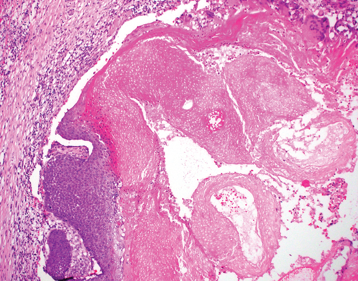
Pilomatricoma, also known as calcifying epithelioma of Malherbe, is a benign pilar tumor that presents as a slowly growing nodule on the head or neck area or arms.13,14 Most pilomatricomas develop by the second decade of life. Multiple lesions may be present in association with myotonic dystrophy or Gardner syndrome among other syndromes.15-17 Similar to PPT, pilomatricomas present as large dermal nodules; however, they tend to be circumscribed and have a trabecular network that consists of basophilic cells and eosinophilic keratinized shadow cells (Figure 1).18 Calcification may be seen and bone formation subsequently may occur.19
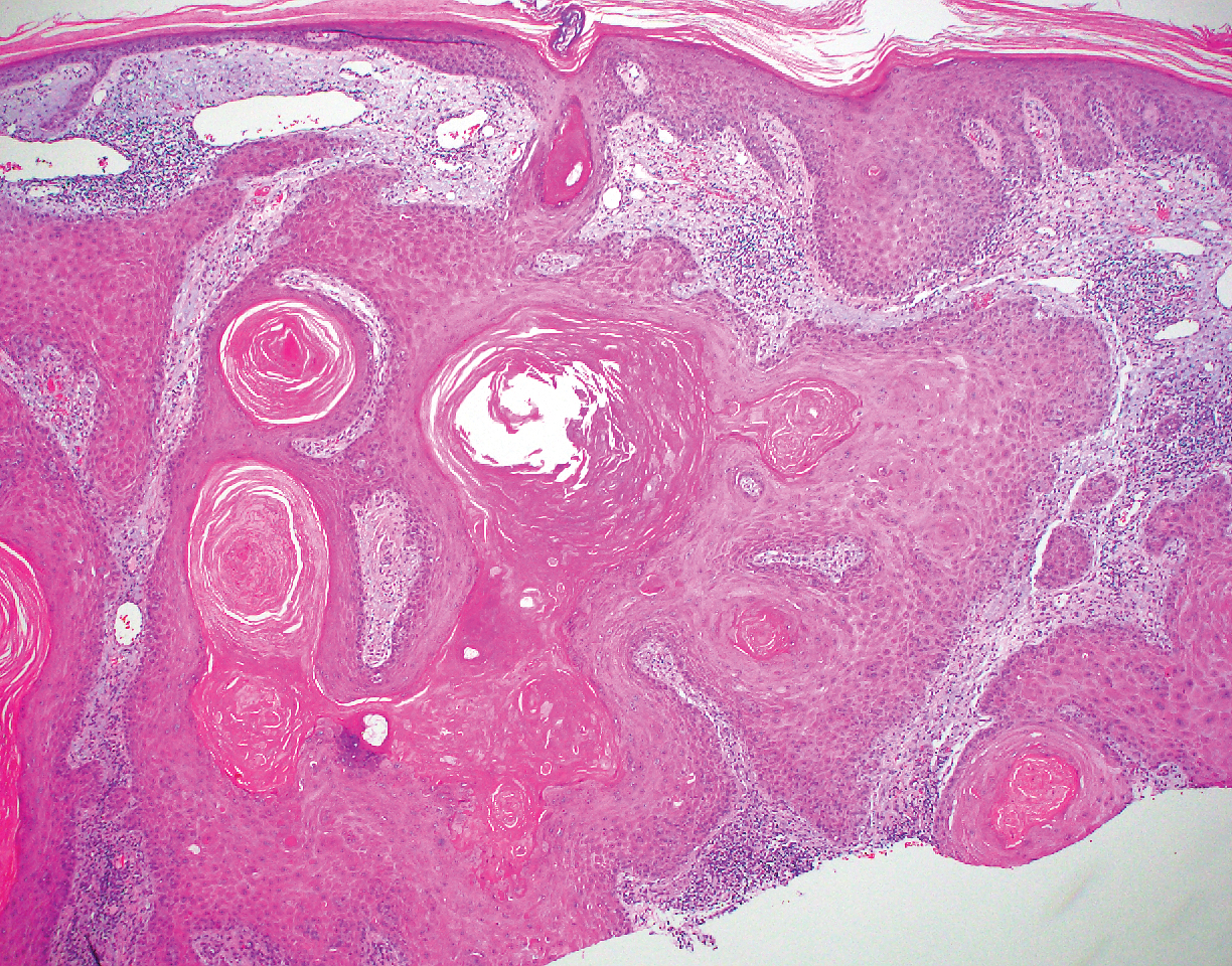
Most sources now consider keratoacanthoma (KA) as a well-differentiated SCC.20 The typical presentation consists of a rapidly growing erythematous to flesh-colored nodule with a central keratinous plug that develops over a period of weeks. If untreated, KAs may resolve over a period of months and leave a depressed scar. Local destruction can result from KAs, and they have the potential to transform into a more aggressive SCC. Accordingly, most clinicians use tissue destructive methods, excision, or Mohs micrographic surgery for treatment based on location. Histologically, a well-circumscribed proliferation of glassy cytoplasm is noted. A depressed keratin-filled center is surrounded by a lip of epithelium extending over the lesion (Figure 2).20,21 Pseudoepitheliomatous hyperplasia accompanied by hypergranulosis is seen in the center of KAs rather than at the periphery, which is typical of non-KA SCCs. Typical KAs lack acantholysis, a feature suggesting a non-KA type of SCC. Neutrophilic microabscesses and eosinophils commonly are seen in KAs.20,21
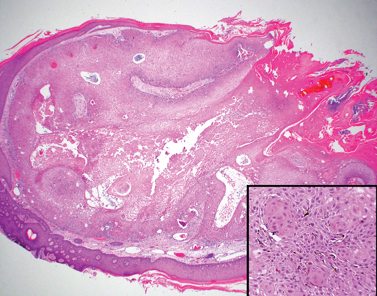
Inverted follicular keratosis is a benign tumor that gained traction as its own entity in the 1960s.22 These lesions typically develop from the follicular infundibulum, but some consider them a version of a wart or seborrheic keratosis.23 They generally are flesh-colored nodules on the upper cutaneous lip or face. Treatment usually consists of complete excision. There are many different growth patterns described, but they typically are endophytic tumors with eosinophilic squamous cells in the center and more basophilic cells at the periphery (Figure 3).24 Characteristically, there are squamous eddies throughout the tumor (Figure 3 [inset]). There also may be a scant lymphohistiocytic infiltrate within the dermis surrounding the lesion.
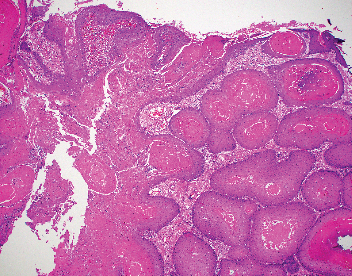
Trichilemmomas are flesh-colored adnexal neoplasms that may present as a solitary lesion or in clusters on the face. They have been reported to occur on all nonglabrous skin sites.25 Multiple lesions may occur in association with Cowden syndrome or with nevus sebaceous.26 A desmoplastic variant of trichilemmomas has been reported.27 Desmoplastic trichilemmomas appear as well-circumscribed tumors of outer root sheath differentiation with lobules extending down into the dermis.28 Vacuolated glycogen-filled keratinocytes are scattered throughout the lesion but are most prominent at the base. At the periphery of the lobules, peripheral palisading of basaloid cells is accompanied by a thickened eosinophilic basement membrane that is periodic acid-Schiff positive. Typical trichilemmomas also can display these features; however, the main differentiating feature of a desmoplastic trichilemmoma is the pink hyalinized stroma separating small islets of basophilic cells (Figure 4). Differentiation from an invasive malignant carcinoma sometimes can be challenging without a focus of typical trichilemmoma or if the biopsy specimen is too superficial.29

Pilar cysts are common tumors that typically arise on the scalp and sometimes are proliferating. Proliferating pilar tumor should be kept on the differential when secondary changes such as ulceration occur in the primary lesion of the scalp. Microscopically, and sometimes clinically, PPT can be difficult to differentiate from other mimickers.
- Wilson-Jones E. Proliferating epidermoid cysts. Arch Dermatol. 1966;94:11-19.
- Saida T, Oohara K, Hori Y, et al. Development of a malignant proliferating trichilemmal cyst in a patient with multiple trichilemmal cysts. Dermatologica. 1983;166:203-208.
- Ye J, Nappi O, Swanson PE, et al. Proliferating pilar tumours: a clinicopathological study of 76 cases with a proposal for definition of benign and malignant variants. Am J Clin Pathol. 2004;122:566-574.
- Garg PK, Dangi A, Khurana N, et al. Malignant proliferating trichilemmal cyst: a case report with review of literature. Malaysian J Pathol. 2009;31:71-76.
- Herrero J, Monteagudo C, Ruiz A, et al. Malignant proliferating trichilemmal tumors: a histopathological and immunohistochemical study of three cases with DNA ploidy and morphometric evaluation. Histopathology. 1998;33:542-546.
- Haas N, Audring H, Sterry W. Carcinoma arising in a proliferating trichilemmal cyst expresses fetal and trichilemmal hair phenotype. Am J Dermatopathol. 2002;24:340-344.
- Rutty GN, Richman PI, Laing JH. Malignant change in trichilemmal cysts: a study of cell proliferation and DNA content. Histopathology. 1992;21:465-468.
- Brownstein MH, Arluk DJ. Proliferating trichilemmal cyst: a simulant of squamous cell carcinoma. Cancer. 1981;48:1207-1214.
- Misago N, Ackerman AB. Tricholemmal carcinoma? Dermatopathol Pract Concept. 1999;5:205-206.
- Misago N, Narisawa Y. Tricholemmal carcinoma in continuity with trichoblastoma within nevus sebaceous. Am J Dermatopathol. 2002;24:149-155.
- Liang H, Wu H, Giorgadze TA, et al. Podoplanin is a highly sensitive and specific marker to distinguish primary skin adnexal carcinomas from adenocarcinomas metastatic to skin. Am J Surg Pathol. 2007;31:304-310.
- Swanson PE, Marrogi AJ, Williams DJ, et al. Trichilemmal carcinoma: clinicopathologic study of 10 cases. J Cutan Pathol. 1992;19:100-109.
- Mehregan AH. Hair follicle tumors of the skin. J Cutan Pathol. 1985;12:189-195.
- Julian CG, Bowers PW. A clinical review of 209 pilomatricomas. J Am Acad Dermatol. 1998;39:191-195.
- Marrogi AJ, Wick MR, Dehner LP. Pilomatrical neoplasms in children and young adults. Am J Dermatopathol. 1992;14:87-94.
- Berberian BJ, Colonna TM, Battaglia M, et al. Multiple pilomatricomas in association with myotonic dystrophy and a family history of melanoma. J Am Acad Dermatol. 1997;37:268-269.
- Cooper PH, Fechner RE. Pilomatricoma-like changes in the epidermal cysts of Gardner's syndrome. J Am Acad Dermatol. 1983;8:639-644.
- Kaddu S, Soyer HP, Cerroni L, et al. Clinical and histopathologic spectrum of pilomatricomas in adults. Int J Dermatol. 1994;33:705-708.
- Sano Y, Mihara M, Miyamoto T, et al. Simultaneous occurrence of calcification and amyloid deposit in pilomatricoma. Acta Derm Venereol. 1990;70:256-259.
- Schwartz RA. Keratoacanthoma. J Am Acad Dermatol. 1994;30:1-19.
- Kwiek B, Schwartz RA. Keratoacanthoma (KA): an update and review. J Am Acad Dermatol. 2016;74:1220-1233.
- Mehregan AH. Inverted follicular keratosis. Arch Dermatol. 1964;89:117-123.
- Spielvogel RL, Austin C, Ackerman AB. Inverted follicular keratosis is not a specific keratosis but a verruca vulgaris (or seborrheic keratosis) with squamous eddies. Am J Dermatopathol. 1983;5:427-445.
- Mehregan AH. Inverted follicular keratosis is a distinct follicular tumor. Am J Dermatopathol. 1983;5:467-470.
- Brownstein MH. Trichilemmoma. benign follicular tumor or viral wart? Am J Dermatopathol. 1980;2:229-231.
- Brownstein MH. Multiple trichilemmomas in Cowden's syndrome. Arch Dermatol. 1979;115:111.
- Roson E, Gomez Centeno P, Sanchez Aguilar D, et al. Desmoplastic trichilemmoma arising within a nevus sebaceous. Am J Dermatopathol. 1998;20:495-497.
- Tellechea O, Reis JP, Baptista AP. Desmoplastic trichilemmoma. Am J Dermatopathol. 1992;14:107-114.
- Sharma R, Sirohi D, Sengupta P, et al. Desmoplastic trichilemmoma of the facial region mimicking invasive carcinoma. J Maxillofac Oral Surg. 2010;10:71-73.
The Diagnosis: Proliferating Pilar Tumor
Proliferating pilar tumor (PPT), or cyst, is a neoplasm of trichilemmal keratinization first described by Wilson-Jones1 in 1966. Proliferating pilar tumors lie on a spectrum with malignant PPT, which is a rare adnexal neoplasm first described by Saida et al2 in 1983. The incidence of PPT is unknown given the paucity of cases and the possible misdiagnosis as squamous cell carcinoma (SCC). Proliferating pilar tumors tend to present on the head and neck of older females as a multilobular and sometimes ulcerating nodule.3 Although PPT can occur de novo, the majority of cases are thought to develop progressively from a benign pilar cyst. Histopathologically, PPT is characterized by cords and nests of squamous cells that display trichilemmal keratinization (quiz images).
Classification of PPT as benign or malignant is challenging, though criteria have been proposed.3-7 Lesions with minimal infiltration into the surrounding dermis and scant mitosis typically behave in a benign manner, while lesions showing nuclear atypia, atypical mitosis, and irregular infiltration into the surrounding dermis can have up to a 50% locoregional recurrence rate.3 In addition, distinguishing a PPT from an SCC or trichilemmal carcinoma also can be difficult; however, SCC is favored when there is a lack of trichilemmal keratinization or when squamous atypia is present in the adjacent epidermis.8 Trichilemmal carcinoma is a rare tumor that has been questioned as a distinct entity.9-12
Pilomatricoma, also known as calcifying epithelioma of Malherbe, is a benign pilar tumor that presents as a slowly growing nodule on the head or neck area or arms.13,14 Most pilomatricomas develop by the second decade of life. Multiple lesions may be present in association with myotonic dystrophy or Gardner syndrome among other syndromes.15-17 Similar to PPT, pilomatricomas present as large dermal nodules; however, they tend to be circumscribed and have a trabecular network that consists of basophilic cells and eosinophilic keratinized shadow cells (Figure 1).18 Calcification may be seen and bone formation subsequently may occur.19
Most sources now consider keratoacanthoma (KA) as a well-differentiated SCC.20 The typical presentation consists of a rapidly growing erythematous to flesh-colored nodule with a central keratinous plug that develops over a period of weeks. If untreated, KAs may resolve over a period of months and leave a depressed scar. Local destruction can result from KAs, and they have the potential to transform into a more aggressive SCC. Accordingly, most clinicians use tissue destructive methods, excision, or Mohs micrographic surgery for treatment based on location. Histologically, a well-circumscribed proliferation of glassy cytoplasm is noted. A depressed keratin-filled center is surrounded by a lip of epithelium extending over the lesion (Figure 2).20,21 Pseudoepitheliomatous hyperplasia accompanied by hypergranulosis is seen in the center of KAs rather than at the periphery, which is typical of non-KA SCCs. Typical KAs lack acantholysis, a feature suggesting a non-KA type of SCC. Neutrophilic microabscesses and eosinophils commonly are seen in KAs.20,21
Inverted follicular keratosis is a benign tumor that gained traction as its own entity in the 1960s.22 These lesions typically develop from the follicular infundibulum, but some consider them a version of a wart or seborrheic keratosis.23 They generally are flesh-colored nodules on the upper cutaneous lip or face. Treatment usually consists of complete excision. There are many different growth patterns described, but they typically are endophytic tumors with eosinophilic squamous cells in the center and more basophilic cells at the periphery (Figure 3).24 Characteristically, there are squamous eddies throughout the tumor (Figure 3 [inset]). There also may be a scant lymphohistiocytic infiltrate within the dermis surrounding the lesion.
Trichilemmomas are flesh-colored adnexal neoplasms that may present as a solitary lesion or in clusters on the face. They have been reported to occur on all nonglabrous skin sites.25 Multiple lesions may occur in association with Cowden syndrome or with nevus sebaceous.26 A desmoplastic variant of trichilemmomas has been reported.27 Desmoplastic trichilemmomas appear as well-circumscribed tumors of outer root sheath differentiation with lobules extending down into the dermis.28 Vacuolated glycogen-filled keratinocytes are scattered throughout the lesion but are most prominent at the base. At the periphery of the lobules, peripheral palisading of basaloid cells is accompanied by a thickened eosinophilic basement membrane that is periodic acid-Schiff positive. Typical trichilemmomas also can display these features; however, the main differentiating feature of a desmoplastic trichilemmoma is the pink hyalinized stroma separating small islets of basophilic cells (Figure 4). Differentiation from an invasive malignant carcinoma sometimes can be challenging without a focus of typical trichilemmoma or if the biopsy specimen is too superficial.29
Pilar cysts are common tumors that typically arise on the scalp and sometimes are proliferating. Proliferating pilar tumor should be kept on the differential when secondary changes such as ulceration occur in the primary lesion of the scalp. Microscopically, and sometimes clinically, PPT can be difficult to differentiate from other mimickers.
The Diagnosis: Proliferating Pilar Tumor
Proliferating pilar tumor (PPT), or cyst, is a neoplasm of trichilemmal keratinization first described by Wilson-Jones1 in 1966. Proliferating pilar tumors lie on a spectrum with malignant PPT, which is a rare adnexal neoplasm first described by Saida et al2 in 1983. The incidence of PPT is unknown given the paucity of cases and the possible misdiagnosis as squamous cell carcinoma (SCC). Proliferating pilar tumors tend to present on the head and neck of older females as a multilobular and sometimes ulcerating nodule.3 Although PPT can occur de novo, the majority of cases are thought to develop progressively from a benign pilar cyst. Histopathologically, PPT is characterized by cords and nests of squamous cells that display trichilemmal keratinization (quiz images).
Classification of PPT as benign or malignant is challenging, though criteria have been proposed.3-7 Lesions with minimal infiltration into the surrounding dermis and scant mitosis typically behave in a benign manner, while lesions showing nuclear atypia, atypical mitosis, and irregular infiltration into the surrounding dermis can have up to a 50% locoregional recurrence rate.3 In addition, distinguishing a PPT from an SCC or trichilemmal carcinoma also can be difficult; however, SCC is favored when there is a lack of trichilemmal keratinization or when squamous atypia is present in the adjacent epidermis.8 Trichilemmal carcinoma is a rare tumor that has been questioned as a distinct entity.9-12
Pilomatricoma, also known as calcifying epithelioma of Malherbe, is a benign pilar tumor that presents as a slowly growing nodule on the head or neck area or arms.13,14 Most pilomatricomas develop by the second decade of life. Multiple lesions may be present in association with myotonic dystrophy or Gardner syndrome among other syndromes.15-17 Similar to PPT, pilomatricomas present as large dermal nodules; however, they tend to be circumscribed and have a trabecular network that consists of basophilic cells and eosinophilic keratinized shadow cells (Figure 1).18 Calcification may be seen and bone formation subsequently may occur.19
Most sources now consider keratoacanthoma (KA) as a well-differentiated SCC.20 The typical presentation consists of a rapidly growing erythematous to flesh-colored nodule with a central keratinous plug that develops over a period of weeks. If untreated, KAs may resolve over a period of months and leave a depressed scar. Local destruction can result from KAs, and they have the potential to transform into a more aggressive SCC. Accordingly, most clinicians use tissue destructive methods, excision, or Mohs micrographic surgery for treatment based on location. Histologically, a well-circumscribed proliferation of glassy cytoplasm is noted. A depressed keratin-filled center is surrounded by a lip of epithelium extending over the lesion (Figure 2).20,21 Pseudoepitheliomatous hyperplasia accompanied by hypergranulosis is seen in the center of KAs rather than at the periphery, which is typical of non-KA SCCs. Typical KAs lack acantholysis, a feature suggesting a non-KA type of SCC. Neutrophilic microabscesses and eosinophils commonly are seen in KAs.20,21
Inverted follicular keratosis is a benign tumor that gained traction as its own entity in the 1960s.22 These lesions typically develop from the follicular infundibulum, but some consider them a version of a wart or seborrheic keratosis.23 They generally are flesh-colored nodules on the upper cutaneous lip or face. Treatment usually consists of complete excision. There are many different growth patterns described, but they typically are endophytic tumors with eosinophilic squamous cells in the center and more basophilic cells at the periphery (Figure 3).24 Characteristically, there are squamous eddies throughout the tumor (Figure 3 [inset]). There also may be a scant lymphohistiocytic infiltrate within the dermis surrounding the lesion.
Trichilemmomas are flesh-colored adnexal neoplasms that may present as a solitary lesion or in clusters on the face. They have been reported to occur on all nonglabrous skin sites.25 Multiple lesions may occur in association with Cowden syndrome or with nevus sebaceous.26 A desmoplastic variant of trichilemmomas has been reported.27 Desmoplastic trichilemmomas appear as well-circumscribed tumors of outer root sheath differentiation with lobules extending down into the dermis.28 Vacuolated glycogen-filled keratinocytes are scattered throughout the lesion but are most prominent at the base. At the periphery of the lobules, peripheral palisading of basaloid cells is accompanied by a thickened eosinophilic basement membrane that is periodic acid-Schiff positive. Typical trichilemmomas also can display these features; however, the main differentiating feature of a desmoplastic trichilemmoma is the pink hyalinized stroma separating small islets of basophilic cells (Figure 4). Differentiation from an invasive malignant carcinoma sometimes can be challenging without a focus of typical trichilemmoma or if the biopsy specimen is too superficial.29
Pilar cysts are common tumors that typically arise on the scalp and sometimes are proliferating. Proliferating pilar tumor should be kept on the differential when secondary changes such as ulceration occur in the primary lesion of the scalp. Microscopically, and sometimes clinically, PPT can be difficult to differentiate from other mimickers.
- Wilson-Jones E. Proliferating epidermoid cysts. Arch Dermatol. 1966;94:11-19.
- Saida T, Oohara K, Hori Y, et al. Development of a malignant proliferating trichilemmal cyst in a patient with multiple trichilemmal cysts. Dermatologica. 1983;166:203-208.
- Ye J, Nappi O, Swanson PE, et al. Proliferating pilar tumours: a clinicopathological study of 76 cases with a proposal for definition of benign and malignant variants. Am J Clin Pathol. 2004;122:566-574.
- Garg PK, Dangi A, Khurana N, et al. Malignant proliferating trichilemmal cyst: a case report with review of literature. Malaysian J Pathol. 2009;31:71-76.
- Herrero J, Monteagudo C, Ruiz A, et al. Malignant proliferating trichilemmal tumors: a histopathological and immunohistochemical study of three cases with DNA ploidy and morphometric evaluation. Histopathology. 1998;33:542-546.
- Haas N, Audring H, Sterry W. Carcinoma arising in a proliferating trichilemmal cyst expresses fetal and trichilemmal hair phenotype. Am J Dermatopathol. 2002;24:340-344.
- Rutty GN, Richman PI, Laing JH. Malignant change in trichilemmal cysts: a study of cell proliferation and DNA content. Histopathology. 1992;21:465-468.
- Brownstein MH, Arluk DJ. Proliferating trichilemmal cyst: a simulant of squamous cell carcinoma. Cancer. 1981;48:1207-1214.
- Misago N, Ackerman AB. Tricholemmal carcinoma? Dermatopathol Pract Concept. 1999;5:205-206.
- Misago N, Narisawa Y. Tricholemmal carcinoma in continuity with trichoblastoma within nevus sebaceous. Am J Dermatopathol. 2002;24:149-155.
- Liang H, Wu H, Giorgadze TA, et al. Podoplanin is a highly sensitive and specific marker to distinguish primary skin adnexal carcinomas from adenocarcinomas metastatic to skin. Am J Surg Pathol. 2007;31:304-310.
- Swanson PE, Marrogi AJ, Williams DJ, et al. Trichilemmal carcinoma: clinicopathologic study of 10 cases. J Cutan Pathol. 1992;19:100-109.
- Mehregan AH. Hair follicle tumors of the skin. J Cutan Pathol. 1985;12:189-195.
- Julian CG, Bowers PW. A clinical review of 209 pilomatricomas. J Am Acad Dermatol. 1998;39:191-195.
- Marrogi AJ, Wick MR, Dehner LP. Pilomatrical neoplasms in children and young adults. Am J Dermatopathol. 1992;14:87-94.
- Berberian BJ, Colonna TM, Battaglia M, et al. Multiple pilomatricomas in association with myotonic dystrophy and a family history of melanoma. J Am Acad Dermatol. 1997;37:268-269.
- Cooper PH, Fechner RE. Pilomatricoma-like changes in the epidermal cysts of Gardner's syndrome. J Am Acad Dermatol. 1983;8:639-644.
- Kaddu S, Soyer HP, Cerroni L, et al. Clinical and histopathologic spectrum of pilomatricomas in adults. Int J Dermatol. 1994;33:705-708.
- Sano Y, Mihara M, Miyamoto T, et al. Simultaneous occurrence of calcification and amyloid deposit in pilomatricoma. Acta Derm Venereol. 1990;70:256-259.
- Schwartz RA. Keratoacanthoma. J Am Acad Dermatol. 1994;30:1-19.
- Kwiek B, Schwartz RA. Keratoacanthoma (KA): an update and review. J Am Acad Dermatol. 2016;74:1220-1233.
- Mehregan AH. Inverted follicular keratosis. Arch Dermatol. 1964;89:117-123.
- Spielvogel RL, Austin C, Ackerman AB. Inverted follicular keratosis is not a specific keratosis but a verruca vulgaris (or seborrheic keratosis) with squamous eddies. Am J Dermatopathol. 1983;5:427-445.
- Mehregan AH. Inverted follicular keratosis is a distinct follicular tumor. Am J Dermatopathol. 1983;5:467-470.
- Brownstein MH. Trichilemmoma. benign follicular tumor or viral wart? Am J Dermatopathol. 1980;2:229-231.
- Brownstein MH. Multiple trichilemmomas in Cowden's syndrome. Arch Dermatol. 1979;115:111.
- Roson E, Gomez Centeno P, Sanchez Aguilar D, et al. Desmoplastic trichilemmoma arising within a nevus sebaceous. Am J Dermatopathol. 1998;20:495-497.
- Tellechea O, Reis JP, Baptista AP. Desmoplastic trichilemmoma. Am J Dermatopathol. 1992;14:107-114.
- Sharma R, Sirohi D, Sengupta P, et al. Desmoplastic trichilemmoma of the facial region mimicking invasive carcinoma. J Maxillofac Oral Surg. 2010;10:71-73.
- Wilson-Jones E. Proliferating epidermoid cysts. Arch Dermatol. 1966;94:11-19.
- Saida T, Oohara K, Hori Y, et al. Development of a malignant proliferating trichilemmal cyst in a patient with multiple trichilemmal cysts. Dermatologica. 1983;166:203-208.
- Ye J, Nappi O, Swanson PE, et al. Proliferating pilar tumours: a clinicopathological study of 76 cases with a proposal for definition of benign and malignant variants. Am J Clin Pathol. 2004;122:566-574.
- Garg PK, Dangi A, Khurana N, et al. Malignant proliferating trichilemmal cyst: a case report with review of literature. Malaysian J Pathol. 2009;31:71-76.
- Herrero J, Monteagudo C, Ruiz A, et al. Malignant proliferating trichilemmal tumors: a histopathological and immunohistochemical study of three cases with DNA ploidy and morphometric evaluation. Histopathology. 1998;33:542-546.
- Haas N, Audring H, Sterry W. Carcinoma arising in a proliferating trichilemmal cyst expresses fetal and trichilemmal hair phenotype. Am J Dermatopathol. 2002;24:340-344.
- Rutty GN, Richman PI, Laing JH. Malignant change in trichilemmal cysts: a study of cell proliferation and DNA content. Histopathology. 1992;21:465-468.
- Brownstein MH, Arluk DJ. Proliferating trichilemmal cyst: a simulant of squamous cell carcinoma. Cancer. 1981;48:1207-1214.
- Misago N, Ackerman AB. Tricholemmal carcinoma? Dermatopathol Pract Concept. 1999;5:205-206.
- Misago N, Narisawa Y. Tricholemmal carcinoma in continuity with trichoblastoma within nevus sebaceous. Am J Dermatopathol. 2002;24:149-155.
- Liang H, Wu H, Giorgadze TA, et al. Podoplanin is a highly sensitive and specific marker to distinguish primary skin adnexal carcinomas from adenocarcinomas metastatic to skin. Am J Surg Pathol. 2007;31:304-310.
- Swanson PE, Marrogi AJ, Williams DJ, et al. Trichilemmal carcinoma: clinicopathologic study of 10 cases. J Cutan Pathol. 1992;19:100-109.
- Mehregan AH. Hair follicle tumors of the skin. J Cutan Pathol. 1985;12:189-195.
- Julian CG, Bowers PW. A clinical review of 209 pilomatricomas. J Am Acad Dermatol. 1998;39:191-195.
- Marrogi AJ, Wick MR, Dehner LP. Pilomatrical neoplasms in children and young adults. Am J Dermatopathol. 1992;14:87-94.
- Berberian BJ, Colonna TM, Battaglia M, et al. Multiple pilomatricomas in association with myotonic dystrophy and a family history of melanoma. J Am Acad Dermatol. 1997;37:268-269.
- Cooper PH, Fechner RE. Pilomatricoma-like changes in the epidermal cysts of Gardner's syndrome. J Am Acad Dermatol. 1983;8:639-644.
- Kaddu S, Soyer HP, Cerroni L, et al. Clinical and histopathologic spectrum of pilomatricomas in adults. Int J Dermatol. 1994;33:705-708.
- Sano Y, Mihara M, Miyamoto T, et al. Simultaneous occurrence of calcification and amyloid deposit in pilomatricoma. Acta Derm Venereol. 1990;70:256-259.
- Schwartz RA. Keratoacanthoma. J Am Acad Dermatol. 1994;30:1-19.
- Kwiek B, Schwartz RA. Keratoacanthoma (KA): an update and review. J Am Acad Dermatol. 2016;74:1220-1233.
- Mehregan AH. Inverted follicular keratosis. Arch Dermatol. 1964;89:117-123.
- Spielvogel RL, Austin C, Ackerman AB. Inverted follicular keratosis is not a specific keratosis but a verruca vulgaris (or seborrheic keratosis) with squamous eddies. Am J Dermatopathol. 1983;5:427-445.
- Mehregan AH. Inverted follicular keratosis is a distinct follicular tumor. Am J Dermatopathol. 1983;5:467-470.
- Brownstein MH. Trichilemmoma. benign follicular tumor or viral wart? Am J Dermatopathol. 1980;2:229-231.
- Brownstein MH. Multiple trichilemmomas in Cowden's syndrome. Arch Dermatol. 1979;115:111.
- Roson E, Gomez Centeno P, Sanchez Aguilar D, et al. Desmoplastic trichilemmoma arising within a nevus sebaceous. Am J Dermatopathol. 1998;20:495-497.
- Tellechea O, Reis JP, Baptista AP. Desmoplastic trichilemmoma. Am J Dermatopathol. 1992;14:107-114.
- Sharma R, Sirohi D, Sengupta P, et al. Desmoplastic trichilemmoma of the facial region mimicking invasive carcinoma. J Maxillofac Oral Surg. 2010;10:71-73.
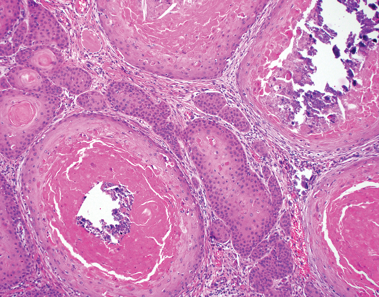
A 66-year-old woman presented to the dermatology clinic with a rapidly enlarging, draining lesion on the scalp. The lesion seemed to enlarge over the last 3 months from a lesion that had been there for years. Physical examination revealed a 2.2-cm ulcerated nodule on the right parietal scalp. A shave biopsy was obtained.
A System View of Primary Care and the Role of the Family Physician
Diabetes boosts bariatric surgery complications
LAS VEGAS – compared with patients without diabetes, in an analysis of more than 550,000 patients who underwent this surgery.

The analysis also showed that among patients with diabetes undergoing bariatric surgery, those with insulin-dependent diabetes had significantly higher rates of these and other complications, compared with patients with diabetes but no insulin dependence, Andrew A. Wheeler, MD, said at a meeting presented by The Obesity Society and the American Society for Metabolic and Bariatric Surgery.
The specific factors driving these observations remain unknown, but may relate at least in part to the efficacy of glycemic control in patients during the months and weeks before surgery, said Dr. Wheeler, chief of metabolic and bariatric surgery at the University of Missouri in Columbia.
“Perioperative blood glucose control reflected in hemoglobin A1c may help us understand if certain patients with diabetes are at increased risk of surgical complications,” Dr. Wheeler said during his talk. “We need to see whether preoperative hemoglobin A1c tracks with the rate of complications,” he added in an interview. “Some surgeons will defer surgery until the level goes down, but right now, everyone uses a different level.”
The study by Dr. Wheeler and associates used data collected from essentially all the bariatric surgeries done at the roughly 840 U.S. centers accredited to do the surgery during 2015-2017 – a total of 555,239 patients – in the Metabolic and Bariatric Surgery Accreditation and Quality Improvement Program run by the American College of Surgeons and the American Society for Metabolic and Bariatric Surgery. The cohort included 413,239 patients without diabetes and 136,010 with diabetes, with the remainder having an unknown status. Nearly all patients underwent either sleeve gastrectomy or gastric bypass, with significantly more patients, 74%, having sleeve gastrectomy in the group without diabetes, and 62% of those with diabetes undergoing sleeve gastrectomy.
Patients with diabetes uniformly had significantly higher rates of complications as measured by several parameters, and among those with diabetes, the complication rates were highest in those with insulin dependence. The researchers ran a multivariate analysis that controlled for many demographic and clinical variables, and found that despite these corrections, patients with diabetes, and especially those with insulin dependence, had higher relative rates of many complications that were statistically significant. In addition to the increased rates of deep surgical-site infections and 30-day ICU admissions, patients with diabetes had a 54% relatively increased rate of wound disruption, and a 29% increased relative rate of superficial surgical-site infections, compared with those without diabetes. And among patients with diabetes, those with insulin dependence had a 40% increased rate of surgical-site infections, a 74% increase in progressive renal failure, and a 39% increased rate of hospital readmission relative to patients with diabetes that was not insulin dependent, Dr. Wheeler reported.
The absolute complication rates were, in general, low. For example, the total rate of superficial plus deep surgical-site infections was less than 1% both in patients with diabetes and those without, although the rate rose above 1% in patients with insulin-dependent diabetes. The most common complication reported by Dr. Wheeler was 30-day hospital readmission, which was 4.0% in patients without diabetes, 4.8% in those with diabetes, and 6.4% in patients with insulin-dependent diabetes. The data also showed that gastric bypass led to more complications than did sleeve gastrectomy, but complications with both types of surgeries increased in patients with diabetes, compared with those without diabetes.
Dr. Wheeler had no disclosures.
SOURCE: Wheeler AA et al. Obesity Week 2019, Abstract A133.
Most bariatric surgeons believe that patients with diabetes who undergo this surgery will have more complications than will patients without diabetes, based on their anecdotal experience and findings from previous, smaller studies. But in Dr. Wheeler’s report, we see this demonstrated in an incredibly large number of patients. This is one of the first studies to use 3 years of data from the Metabolic and Bariatric Surgery Accreditation and Quality Improvement Program, which included more than half a million patients.

A randomized study involving more than 18,000 cases showed that putting bariatric surgery patients on an “enhanced recovery program” before surgery and including measures that minimized insulin resistance and catabolism perioperatively led to a substantial reduction in extended length of stay (Surg Obes Relat Dis. 2019 Nov;15[11]:1977-89). In routine care, bariatric surgeons are now often routinely using “prehabilitation” to prepare patients for surgery.
Corrigan McBride, MD , is professor of surgery and director of bariatric surgery at the University of Nebraska Medical Center in Omaha. She had no disclosures. She made these comments in an interview.
Most bariatric surgeons believe that patients with diabetes who undergo this surgery will have more complications than will patients without diabetes, based on their anecdotal experience and findings from previous, smaller studies. But in Dr. Wheeler’s report, we see this demonstrated in an incredibly large number of patients. This is one of the first studies to use 3 years of data from the Metabolic and Bariatric Surgery Accreditation and Quality Improvement Program, which included more than half a million patients.
A randomized study involving more than 18,000 cases showed that putting bariatric surgery patients on an “enhanced recovery program” before surgery and including measures that minimized insulin resistance and catabolism perioperatively led to a substantial reduction in extended length of stay (Surg Obes Relat Dis. 2019 Nov;15[11]:1977-89). In routine care, bariatric surgeons are now often routinely using “prehabilitation” to prepare patients for surgery.
Corrigan McBride, MD , is professor of surgery and director of bariatric surgery at the University of Nebraska Medical Center in Omaha. She had no disclosures. She made these comments in an interview.
Most bariatric surgeons believe that patients with diabetes who undergo this surgery will have more complications than will patients without diabetes, based on their anecdotal experience and findings from previous, smaller studies. But in Dr. Wheeler’s report, we see this demonstrated in an incredibly large number of patients. This is one of the first studies to use 3 years of data from the Metabolic and Bariatric Surgery Accreditation and Quality Improvement Program, which included more than half a million patients.
A randomized study involving more than 18,000 cases showed that putting bariatric surgery patients on an “enhanced recovery program” before surgery and including measures that minimized insulin resistance and catabolism perioperatively led to a substantial reduction in extended length of stay (Surg Obes Relat Dis. 2019 Nov;15[11]:1977-89). In routine care, bariatric surgeons are now often routinely using “prehabilitation” to prepare patients for surgery.
Corrigan McBride, MD , is professor of surgery and director of bariatric surgery at the University of Nebraska Medical Center in Omaha. She had no disclosures. She made these comments in an interview.
LAS VEGAS – compared with patients without diabetes, in an analysis of more than 550,000 patients who underwent this surgery.
The analysis also showed that among patients with diabetes undergoing bariatric surgery, those with insulin-dependent diabetes had significantly higher rates of these and other complications, compared with patients with diabetes but no insulin dependence, Andrew A. Wheeler, MD, said at a meeting presented by The Obesity Society and the American Society for Metabolic and Bariatric Surgery.
The specific factors driving these observations remain unknown, but may relate at least in part to the efficacy of glycemic control in patients during the months and weeks before surgery, said Dr. Wheeler, chief of metabolic and bariatric surgery at the University of Missouri in Columbia.
“Perioperative blood glucose control reflected in hemoglobin A1c may help us understand if certain patients with diabetes are at increased risk of surgical complications,” Dr. Wheeler said during his talk. “We need to see whether preoperative hemoglobin A1c tracks with the rate of complications,” he added in an interview. “Some surgeons will defer surgery until the level goes down, but right now, everyone uses a different level.”
The study by Dr. Wheeler and associates used data collected from essentially all the bariatric surgeries done at the roughly 840 U.S. centers accredited to do the surgery during 2015-2017 – a total of 555,239 patients – in the Metabolic and Bariatric Surgery Accreditation and Quality Improvement Program run by the American College of Surgeons and the American Society for Metabolic and Bariatric Surgery. The cohort included 413,239 patients without diabetes and 136,010 with diabetes, with the remainder having an unknown status. Nearly all patients underwent either sleeve gastrectomy or gastric bypass, with significantly more patients, 74%, having sleeve gastrectomy in the group without diabetes, and 62% of those with diabetes undergoing sleeve gastrectomy.
Patients with diabetes uniformly had significantly higher rates of complications as measured by several parameters, and among those with diabetes, the complication rates were highest in those with insulin dependence. The researchers ran a multivariate analysis that controlled for many demographic and clinical variables, and found that despite these corrections, patients with diabetes, and especially those with insulin dependence, had higher relative rates of many complications that were statistically significant. In addition to the increased rates of deep surgical-site infections and 30-day ICU admissions, patients with diabetes had a 54% relatively increased rate of wound disruption, and a 29% increased relative rate of superficial surgical-site infections, compared with those without diabetes. And among patients with diabetes, those with insulin dependence had a 40% increased rate of surgical-site infections, a 74% increase in progressive renal failure, and a 39% increased rate of hospital readmission relative to patients with diabetes that was not insulin dependent, Dr. Wheeler reported.
The absolute complication rates were, in general, low. For example, the total rate of superficial plus deep surgical-site infections was less than 1% both in patients with diabetes and those without, although the rate rose above 1% in patients with insulin-dependent diabetes. The most common complication reported by Dr. Wheeler was 30-day hospital readmission, which was 4.0% in patients without diabetes, 4.8% in those with diabetes, and 6.4% in patients with insulin-dependent diabetes. The data also showed that gastric bypass led to more complications than did sleeve gastrectomy, but complications with both types of surgeries increased in patients with diabetes, compared with those without diabetes.
Dr. Wheeler had no disclosures.
SOURCE: Wheeler AA et al. Obesity Week 2019, Abstract A133.
LAS VEGAS – compared with patients without diabetes, in an analysis of more than 550,000 patients who underwent this surgery.
The analysis also showed that among patients with diabetes undergoing bariatric surgery, those with insulin-dependent diabetes had significantly higher rates of these and other complications, compared with patients with diabetes but no insulin dependence, Andrew A. Wheeler, MD, said at a meeting presented by The Obesity Society and the American Society for Metabolic and Bariatric Surgery.
The specific factors driving these observations remain unknown, but may relate at least in part to the efficacy of glycemic control in patients during the months and weeks before surgery, said Dr. Wheeler, chief of metabolic and bariatric surgery at the University of Missouri in Columbia.
“Perioperative blood glucose control reflected in hemoglobin A1c may help us understand if certain patients with diabetes are at increased risk of surgical complications,” Dr. Wheeler said during his talk. “We need to see whether preoperative hemoglobin A1c tracks with the rate of complications,” he added in an interview. “Some surgeons will defer surgery until the level goes down, but right now, everyone uses a different level.”
The study by Dr. Wheeler and associates used data collected from essentially all the bariatric surgeries done at the roughly 840 U.S. centers accredited to do the surgery during 2015-2017 – a total of 555,239 patients – in the Metabolic and Bariatric Surgery Accreditation and Quality Improvement Program run by the American College of Surgeons and the American Society for Metabolic and Bariatric Surgery. The cohort included 413,239 patients without diabetes and 136,010 with diabetes, with the remainder having an unknown status. Nearly all patients underwent either sleeve gastrectomy or gastric bypass, with significantly more patients, 74%, having sleeve gastrectomy in the group without diabetes, and 62% of those with diabetes undergoing sleeve gastrectomy.
Patients with diabetes uniformly had significantly higher rates of complications as measured by several parameters, and among those with diabetes, the complication rates were highest in those with insulin dependence. The researchers ran a multivariate analysis that controlled for many demographic and clinical variables, and found that despite these corrections, patients with diabetes, and especially those with insulin dependence, had higher relative rates of many complications that were statistically significant. In addition to the increased rates of deep surgical-site infections and 30-day ICU admissions, patients with diabetes had a 54% relatively increased rate of wound disruption, and a 29% increased relative rate of superficial surgical-site infections, compared with those without diabetes. And among patients with diabetes, those with insulin dependence had a 40% increased rate of surgical-site infections, a 74% increase in progressive renal failure, and a 39% increased rate of hospital readmission relative to patients with diabetes that was not insulin dependent, Dr. Wheeler reported.
The absolute complication rates were, in general, low. For example, the total rate of superficial plus deep surgical-site infections was less than 1% both in patients with diabetes and those without, although the rate rose above 1% in patients with insulin-dependent diabetes. The most common complication reported by Dr. Wheeler was 30-day hospital readmission, which was 4.0% in patients without diabetes, 4.8% in those with diabetes, and 6.4% in patients with insulin-dependent diabetes. The data also showed that gastric bypass led to more complications than did sleeve gastrectomy, but complications with both types of surgeries increased in patients with diabetes, compared with those without diabetes.
Dr. Wheeler had no disclosures.
SOURCE: Wheeler AA et al. Obesity Week 2019, Abstract A133.
REPORTING FROM OBESITY WEEK 2019
AGA News
AGA’s flagship research grant goes to ...
The AGA Research Scholar Award, funded by the AGA Research Foundation, is our premier funding mechanism, providing $100,000 per year for 3 years to early-career faculty working toward independent careers in digestive disease research. Our AGA Research Scholar Award recipients have a proven track record of receiving substantial funding and leadership roles in GI following the receipt of their AGA award. Read about our most recent class of RSA recipients – we’re confident they are future leaders in our field. Learn more about the AGA Research Foundation at www.gastro.org/foundation.
Parambir Dulai, MD
University of California, San Diego

Project title: Development and validation of machine learning optimized predictive models for response to different biologic agents in patients with Crohn’s disease and ulcerative colitis.
Dr. Dulai is using his grant to build and refine a decision-support platform to help providers and patients navigate the complex landscape of choosing between available biologics for the treatment of inflammatory bowel disease (IBD).
Amy Hemperly, DO
University of California, San Diego
AGA-Rady Children’s Institute for Genomic Medicine Research Scholar Award in Pediatric Genomics

Project title: Integration of pharmacogenomics and pharmacometabolomics with pharmacokinetics for biomarker discovery in pediatric inflammatory bowel disease
Dr. Hemperly’s research assesses the influence of genetic variations and metabolic and microbial changes on response to anti–tumor necrosis factor (anti-TNF) therapy in pediatric IBD patients. This work will ultimately elucidate factors that improve a patient’s response to therapy.
Rodney Infante, MD, PhD
University of Texas Southwestern Medical Center, Dallas

Project title: Regulation of gastrointestinal cancer cachexia by a tumor-adipose-hypothalamic axis
Dr. Infante and his lab will use the AGA grant to improve our understanding of the mechanism and clinical relevance of cachexia-associated anorexia and tissue wasting in order to identify effective therapeutic targets.
Suraj Patel, MD, PhD
Massachusetts General Hospital, Boston

Project title: Hepatic IRF3 is a transcriptional regulator of steatosis and insulin resistance in NAFLD
Dr. Patel’s research focuses on the role of innate immunity in cellular metabolism and insulin resistance. Specifically, he’s interested in determining how chronic inflammation fuels the genetic and epigenetic changes we see in overnutritional states such as nonalcoholic fatty liver disease (NAFLD).
Jason Pitarresi, PhD
University of Pennsylvania Health System, Philadelphia
AGA-Bern Schwartz Family Fund Research Scholar Award in Pancreatic Cancer
Project title: PTHLH drives epithelial-to-mesenchymal transition and metastasis in pancreatic cancer
With this funding, Dr. Pitarresi will continue on his quest to identify novel drivers of pancreatic cancer development and metastasis with use of genetically engineered mouse models and patient-derived 3D organoids. Dr. Pitarresi is hoping that anti-PTHLH may fill a treatment void and ultimately increase the quality of life in these patients.
Eric Shah, MD, MBA
Dartmouth-Hitchcock Medical Center, Lebanon, N.H.
AGA-Shire Research Scholar Award in Functional GI and Motility Disorders*

Project title: Office-based anorectal testing to diagnose evacuation disorders and predict outcomes with biofeedback therapy: The rectal expulsion device (RED)
Dr. Shah’s research aims to validate a diagnostic test to triage patients with chronic constipation to the most effective treatment in general GI practice. This work will ultimately help patients with motility and functional bowel conditions and their providers reach a confident diagnosis and understand their treatment options.
*Funded by Shire Plc, now part of Takeda
Shailja Shah, MD, MPH
Vanderbilt University Medical Center, Nashville, Tenn.

Project title: Defining host-specific genetic and non-genetic determinants of Helicobacter pylori eradication failure using a large prospective cohort and genomic biobank
Dr. Shah’s research is focused on personalizing the clinical management of H. pylori such that eradication efforts can be optimized and targeted to the less than 1-3% of the estimated 4.4 billion individuals infected with H. pylori who are most at risk for complications, such as gastric cancer, and avoided in those who are unlikely to benefit and may even experience harm from eradication therapy.
Xiao Tan, MD, PhD
Massachusetts General Hospital, Boston
AGA-Takeda Pharmaceuticals Research Scholar Award in Inflammatory Bowel Disease

Project title: Paper-based diagnostics of microbial and host biomarkers to predict responsiveness to IBD therapy
Dr. Tan will develop low-cost, point-of-care microbiome diagnostics to ultimately help physicians’ make diagnoses, monitor, and select treatment for patients with IBD.
Michael Thompson, MD, PhD
Washington University, Saint Louis, Mo.
Project title: Mechanisms of altered bile acid homeostasis and non-alcoholic fatty liver disease in offspring exposed to maternal obesity
Dr. Thompson’s research is focused on how perinatal exposures impact risk for metabolic liver disease in offspring.
My day on Capitol Hill
By Richard K. Sterling, MD, MSC, AGAF
When initially asked to represent AGA on Capitol Hill for the Global Liver Institute (GLI) congressional briefing on liver cancer and the LIVER Act on Oct. 31, I felt both honored and somewhat frightened. Honored that AGA thought enough of me as a hepatologist to represent them and frightened, not because it was Halloween, but because I would be speaking to members of Congress and their staff on issues that may impact policy and thousands if not millions of Americans. Along with myself, were Donna Cryer, founder and CEO of the GLI; John Groopman, PhD, an epidemiologist from Johns Hopkins focusing on liver disease; and two patients with liver disease who had a compelling story to tell. In addition, our briefing and Capitol Hill advocacy day included patients with a history of liver cancer and members of the Hepatitis B Foundation.

In preparation, Andrew Scott from the GLI helped me in identifying the target audience and in developing slides to present to Congress members and their aides that would show those at risk for liver cancer, the increasing incidence of the disease, and the importance of diagnosis at an early stage when curative treatment options are readily available. Travel and hotel logistics were taken care of by Kathleen Teixeira and AGA staff, and it was comforting to see them in the audience.
The briefing took place in the Cannon office building and was standing room only. After a brief introduction by Andrew Scott, I was the first speaker followed by our patient advocates and Dr. Groopman. The LIVER Act (H.R. 3016) is sponsored by Congresswomen Nydia Velazquez (D-NY) and would drive several public health initiatives that would help people of all ages, lifestyles, and ethnic backgrounds to reduce their risk for liver cancer and related illnesses by enhancing the federal government’s prevention, education, and disease surveillance capabilities while empowering local entities to promote treatment and raise awareness. It also supports increased funding to both the Centers for Disease Control and Prevention and the National Institutes of Health for liver disease and liver cancer research.
We had plenty of time for questions from the audience and I saw a lot of nodding from many present acknowledging that they had friends or family who had liver disease. Although our briefing was happening at the same time as the vote on formalizing the impeachment inquiry (you can hear the buzzing going off and the red lights flashing that the vote was about to happen; see Facebook; HepBFoundation video), congressional staff did not leave.
After the meeting, the patient advocates along with members of the GLI, Hepatitis B Foundation, and others met one-on-one with additional members of congress and their staff. While on the train home, I had time to reflect on the day and hoped that our message would be advanced through congress.
AGA, along with our sister societies (American Association for the Study of Liver Diseases and American College of Gastroenterology) are our voice and advocates for advancing legislation through congress. Days like today allow our members to get involved. It is an exciting way to help our congressional representatives take action on what matters most to us: improved patient care, supporting research, promoting education, and reducing the overall burden to accomplish these important goals.
While some say Virginia is for Lovers, I say Virginia is for Livers (#LoveYourLiver). For more on this and the Liver Biliary Council offerings at Digestive Disease Week, follow me on twitter (@RichSterlingMD).
Dr. Sterling is professor of medicine, chief of hepatology, division of gastroenterology, hepatology and nutrition, Virginia Commonwealth University, Richmond; vice-chair, AGA Liver Biliary Section, DDW Council.
New AGA guideline: Management of GIM
AGA released a new clinical practice guideline in Gastroenterology with recommendations for the management of patients with gastric intestinal metaplasia (GIM) detected as part of routine upper endoscopy for reasons including work up of endoscopically identified gastropathy/presumed gastritis, dyspepsia, or exclusion of Helicobacter pylori.
Guideline recommendations
1. In patients with GIM, AGA recommends testing for H. pylori followed by eradication over no testing and eradication. (Strong recommendation: moderate-quality evidence)
2. In patients with GIM, AGA suggests against routine use of endoscopic surveillance. (Conditional recommendation: very-low-quality evidence)
Comment: Patients with GIM at higher risk for gastric cancer who put a high value on potential but uncertain reduction in gastric cancer mortality, and who put a low value on potential risks of surveillance endoscopies, may reasonably elect for surveillance.
Patients with GIM specifically at higher risk of gastric cancer include those with the following:
- Incomplete versus complete GIM.
- Extensive versus limited GIM.
- Family history of gastric cancer.
Patients at overall increased risk for gastric cancer include the following:
- Racial/ethnic minorities.
- Immigrants from high incidence regions.
3. In patients with GIM, AGA suggests against routine repeat short-interval endoscopy with biopsies for the purpose of risk stratification. (Conditional recommendation: very-low-quality evidence)
Comment: Based on shared decision making, patients with GIM and high-risk stigmata, those with concerns about completeness of baseline endoscopy, and/or those who are at overall increased risk for gastric cancer (racial/ethnic minorities, immigrants from regions with high gastric cancer incidence, or individuals with family history of first-degree relative with gastric cancer) may reasonably elect for repeat endoscopy within 1 year for risk stratification.
This guideline will be published in the February print issue of Gastroenterology with additional resources to help you implement in your practice.
A GI society update on MOC reform
Our work was suspended when American Board of Internal Medicine (ABIM) announced the creation of a new longitudinal assessment option for maintenance of certification across all specialties.
GI society leaders are in touch with ABIM. Here’s an update on what we know: The ABIM board of directors committed to evolve its program to provide a longitudinal assessment option for Maintenance of Certification (MOC), offering a self-paced pathway for physicians to acquire and demonstrate ongoing knowledge. The traditional, long-form assessment will also remain an option because some physicians have expressed a preference for a point-in-time exam taken less frequently.
Our next steps include seeking clarity from ABIM including the following:
1. The milestones in the process to create the new pathway.
2. When the new pathway will be available to diplomates.
3. Consideration and integration of the GI societies’ principles in the development of the new pathway for recertification, including these considerations:
- MOC needs to be simpler, less intrusive, and less expensive.
- We continue to support alternatives to the high-stakes, every-10-year recertification exam.
- We do not support single source or time-limited assessments because they do not represent the current realities of medicine in the digital age.
- We support the concept that, for the many diplomates who specialize within certain areas of gastroenterology and hepatology, MOC should not include high-stakes assessments of areas in which the diplomate may not practice.
- We support the principles of lifelong learning, as evidenced by ongoing CME activities, rather than lifelong testing.
4. The role the GI societies, as representatives for thousands of U.S. members who are ABIM diplomates, play in the creation and implementation of the new pathway.
AASLD, ACG, AGA, and American Society for Gastrointestinal Endoscopy want to be fully informed and fully respected partners in an endeavor that touches upon one of the toughest challenges facing our members and the single issue we hear about most often requesting our help.
We will continue to update our members as we learn the answers to these questions from ABIM.
Together, our first priority on the MOC issue remains ensuring that GI diplomates have a pathway for recertification that meets your needs.
Watch your step (therapy) – understanding ‘fail first’
Sometimes known as “fail first,” step therapy is a tool used by insurance companies that requires patients to fail medications before agreeing to cover a health care provider’s initial treatment recommendation.
Often affecting patients with inflammatory bowel disease (IBD), step therapy focuses on the use of insurer-preferred treatments rather than effective, patient-centric therapies. In addition to causing many patient hardships, this protocol allows insurance companies to come between the provider-patient relationship and dictate a patient’s course of treatment.
To help clinicians navigate this challenging landscape, AGA is pleased to offer a new step therapy webpage that details the step therapy protocol and opportunities to advocate for patient protections.
Additional education modules – including videos, podcasts and other resources – are also available for several states that have implemented safe step therapy laws, including Illinois, New York, and Texas.
Visit the Navigating State Step Therapy Laws program page to learn more about the following:
- What is the step therapy protocol?
- How does step therapy impact a health care provider’s ability to provide patient care?
- Which states have implemented step therapy laws?
- How do state step therapy laws provide physician rights and patient protection?
- Tips to share with your patients.
- What are AGA’s advocacy efforts – and how can I help?
Education modules for additional states will be available in early 2020.
AGA’s Navigating State Step Therapy Laws program is funded by an unrestricted educational grant from Takeda and Pfizer.
AGA’s flagship research grant goes to ...
The AGA Research Scholar Award, funded by the AGA Research Foundation, is our premier funding mechanism, providing $100,000 per year for 3 years to early-career faculty working toward independent careers in digestive disease research. Our AGA Research Scholar Award recipients have a proven track record of receiving substantial funding and leadership roles in GI following the receipt of their AGA award. Read about our most recent class of RSA recipients – we’re confident they are future leaders in our field. Learn more about the AGA Research Foundation at www.gastro.org/foundation.
Parambir Dulai, MD
University of California, San Diego
Project title: Development and validation of machine learning optimized predictive models for response to different biologic agents in patients with Crohn’s disease and ulcerative colitis.
Dr. Dulai is using his grant to build and refine a decision-support platform to help providers and patients navigate the complex landscape of choosing between available biologics for the treatment of inflammatory bowel disease (IBD).
Amy Hemperly, DO
University of California, San Diego
AGA-Rady Children’s Institute for Genomic Medicine Research Scholar Award in Pediatric Genomics
Project title: Integration of pharmacogenomics and pharmacometabolomics with pharmacokinetics for biomarker discovery in pediatric inflammatory bowel disease
Dr. Hemperly’s research assesses the influence of genetic variations and metabolic and microbial changes on response to anti–tumor necrosis factor (anti-TNF) therapy in pediatric IBD patients. This work will ultimately elucidate factors that improve a patient’s response to therapy.
Rodney Infante, MD, PhD
University of Texas Southwestern Medical Center, Dallas
Project title: Regulation of gastrointestinal cancer cachexia by a tumor-adipose-hypothalamic axis
Dr. Infante and his lab will use the AGA grant to improve our understanding of the mechanism and clinical relevance of cachexia-associated anorexia and tissue wasting in order to identify effective therapeutic targets.
Suraj Patel, MD, PhD
Massachusetts General Hospital, Boston
Project title: Hepatic IRF3 is a transcriptional regulator of steatosis and insulin resistance in NAFLD
Dr. Patel’s research focuses on the role of innate immunity in cellular metabolism and insulin resistance. Specifically, he’s interested in determining how chronic inflammation fuels the genetic and epigenetic changes we see in overnutritional states such as nonalcoholic fatty liver disease (NAFLD).
Jason Pitarresi, PhD
University of Pennsylvania Health System, Philadelphia
AGA-Bern Schwartz Family Fund Research Scholar Award in Pancreatic Cancer
Project title: PTHLH drives epithelial-to-mesenchymal transition and metastasis in pancreatic cancer
With this funding, Dr. Pitarresi will continue on his quest to identify novel drivers of pancreatic cancer development and metastasis with use of genetically engineered mouse models and patient-derived 3D organoids. Dr. Pitarresi is hoping that anti-PTHLH may fill a treatment void and ultimately increase the quality of life in these patients.
Eric Shah, MD, MBA
Dartmouth-Hitchcock Medical Center, Lebanon, N.H.
AGA-Shire Research Scholar Award in Functional GI and Motility Disorders*
Project title: Office-based anorectal testing to diagnose evacuation disorders and predict outcomes with biofeedback therapy: The rectal expulsion device (RED)
Dr. Shah’s research aims to validate a diagnostic test to triage patients with chronic constipation to the most effective treatment in general GI practice. This work will ultimately help patients with motility and functional bowel conditions and their providers reach a confident diagnosis and understand their treatment options.
*Funded by Shire Plc, now part of Takeda
Shailja Shah, MD, MPH
Vanderbilt University Medical Center, Nashville, Tenn.
Project title: Defining host-specific genetic and non-genetic determinants of Helicobacter pylori eradication failure using a large prospective cohort and genomic biobank
Dr. Shah’s research is focused on personalizing the clinical management of H. pylori such that eradication efforts can be optimized and targeted to the less than 1-3% of the estimated 4.4 billion individuals infected with H. pylori who are most at risk for complications, such as gastric cancer, and avoided in those who are unlikely to benefit and may even experience harm from eradication therapy.
Xiao Tan, MD, PhD
Massachusetts General Hospital, Boston
AGA-Takeda Pharmaceuticals Research Scholar Award in Inflammatory Bowel Disease
Project title: Paper-based diagnostics of microbial and host biomarkers to predict responsiveness to IBD therapy
Dr. Tan will develop low-cost, point-of-care microbiome diagnostics to ultimately help physicians’ make diagnoses, monitor, and select treatment for patients with IBD.
Michael Thompson, MD, PhD
Washington University, Saint Louis, Mo.
Project title: Mechanisms of altered bile acid homeostasis and non-alcoholic fatty liver disease in offspring exposed to maternal obesity
Dr. Thompson’s research is focused on how perinatal exposures impact risk for metabolic liver disease in offspring.
My day on Capitol Hill
By Richard K. Sterling, MD, MSC, AGAF
When initially asked to represent AGA on Capitol Hill for the Global Liver Institute (GLI) congressional briefing on liver cancer and the LIVER Act on Oct. 31, I felt both honored and somewhat frightened. Honored that AGA thought enough of me as a hepatologist to represent them and frightened, not because it was Halloween, but because I would be speaking to members of Congress and their staff on issues that may impact policy and thousands if not millions of Americans. Along with myself, were Donna Cryer, founder and CEO of the GLI; John Groopman, PhD, an epidemiologist from Johns Hopkins focusing on liver disease; and two patients with liver disease who had a compelling story to tell. In addition, our briefing and Capitol Hill advocacy day included patients with a history of liver cancer and members of the Hepatitis B Foundation.
In preparation, Andrew Scott from the GLI helped me in identifying the target audience and in developing slides to present to Congress members and their aides that would show those at risk for liver cancer, the increasing incidence of the disease, and the importance of diagnosis at an early stage when curative treatment options are readily available. Travel and hotel logistics were taken care of by Kathleen Teixeira and AGA staff, and it was comforting to see them in the audience.
The briefing took place in the Cannon office building and was standing room only. After a brief introduction by Andrew Scott, I was the first speaker followed by our patient advocates and Dr. Groopman. The LIVER Act (H.R. 3016) is sponsored by Congresswomen Nydia Velazquez (D-NY) and would drive several public health initiatives that would help people of all ages, lifestyles, and ethnic backgrounds to reduce their risk for liver cancer and related illnesses by enhancing the federal government’s prevention, education, and disease surveillance capabilities while empowering local entities to promote treatment and raise awareness. It also supports increased funding to both the Centers for Disease Control and Prevention and the National Institutes of Health for liver disease and liver cancer research.
We had plenty of time for questions from the audience and I saw a lot of nodding from many present acknowledging that they had friends or family who had liver disease. Although our briefing was happening at the same time as the vote on formalizing the impeachment inquiry (you can hear the buzzing going off and the red lights flashing that the vote was about to happen; see Facebook; HepBFoundation video), congressional staff did not leave.
After the meeting, the patient advocates along with members of the GLI, Hepatitis B Foundation, and others met one-on-one with additional members of congress and their staff. While on the train home, I had time to reflect on the day and hoped that our message would be advanced through congress.
AGA, along with our sister societies (American Association for the Study of Liver Diseases and American College of Gastroenterology) are our voice and advocates for advancing legislation through congress. Days like today allow our members to get involved. It is an exciting way to help our congressional representatives take action on what matters most to us: improved patient care, supporting research, promoting education, and reducing the overall burden to accomplish these important goals.
While some say Virginia is for Lovers, I say Virginia is for Livers (#LoveYourLiver). For more on this and the Liver Biliary Council offerings at Digestive Disease Week, follow me on twitter (@RichSterlingMD).
Dr. Sterling is professor of medicine, chief of hepatology, division of gastroenterology, hepatology and nutrition, Virginia Commonwealth University, Richmond; vice-chair, AGA Liver Biliary Section, DDW Council.
New AGA guideline: Management of GIM
AGA released a new clinical practice guideline in Gastroenterology with recommendations for the management of patients with gastric intestinal metaplasia (GIM) detected as part of routine upper endoscopy for reasons including work up of endoscopically identified gastropathy/presumed gastritis, dyspepsia, or exclusion of Helicobacter pylori.
Guideline recommendations
1. In patients with GIM, AGA recommends testing for H. pylori followed by eradication over no testing and eradication. (Strong recommendation: moderate-quality evidence)
2. In patients with GIM, AGA suggests against routine use of endoscopic surveillance. (Conditional recommendation: very-low-quality evidence)
Comment: Patients with GIM at higher risk for gastric cancer who put a high value on potential but uncertain reduction in gastric cancer mortality, and who put a low value on potential risks of surveillance endoscopies, may reasonably elect for surveillance.
Patients with GIM specifically at higher risk of gastric cancer include those with the following:
- Incomplete versus complete GIM.
- Extensive versus limited GIM.
- Family history of gastric cancer.
Patients at overall increased risk for gastric cancer include the following:
- Racial/ethnic minorities.
- Immigrants from high incidence regions.
3. In patients with GIM, AGA suggests against routine repeat short-interval endoscopy with biopsies for the purpose of risk stratification. (Conditional recommendation: very-low-quality evidence)
Comment: Based on shared decision making, patients with GIM and high-risk stigmata, those with concerns about completeness of baseline endoscopy, and/or those who are at overall increased risk for gastric cancer (racial/ethnic minorities, immigrants from regions with high gastric cancer incidence, or individuals with family history of first-degree relative with gastric cancer) may reasonably elect for repeat endoscopy within 1 year for risk stratification.
This guideline will be published in the February print issue of Gastroenterology with additional resources to help you implement in your practice.
A GI society update on MOC reform
Our work was suspended when American Board of Internal Medicine (ABIM) announced the creation of a new longitudinal assessment option for maintenance of certification across all specialties.
GI society leaders are in touch with ABIM. Here’s an update on what we know: The ABIM board of directors committed to evolve its program to provide a longitudinal assessment option for Maintenance of Certification (MOC), offering a self-paced pathway for physicians to acquire and demonstrate ongoing knowledge. The traditional, long-form assessment will also remain an option because some physicians have expressed a preference for a point-in-time exam taken less frequently.
Our next steps include seeking clarity from ABIM including the following:
1. The milestones in the process to create the new pathway.
2. When the new pathway will be available to diplomates.
3. Consideration and integration of the GI societies’ principles in the development of the new pathway for recertification, including these considerations:
- MOC needs to be simpler, less intrusive, and less expensive.
- We continue to support alternatives to the high-stakes, every-10-year recertification exam.
- We do not support single source or time-limited assessments because they do not represent the current realities of medicine in the digital age.
- We support the concept that, for the many diplomates who specialize within certain areas of gastroenterology and hepatology, MOC should not include high-stakes assessments of areas in which the diplomate may not practice.
- We support the principles of lifelong learning, as evidenced by ongoing CME activities, rather than lifelong testing.
4. The role the GI societies, as representatives for thousands of U.S. members who are ABIM diplomates, play in the creation and implementation of the new pathway.
AASLD, ACG, AGA, and American Society for Gastrointestinal Endoscopy want to be fully informed and fully respected partners in an endeavor that touches upon one of the toughest challenges facing our members and the single issue we hear about most often requesting our help.
We will continue to update our members as we learn the answers to these questions from ABIM.
Together, our first priority on the MOC issue remains ensuring that GI diplomates have a pathway for recertification that meets your needs.
Watch your step (therapy) – understanding ‘fail first’
Sometimes known as “fail first,” step therapy is a tool used by insurance companies that requires patients to fail medications before agreeing to cover a health care provider’s initial treatment recommendation.
Often affecting patients with inflammatory bowel disease (IBD), step therapy focuses on the use of insurer-preferred treatments rather than effective, patient-centric therapies. In addition to causing many patient hardships, this protocol allows insurance companies to come between the provider-patient relationship and dictate a patient’s course of treatment.
To help clinicians navigate this challenging landscape, AGA is pleased to offer a new step therapy webpage that details the step therapy protocol and opportunities to advocate for patient protections.
Additional education modules – including videos, podcasts and other resources – are also available for several states that have implemented safe step therapy laws, including Illinois, New York, and Texas.
Visit the Navigating State Step Therapy Laws program page to learn more about the following:
- What is the step therapy protocol?
- How does step therapy impact a health care provider’s ability to provide patient care?
- Which states have implemented step therapy laws?
- How do state step therapy laws provide physician rights and patient protection?
- Tips to share with your patients.
- What are AGA’s advocacy efforts – and how can I help?
Education modules for additional states will be available in early 2020.
AGA’s Navigating State Step Therapy Laws program is funded by an unrestricted educational grant from Takeda and Pfizer.
AGA’s flagship research grant goes to ...
The AGA Research Scholar Award, funded by the AGA Research Foundation, is our premier funding mechanism, providing $100,000 per year for 3 years to early-career faculty working toward independent careers in digestive disease research. Our AGA Research Scholar Award recipients have a proven track record of receiving substantial funding and leadership roles in GI following the receipt of their AGA award. Read about our most recent class of RSA recipients – we’re confident they are future leaders in our field. Learn more about the AGA Research Foundation at www.gastro.org/foundation.
Parambir Dulai, MD
University of California, San Diego
Project title: Development and validation of machine learning optimized predictive models for response to different biologic agents in patients with Crohn’s disease and ulcerative colitis.
Dr. Dulai is using his grant to build and refine a decision-support platform to help providers and patients navigate the complex landscape of choosing between available biologics for the treatment of inflammatory bowel disease (IBD).
Amy Hemperly, DO
University of California, San Diego
AGA-Rady Children’s Institute for Genomic Medicine Research Scholar Award in Pediatric Genomics
Project title: Integration of pharmacogenomics and pharmacometabolomics with pharmacokinetics for biomarker discovery in pediatric inflammatory bowel disease
Dr. Hemperly’s research assesses the influence of genetic variations and metabolic and microbial changes on response to anti–tumor necrosis factor (anti-TNF) therapy in pediatric IBD patients. This work will ultimately elucidate factors that improve a patient’s response to therapy.
Rodney Infante, MD, PhD
University of Texas Southwestern Medical Center, Dallas
Project title: Regulation of gastrointestinal cancer cachexia by a tumor-adipose-hypothalamic axis
Dr. Infante and his lab will use the AGA grant to improve our understanding of the mechanism and clinical relevance of cachexia-associated anorexia and tissue wasting in order to identify effective therapeutic targets.
Suraj Patel, MD, PhD
Massachusetts General Hospital, Boston
Project title: Hepatic IRF3 is a transcriptional regulator of steatosis and insulin resistance in NAFLD
Dr. Patel’s research focuses on the role of innate immunity in cellular metabolism and insulin resistance. Specifically, he’s interested in determining how chronic inflammation fuels the genetic and epigenetic changes we see in overnutritional states such as nonalcoholic fatty liver disease (NAFLD).
Jason Pitarresi, PhD
University of Pennsylvania Health System, Philadelphia
AGA-Bern Schwartz Family Fund Research Scholar Award in Pancreatic Cancer
Project title: PTHLH drives epithelial-to-mesenchymal transition and metastasis in pancreatic cancer
With this funding, Dr. Pitarresi will continue on his quest to identify novel drivers of pancreatic cancer development and metastasis with use of genetically engineered mouse models and patient-derived 3D organoids. Dr. Pitarresi is hoping that anti-PTHLH may fill a treatment void and ultimately increase the quality of life in these patients.
Eric Shah, MD, MBA
Dartmouth-Hitchcock Medical Center, Lebanon, N.H.
AGA-Shire Research Scholar Award in Functional GI and Motility Disorders*
Project title: Office-based anorectal testing to diagnose evacuation disorders and predict outcomes with biofeedback therapy: The rectal expulsion device (RED)
Dr. Shah’s research aims to validate a diagnostic test to triage patients with chronic constipation to the most effective treatment in general GI practice. This work will ultimately help patients with motility and functional bowel conditions and their providers reach a confident diagnosis and understand their treatment options.
*Funded by Shire Plc, now part of Takeda
Shailja Shah, MD, MPH
Vanderbilt University Medical Center, Nashville, Tenn.
Project title: Defining host-specific genetic and non-genetic determinants of Helicobacter pylori eradication failure using a large prospective cohort and genomic biobank
Dr. Shah’s research is focused on personalizing the clinical management of H. pylori such that eradication efforts can be optimized and targeted to the less than 1-3% of the estimated 4.4 billion individuals infected with H. pylori who are most at risk for complications, such as gastric cancer, and avoided in those who are unlikely to benefit and may even experience harm from eradication therapy.
Xiao Tan, MD, PhD
Massachusetts General Hospital, Boston
AGA-Takeda Pharmaceuticals Research Scholar Award in Inflammatory Bowel Disease
Project title: Paper-based diagnostics of microbial and host biomarkers to predict responsiveness to IBD therapy
Dr. Tan will develop low-cost, point-of-care microbiome diagnostics to ultimately help physicians’ make diagnoses, monitor, and select treatment for patients with IBD.
Michael Thompson, MD, PhD
Washington University, Saint Louis, Mo.
Project title: Mechanisms of altered bile acid homeostasis and non-alcoholic fatty liver disease in offspring exposed to maternal obesity
Dr. Thompson’s research is focused on how perinatal exposures impact risk for metabolic liver disease in offspring.
My day on Capitol Hill
By Richard K. Sterling, MD, MSC, AGAF
When initially asked to represent AGA on Capitol Hill for the Global Liver Institute (GLI) congressional briefing on liver cancer and the LIVER Act on Oct. 31, I felt both honored and somewhat frightened. Honored that AGA thought enough of me as a hepatologist to represent them and frightened, not because it was Halloween, but because I would be speaking to members of Congress and their staff on issues that may impact policy and thousands if not millions of Americans. Along with myself, were Donna Cryer, founder and CEO of the GLI; John Groopman, PhD, an epidemiologist from Johns Hopkins focusing on liver disease; and two patients with liver disease who had a compelling story to tell. In addition, our briefing and Capitol Hill advocacy day included patients with a history of liver cancer and members of the Hepatitis B Foundation.
In preparation, Andrew Scott from the GLI helped me in identifying the target audience and in developing slides to present to Congress members and their aides that would show those at risk for liver cancer, the increasing incidence of the disease, and the importance of diagnosis at an early stage when curative treatment options are readily available. Travel and hotel logistics were taken care of by Kathleen Teixeira and AGA staff, and it was comforting to see them in the audience.
The briefing took place in the Cannon office building and was standing room only. After a brief introduction by Andrew Scott, I was the first speaker followed by our patient advocates and Dr. Groopman. The LIVER Act (H.R. 3016) is sponsored by Congresswomen Nydia Velazquez (D-NY) and would drive several public health initiatives that would help people of all ages, lifestyles, and ethnic backgrounds to reduce their risk for liver cancer and related illnesses by enhancing the federal government’s prevention, education, and disease surveillance capabilities while empowering local entities to promote treatment and raise awareness. It also supports increased funding to both the Centers for Disease Control and Prevention and the National Institutes of Health for liver disease and liver cancer research.
We had plenty of time for questions from the audience and I saw a lot of nodding from many present acknowledging that they had friends or family who had liver disease. Although our briefing was happening at the same time as the vote on formalizing the impeachment inquiry (you can hear the buzzing going off and the red lights flashing that the vote was about to happen; see Facebook; HepBFoundation video), congressional staff did not leave.
After the meeting, the patient advocates along with members of the GLI, Hepatitis B Foundation, and others met one-on-one with additional members of congress and their staff. While on the train home, I had time to reflect on the day and hoped that our message would be advanced through congress.
AGA, along with our sister societies (American Association for the Study of Liver Diseases and American College of Gastroenterology) are our voice and advocates for advancing legislation through congress. Days like today allow our members to get involved. It is an exciting way to help our congressional representatives take action on what matters most to us: improved patient care, supporting research, promoting education, and reducing the overall burden to accomplish these important goals.
While some say Virginia is for Lovers, I say Virginia is for Livers (#LoveYourLiver). For more on this and the Liver Biliary Council offerings at Digestive Disease Week, follow me on twitter (@RichSterlingMD).
Dr. Sterling is professor of medicine, chief of hepatology, division of gastroenterology, hepatology and nutrition, Virginia Commonwealth University, Richmond; vice-chair, AGA Liver Biliary Section, DDW Council.
New AGA guideline: Management of GIM
AGA released a new clinical practice guideline in Gastroenterology with recommendations for the management of patients with gastric intestinal metaplasia (GIM) detected as part of routine upper endoscopy for reasons including work up of endoscopically identified gastropathy/presumed gastritis, dyspepsia, or exclusion of Helicobacter pylori.
Guideline recommendations
1. In patients with GIM, AGA recommends testing for H. pylori followed by eradication over no testing and eradication. (Strong recommendation: moderate-quality evidence)
2. In patients with GIM, AGA suggests against routine use of endoscopic surveillance. (Conditional recommendation: very-low-quality evidence)
Comment: Patients with GIM at higher risk for gastric cancer who put a high value on potential but uncertain reduction in gastric cancer mortality, and who put a low value on potential risks of surveillance endoscopies, may reasonably elect for surveillance.
Patients with GIM specifically at higher risk of gastric cancer include those with the following:
- Incomplete versus complete GIM.
- Extensive versus limited GIM.
- Family history of gastric cancer.
Patients at overall increased risk for gastric cancer include the following:
- Racial/ethnic minorities.
- Immigrants from high incidence regions.
3. In patients with GIM, AGA suggests against routine repeat short-interval endoscopy with biopsies for the purpose of risk stratification. (Conditional recommendation: very-low-quality evidence)
Comment: Based on shared decision making, patients with GIM and high-risk stigmata, those with concerns about completeness of baseline endoscopy, and/or those who are at overall increased risk for gastric cancer (racial/ethnic minorities, immigrants from regions with high gastric cancer incidence, or individuals with family history of first-degree relative with gastric cancer) may reasonably elect for repeat endoscopy within 1 year for risk stratification.
This guideline will be published in the February print issue of Gastroenterology with additional resources to help you implement in your practice.
A GI society update on MOC reform
Our work was suspended when American Board of Internal Medicine (ABIM) announced the creation of a new longitudinal assessment option for maintenance of certification across all specialties.
GI society leaders are in touch with ABIM. Here’s an update on what we know: The ABIM board of directors committed to evolve its program to provide a longitudinal assessment option for Maintenance of Certification (MOC), offering a self-paced pathway for physicians to acquire and demonstrate ongoing knowledge. The traditional, long-form assessment will also remain an option because some physicians have expressed a preference for a point-in-time exam taken less frequently.
Our next steps include seeking clarity from ABIM including the following:
1. The milestones in the process to create the new pathway.
2. When the new pathway will be available to diplomates.
3. Consideration and integration of the GI societies’ principles in the development of the new pathway for recertification, including these considerations:
- MOC needs to be simpler, less intrusive, and less expensive.
- We continue to support alternatives to the high-stakes, every-10-year recertification exam.
- We do not support single source or time-limited assessments because they do not represent the current realities of medicine in the digital age.
- We support the concept that, for the many diplomates who specialize within certain areas of gastroenterology and hepatology, MOC should not include high-stakes assessments of areas in which the diplomate may not practice.
- We support the principles of lifelong learning, as evidenced by ongoing CME activities, rather than lifelong testing.
4. The role the GI societies, as representatives for thousands of U.S. members who are ABIM diplomates, play in the creation and implementation of the new pathway.
AASLD, ACG, AGA, and American Society for Gastrointestinal Endoscopy want to be fully informed and fully respected partners in an endeavor that touches upon one of the toughest challenges facing our members and the single issue we hear about most often requesting our help.
We will continue to update our members as we learn the answers to these questions from ABIM.
Together, our first priority on the MOC issue remains ensuring that GI diplomates have a pathway for recertification that meets your needs.
Watch your step (therapy) – understanding ‘fail first’
Sometimes known as “fail first,” step therapy is a tool used by insurance companies that requires patients to fail medications before agreeing to cover a health care provider’s initial treatment recommendation.
Often affecting patients with inflammatory bowel disease (IBD), step therapy focuses on the use of insurer-preferred treatments rather than effective, patient-centric therapies. In addition to causing many patient hardships, this protocol allows insurance companies to come between the provider-patient relationship and dictate a patient’s course of treatment.
To help clinicians navigate this challenging landscape, AGA is pleased to offer a new step therapy webpage that details the step therapy protocol and opportunities to advocate for patient protections.
Additional education modules – including videos, podcasts and other resources – are also available for several states that have implemented safe step therapy laws, including Illinois, New York, and Texas.
Visit the Navigating State Step Therapy Laws program page to learn more about the following:
- What is the step therapy protocol?
- How does step therapy impact a health care provider’s ability to provide patient care?
- Which states have implemented step therapy laws?
- How do state step therapy laws provide physician rights and patient protection?
- Tips to share with your patients.
- What are AGA’s advocacy efforts – and how can I help?
Education modules for additional states will be available in early 2020.
AGA’s Navigating State Step Therapy Laws program is funded by an unrestricted educational grant from Takeda and Pfizer.

Calendar
For more information about upcoming events and award deadlines, please visit http://agau.gastro.org and http://www.gastro.org/research-funding.
UPCOMING EVENTS
Feb 20; Mar. 24, 2020
Coding and Reimbursement Solutions by McVey Associates, Inc.
Improve the efficiency and performance of your practice by staying current on the latest reimbursement, coding, and compliance changes.
Knoxville, Tenn. (2/20); Birmingham, Ala. (3/24)
Mar. 7-8, 2020
Gut Microbiota for Health World Summit 2020
The focus of the 2020 program will include dietary and nondietary factors shaping the gut microbiome, the microbiome as orchestrator for the immune system, and drug interactions and the microbiome. The summit is sponsored by the European Society for Neurogastroenterology & Motility and the American Gastroenterological Association.
Madrid, Spain

Mar. 10-11; 11-12; 25-26; Apr. 15-16; May 13-14, 2020
Two-Day, In-Depth Coding Seminar by McVey Associates, Inc.
Become a certified GI coder with a two-day, in-depth training course provided by McVey Associates, Inc.
Orlando, Fla. (3/10-11); Novi, Mich. (3/11-12); Charlotte, N.C. (3/25-26); Columbus, Ohio (4/15-16); Chicago, Ill. (5/13-14)
Mar. 21; Apr. 15, 2020
Regional Practice Skills Workshop
AGA Regional Practice Skills workshops are free in-person, half-day courses that provide trainees and early-career gastroenterologists with practical insights about GI business issues that will shape their professional development. Faculty will discuss employment models, reimbursement strategies, health economics and policy, billing issues, contract negotiations, and other subjects to help attendees navigate the rapidly shifting GI business environment. All workshops are open to both members and nonmembers.
Ann Arbor, Mich. (3/21); Philadelphia, Penn. (4/15)
May 2-5, 2020
Digestive Disease Week® (DDW)
Digestive Disease Week® (DDW) is the world’s leading educational forum for academicians, clinicians, researchers, students, and trainees working in gastroenterology, hepatology, GI endoscopy, gastrointestinal surgery, and related fields. Whether you work in patient care, research, education, or administration, the DDW program offers something for you.
Chicago, Ill.
May 2-3, 2020
2020 AGA Postgraduate Course
The AGA Postgraduate Course is a comprehensive 1.5-day program highlighting groundbreaking advances in the delivery of high-quality, patient-centered GI care. Offering general and breakout sessions, learning lunches, and case-based and panel discussions, attendees will gain a deeper understanding of how to diagnose and treat a variety of disease states and digestive disorders.
June 3-6, 2020
2020 AGA Tech Summit
Visit https://techsummit.gastro.org/ for more details.
San Francisco, Calif.
Aug. 14-15, 2020
James W. Freston Single-Topic Conference: Gastrointestinal Organoids and Engineered Organ Systems
The 2020 Freston Conference will focus on GI organoids and engineered organ systems.
Chicago, Ill.
Aug. 14-16, 2020
2020 Principles of GI for the NP and PA
Principles of GI is designed by leading advanced practice providers, GI experts, and physicians to mirror real-life settings for nurse practitioners and physician assistants that lead to GI clinical success.
Denver, Colo.
AWARDS DEADLINES
AGA Fellow Abstract Award
This $500 travel award supports recipients who are MD, PhD, or equivalent fellows giving abstract-based oral or poster presentations at Digestive Disease Week® (DDW). The top-scoring abstract will be designated the Fellow Abstract of the Year and receive a $1,000 award.
Application Deadline: Feb. 26, 2020
AGA Student Abstract Award
This $500 travel award supports recipients who are graduate students, medical students, or medical residents (residents up to postgraduate year three) giving abstract-based oral or poster presentations at Digestive Disease Week® (DDW).
Application Deadline: Feb. 26, 2020
AGA-Moti L. & Kamla Rustgi International Travel Awards
This $750 travel award supports recipients who are young (i.e., 35 years of age or younger at the time of DDW) basic, translational, or clinical investigators residing outside North America to support travel and related expenses to attend Digestive Disease Week® (DDW).
Application Deadline: Feb. 26, 2020
For more information about upcoming events and award deadlines, please visit http://agau.gastro.org and http://www.gastro.org/research-funding.
UPCOMING EVENTS
Feb 20; Mar. 24, 2020
Coding and Reimbursement Solutions by McVey Associates, Inc.
Improve the efficiency and performance of your practice by staying current on the latest reimbursement, coding, and compliance changes.
Knoxville, Tenn. (2/20); Birmingham, Ala. (3/24)
Mar. 7-8, 2020
Gut Microbiota for Health World Summit 2020
The focus of the 2020 program will include dietary and nondietary factors shaping the gut microbiome, the microbiome as orchestrator for the immune system, and drug interactions and the microbiome. The summit is sponsored by the European Society for Neurogastroenterology & Motility and the American Gastroenterological Association.
Madrid, Spain
Mar. 10-11; 11-12; 25-26; Apr. 15-16; May 13-14, 2020
Two-Day, In-Depth Coding Seminar by McVey Associates, Inc.
Become a certified GI coder with a two-day, in-depth training course provided by McVey Associates, Inc.
Orlando, Fla. (3/10-11); Novi, Mich. (3/11-12); Charlotte, N.C. (3/25-26); Columbus, Ohio (4/15-16); Chicago, Ill. (5/13-14)
Mar. 21; Apr. 15, 2020
Regional Practice Skills Workshop
AGA Regional Practice Skills workshops are free in-person, half-day courses that provide trainees and early-career gastroenterologists with practical insights about GI business issues that will shape their professional development. Faculty will discuss employment models, reimbursement strategies, health economics and policy, billing issues, contract negotiations, and other subjects to help attendees navigate the rapidly shifting GI business environment. All workshops are open to both members and nonmembers.
Ann Arbor, Mich. (3/21); Philadelphia, Penn. (4/15)
May 2-5, 2020
Digestive Disease Week® (DDW)
Digestive Disease Week® (DDW) is the world’s leading educational forum for academicians, clinicians, researchers, students, and trainees working in gastroenterology, hepatology, GI endoscopy, gastrointestinal surgery, and related fields. Whether you work in patient care, research, education, or administration, the DDW program offers something for you.
Chicago, Ill.
May 2-3, 2020
2020 AGA Postgraduate Course
The AGA Postgraduate Course is a comprehensive 1.5-day program highlighting groundbreaking advances in the delivery of high-quality, patient-centered GI care. Offering general and breakout sessions, learning lunches, and case-based and panel discussions, attendees will gain a deeper understanding of how to diagnose and treat a variety of disease states and digestive disorders.
June 3-6, 2020
2020 AGA Tech Summit
Visit https://techsummit.gastro.org/ for more details.
San Francisco, Calif.
Aug. 14-15, 2020
James W. Freston Single-Topic Conference: Gastrointestinal Organoids and Engineered Organ Systems
The 2020 Freston Conference will focus on GI organoids and engineered organ systems.
Chicago, Ill.
Aug. 14-16, 2020
2020 Principles of GI for the NP and PA
Principles of GI is designed by leading advanced practice providers, GI experts, and physicians to mirror real-life settings for nurse practitioners and physician assistants that lead to GI clinical success.
Denver, Colo.
AWARDS DEADLINES
AGA Fellow Abstract Award
This $500 travel award supports recipients who are MD, PhD, or equivalent fellows giving abstract-based oral or poster presentations at Digestive Disease Week® (DDW). The top-scoring abstract will be designated the Fellow Abstract of the Year and receive a $1,000 award.
Application Deadline: Feb. 26, 2020
AGA Student Abstract Award
This $500 travel award supports recipients who are graduate students, medical students, or medical residents (residents up to postgraduate year three) giving abstract-based oral or poster presentations at Digestive Disease Week® (DDW).
Application Deadline: Feb. 26, 2020
AGA-Moti L. & Kamla Rustgi International Travel Awards
This $750 travel award supports recipients who are young (i.e., 35 years of age or younger at the time of DDW) basic, translational, or clinical investigators residing outside North America to support travel and related expenses to attend Digestive Disease Week® (DDW).
Application Deadline: Feb. 26, 2020
For more information about upcoming events and award deadlines, please visit http://agau.gastro.org and http://www.gastro.org/research-funding.
UPCOMING EVENTS
Feb 20; Mar. 24, 2020
Coding and Reimbursement Solutions by McVey Associates, Inc.
Improve the efficiency and performance of your practice by staying current on the latest reimbursement, coding, and compliance changes.
Knoxville, Tenn. (2/20); Birmingham, Ala. (3/24)
Mar. 7-8, 2020
Gut Microbiota for Health World Summit 2020
The focus of the 2020 program will include dietary and nondietary factors shaping the gut microbiome, the microbiome as orchestrator for the immune system, and drug interactions and the microbiome. The summit is sponsored by the European Society for Neurogastroenterology & Motility and the American Gastroenterological Association.
Madrid, Spain
Mar. 10-11; 11-12; 25-26; Apr. 15-16; May 13-14, 2020
Two-Day, In-Depth Coding Seminar by McVey Associates, Inc.
Become a certified GI coder with a two-day, in-depth training course provided by McVey Associates, Inc.
Orlando, Fla. (3/10-11); Novi, Mich. (3/11-12); Charlotte, N.C. (3/25-26); Columbus, Ohio (4/15-16); Chicago, Ill. (5/13-14)
Mar. 21; Apr. 15, 2020
Regional Practice Skills Workshop
AGA Regional Practice Skills workshops are free in-person, half-day courses that provide trainees and early-career gastroenterologists with practical insights about GI business issues that will shape their professional development. Faculty will discuss employment models, reimbursement strategies, health economics and policy, billing issues, contract negotiations, and other subjects to help attendees navigate the rapidly shifting GI business environment. All workshops are open to both members and nonmembers.
Ann Arbor, Mich. (3/21); Philadelphia, Penn. (4/15)
May 2-5, 2020
Digestive Disease Week® (DDW)
Digestive Disease Week® (DDW) is the world’s leading educational forum for academicians, clinicians, researchers, students, and trainees working in gastroenterology, hepatology, GI endoscopy, gastrointestinal surgery, and related fields. Whether you work in patient care, research, education, or administration, the DDW program offers something for you.
Chicago, Ill.
May 2-3, 2020
2020 AGA Postgraduate Course
The AGA Postgraduate Course is a comprehensive 1.5-day program highlighting groundbreaking advances in the delivery of high-quality, patient-centered GI care. Offering general and breakout sessions, learning lunches, and case-based and panel discussions, attendees will gain a deeper understanding of how to diagnose and treat a variety of disease states and digestive disorders.
June 3-6, 2020
2020 AGA Tech Summit
Visit https://techsummit.gastro.org/ for more details.
San Francisco, Calif.
Aug. 14-15, 2020
James W. Freston Single-Topic Conference: Gastrointestinal Organoids and Engineered Organ Systems
The 2020 Freston Conference will focus on GI organoids and engineered organ systems.
Chicago, Ill.
Aug. 14-16, 2020
2020 Principles of GI for the NP and PA
Principles of GI is designed by leading advanced practice providers, GI experts, and physicians to mirror real-life settings for nurse practitioners and physician assistants that lead to GI clinical success.
Denver, Colo.
AWARDS DEADLINES
AGA Fellow Abstract Award
This $500 travel award supports recipients who are MD, PhD, or equivalent fellows giving abstract-based oral or poster presentations at Digestive Disease Week® (DDW). The top-scoring abstract will be designated the Fellow Abstract of the Year and receive a $1,000 award.
Application Deadline: Feb. 26, 2020
AGA Student Abstract Award
This $500 travel award supports recipients who are graduate students, medical students, or medical residents (residents up to postgraduate year three) giving abstract-based oral or poster presentations at Digestive Disease Week® (DDW).
Application Deadline: Feb. 26, 2020
AGA-Moti L. & Kamla Rustgi International Travel Awards
This $750 travel award supports recipients who are young (i.e., 35 years of age or younger at the time of DDW) basic, translational, or clinical investigators residing outside North America to support travel and related expenses to attend Digestive Disease Week® (DDW).
Application Deadline: Feb. 26, 2020

Why is AOM frequency decreasing in the pneumococcal conjugate vaccine era?
In 2000, pneumococcal conjugate vaccine 7 (PCV7) was introduced in the United States, and in 2010, PCV13 was introduced. When each of those vaccines were used, they reduced acute otitis media (AOM) incidence caused by the pneumococcal types included in the vaccines. In the time frame of those vaccine introductions, about one-third of AOM cases occurred because of pneumococci and half of those cases occurred because of strains expressing the serotypes in the two formulations of the vaccines. Efficacy is about 70% for AOM prevention for PCVs. The math matches clinical trial results that have shown about an 11%-12% reduction of all AOM attributable to PCVs. However, our group continues to do tympanocentesis to track the etiology of AOM, and we have reported that elimination of strains of pneumococci expressing capsular types included in the PCVs has been followed by emergence of replacement strains of pneumococci that express non-PCV capsules. We also have shown that Haemophilus influenzae has increased proportionally as a cause of AOM and is the most frequent cause of recurrent AOM. So what else is going on?

My colleague, Stephen I. Pelton, MD, – another ID Consult columnist – is a coauthor of a paper along with Ron Dagan, MD; Lauren Bakaletz, PhD; and Robert Cohen, MD, (all major figures in pneumococcal disease or AOM) that was published in Lancet Infectious Diseases (Dagan R et al. Lancet Infect Dis. 2016 Apr;16[4]:480-92.). They gathered evidence suggesting that prevention of early AOM episodes caused by pneumococci expressing PCV serotypes resulted in a reduction of subsequent complex cases caused by nonvaccine serotypes and other otopathogens. Thus, PCVs may have an impact on AOM indirectly attributable to vaccination.
However, the American Academy of Pediatrics made several recommendations in the 2004 and 2013 guidelines for diagnosis and management of AOM that had a remarkable impact in reducing the frequency that this infection is diagnosed and treated as well. The recommendations included:
- Stricter diagnostic criteria in 2004 that became more strict in 2013 requiring bulging of the eardrum.
- Introduction of “watchful waiting” as an option in management that possibly led to no antibiotic treatment.
- Introduction of delayed prescription of antibiotic when diagnosis was uncertain that possibly led to no antibiotic treatment.
- Endorsement of specific antibiotics with the greatest anticipated efficacy taking into consideration spectrum of activity, safety, and costs.
In the same general time frame, a second development occurred: The Centers for Disease Control and Prevention launched a national campaign to reduce unnecessary and inappropriate antibiotic use in an effort to reduce rising antibiotic resistance among bacteria. The public media and professional communication campaign emphasized that antibiotic treatment carried with it risks that should be considered by patients and clinicians.
Because of the AAP and CDC recommendations, clinicians diagnosed AOM less frequently, and they treated it less frequently. Parents of children took note of the fact that their children with viral upper respiratory infections suspected to have AOM were diagnosed with AOM less often; even when a diagnosis was made, an antibiotic was prescribed less often. Therefore, parents brought their children to clinicians less often when their child had a viral upper respiratory infections or when they suspected AOM.

In addition, guidelines endorsed specific antibiotics that had better efficacy in treatment of AOM. Therefore, when clinicians did treat the infection with antibiotics, they used more effective drugs resulting in fewer treatment failures. This gives the impression of less-frequent AOM as well.
Both universal PCV use and universal influenza vaccine use have been endorsed in recent years, and uptake of that recommendation has increased over time. Clinical trials have shown that influenza is a common virus associated with secondary bacterial AOM.
Lastly, returning to antibiotic use, we now increasingly appreciate the adverse effect on the natural microbiome of the nasopharynx and gut when antibiotics are given. Natural resistance provided by commensals is disrupted when antibiotics are given. This may allow otopathogens to colonize the nasopharynx more readily, an effect that may last for months after a single antibiotic course. We also appreciate more that the microbiome modulates our immune system favorably, so antibiotics that disrupt the microbiome may have an adverse effect on innate or adaptive immunity as well. These adverse consequences of antibiotic use on microbiome and immunity are reduced when less antibiotics are given to children, as has been occurring over the past 2 decades.
Dr. Pichichero is a specialist in pediatric infectious diseases and director of the Research Institute at Rochester (N.Y.) General Hospital. He said he had no relevent financial disclosures. Email him at pdnews@mdedge.com.
In 2000, pneumococcal conjugate vaccine 7 (PCV7) was introduced in the United States, and in 2010, PCV13 was introduced. When each of those vaccines were used, they reduced acute otitis media (AOM) incidence caused by the pneumococcal types included in the vaccines. In the time frame of those vaccine introductions, about one-third of AOM cases occurred because of pneumococci and half of those cases occurred because of strains expressing the serotypes in the two formulations of the vaccines. Efficacy is about 70% for AOM prevention for PCVs. The math matches clinical trial results that have shown about an 11%-12% reduction of all AOM attributable to PCVs. However, our group continues to do tympanocentesis to track the etiology of AOM, and we have reported that elimination of strains of pneumococci expressing capsular types included in the PCVs has been followed by emergence of replacement strains of pneumococci that express non-PCV capsules. We also have shown that Haemophilus influenzae has increased proportionally as a cause of AOM and is the most frequent cause of recurrent AOM. So what else is going on?
My colleague, Stephen I. Pelton, MD, – another ID Consult columnist – is a coauthor of a paper along with Ron Dagan, MD; Lauren Bakaletz, PhD; and Robert Cohen, MD, (all major figures in pneumococcal disease or AOM) that was published in Lancet Infectious Diseases (Dagan R et al. Lancet Infect Dis. 2016 Apr;16[4]:480-92.). They gathered evidence suggesting that prevention of early AOM episodes caused by pneumococci expressing PCV serotypes resulted in a reduction of subsequent complex cases caused by nonvaccine serotypes and other otopathogens. Thus, PCVs may have an impact on AOM indirectly attributable to vaccination.
However, the American Academy of Pediatrics made several recommendations in the 2004 and 2013 guidelines for diagnosis and management of AOM that had a remarkable impact in reducing the frequency that this infection is diagnosed and treated as well. The recommendations included:
- Stricter diagnostic criteria in 2004 that became more strict in 2013 requiring bulging of the eardrum.
- Introduction of “watchful waiting” as an option in management that possibly led to no antibiotic treatment.
- Introduction of delayed prescription of antibiotic when diagnosis was uncertain that possibly led to no antibiotic treatment.
- Endorsement of specific antibiotics with the greatest anticipated efficacy taking into consideration spectrum of activity, safety, and costs.
In the same general time frame, a second development occurred: The Centers for Disease Control and Prevention launched a national campaign to reduce unnecessary and inappropriate antibiotic use in an effort to reduce rising antibiotic resistance among bacteria. The public media and professional communication campaign emphasized that antibiotic treatment carried with it risks that should be considered by patients and clinicians.
Because of the AAP and CDC recommendations, clinicians diagnosed AOM less frequently, and they treated it less frequently. Parents of children took note of the fact that their children with viral upper respiratory infections suspected to have AOM were diagnosed with AOM less often; even when a diagnosis was made, an antibiotic was prescribed less often. Therefore, parents brought their children to clinicians less often when their child had a viral upper respiratory infections or when they suspected AOM.
In addition, guidelines endorsed specific antibiotics that had better efficacy in treatment of AOM. Therefore, when clinicians did treat the infection with antibiotics, they used more effective drugs resulting in fewer treatment failures. This gives the impression of less-frequent AOM as well.
Both universal PCV use and universal influenza vaccine use have been endorsed in recent years, and uptake of that recommendation has increased over time. Clinical trials have shown that influenza is a common virus associated with secondary bacterial AOM.
Lastly, returning to antibiotic use, we now increasingly appreciate the adverse effect on the natural microbiome of the nasopharynx and gut when antibiotics are given. Natural resistance provided by commensals is disrupted when antibiotics are given. This may allow otopathogens to colonize the nasopharynx more readily, an effect that may last for months after a single antibiotic course. We also appreciate more that the microbiome modulates our immune system favorably, so antibiotics that disrupt the microbiome may have an adverse effect on innate or adaptive immunity as well. These adverse consequences of antibiotic use on microbiome and immunity are reduced when less antibiotics are given to children, as has been occurring over the past 2 decades.
Dr. Pichichero is a specialist in pediatric infectious diseases and director of the Research Institute at Rochester (N.Y.) General Hospital. He said he had no relevent financial disclosures. Email him at pdnews@mdedge.com.
In 2000, pneumococcal conjugate vaccine 7 (PCV7) was introduced in the United States, and in 2010, PCV13 was introduced. When each of those vaccines were used, they reduced acute otitis media (AOM) incidence caused by the pneumococcal types included in the vaccines. In the time frame of those vaccine introductions, about one-third of AOM cases occurred because of pneumococci and half of those cases occurred because of strains expressing the serotypes in the two formulations of the vaccines. Efficacy is about 70% for AOM prevention for PCVs. The math matches clinical trial results that have shown about an 11%-12% reduction of all AOM attributable to PCVs. However, our group continues to do tympanocentesis to track the etiology of AOM, and we have reported that elimination of strains of pneumococci expressing capsular types included in the PCVs has been followed by emergence of replacement strains of pneumococci that express non-PCV capsules. We also have shown that Haemophilus influenzae has increased proportionally as a cause of AOM and is the most frequent cause of recurrent AOM. So what else is going on?
My colleague, Stephen I. Pelton, MD, – another ID Consult columnist – is a coauthor of a paper along with Ron Dagan, MD; Lauren Bakaletz, PhD; and Robert Cohen, MD, (all major figures in pneumococcal disease or AOM) that was published in Lancet Infectious Diseases (Dagan R et al. Lancet Infect Dis. 2016 Apr;16[4]:480-92.). They gathered evidence suggesting that prevention of early AOM episodes caused by pneumococci expressing PCV serotypes resulted in a reduction of subsequent complex cases caused by nonvaccine serotypes and other otopathogens. Thus, PCVs may have an impact on AOM indirectly attributable to vaccination.
However, the American Academy of Pediatrics made several recommendations in the 2004 and 2013 guidelines for diagnosis and management of AOM that had a remarkable impact in reducing the frequency that this infection is diagnosed and treated as well. The recommendations included:
- Stricter diagnostic criteria in 2004 that became more strict in 2013 requiring bulging of the eardrum.
- Introduction of “watchful waiting” as an option in management that possibly led to no antibiotic treatment.
- Introduction of delayed prescription of antibiotic when diagnosis was uncertain that possibly led to no antibiotic treatment.
- Endorsement of specific antibiotics with the greatest anticipated efficacy taking into consideration spectrum of activity, safety, and costs.
In the same general time frame, a second development occurred: The Centers for Disease Control and Prevention launched a national campaign to reduce unnecessary and inappropriate antibiotic use in an effort to reduce rising antibiotic resistance among bacteria. The public media and professional communication campaign emphasized that antibiotic treatment carried with it risks that should be considered by patients and clinicians.
Because of the AAP and CDC recommendations, clinicians diagnosed AOM less frequently, and they treated it less frequently. Parents of children took note of the fact that their children with viral upper respiratory infections suspected to have AOM were diagnosed with AOM less often; even when a diagnosis was made, an antibiotic was prescribed less often. Therefore, parents brought their children to clinicians less often when their child had a viral upper respiratory infections or when they suspected AOM.
In addition, guidelines endorsed specific antibiotics that had better efficacy in treatment of AOM. Therefore, when clinicians did treat the infection with antibiotics, they used more effective drugs resulting in fewer treatment failures. This gives the impression of less-frequent AOM as well.
Both universal PCV use and universal influenza vaccine use have been endorsed in recent years, and uptake of that recommendation has increased over time. Clinical trials have shown that influenza is a common virus associated with secondary bacterial AOM.
Lastly, returning to antibiotic use, we now increasingly appreciate the adverse effect on the natural microbiome of the nasopharynx and gut when antibiotics are given. Natural resistance provided by commensals is disrupted when antibiotics are given. This may allow otopathogens to colonize the nasopharynx more readily, an effect that may last for months after a single antibiotic course. We also appreciate more that the microbiome modulates our immune system favorably, so antibiotics that disrupt the microbiome may have an adverse effect on innate or adaptive immunity as well. These adverse consequences of antibiotic use on microbiome and immunity are reduced when less antibiotics are given to children, as has been occurring over the past 2 decades.
Dr. Pichichero is a specialist in pediatric infectious diseases and director of the Research Institute at Rochester (N.Y.) General Hospital. He said he had no relevent financial disclosures. Email him at pdnews@mdedge.com.