User login
MDedge Daily News: Three daily meals best in type 2 diabetes
Three regular meals a day are best for type 2 diabetes, surgery is more successful for adrenal cortical carcinoma, how thyroid-stimulating hormone affects infertility, and new practice guidelines on testosterone therapy make their debut.
Listen to the MDedge Daily News podcast for all the details on today’s top news.

Stroke Patients May Have a Wider Window of Treatment Opportunity
Thrombectomy is currently approved for use up to 6 hours after symptom onset; the researchers from the Endovascular Therapy Following Imaging Evaluation for the Ischemic Stroke (DEFUSE 3) trial discovered that even 16 hours after symptom onset, the procedure could improve outcomes compared with those of standard medical therapy.
Using automated software to analyze perfusion magnetic resonance imaging or computer tomography scans, the researchers identified patients thought to have salvageable tissue. The patients were randomly assigned to receive endovascular thrombectomy plus standard medical therapy or medical therapy alone.
In the thrombectomy group, 45% of patients achieved functional independence compared with 17% of the control group. Thrombectomy also was associated with improved survival: 14% of the treated group died within 90 days of the study compared with 26% of the control group.
The DEFUSE 3 trial is a large study supported by StrokeNet, a network of hospitals providing research infrastructure for multisite clinical trials, in this case, at 38 centers. The study was ended early because of “overwhelming” evidence of benefit from the clot removal procedure.
“These striking results will have an immediate impact and save people from lifelong disability or death,” said Walter Korshetz, MD, director of the National Institute of Neurological Disorders and Stroke. “I really cannot overstate the size of this effect.” He adds that 1 of 3 stroke patients with at-risk brain tissue improves, and some may walk out of the hospital “saved from what would otherwise have been a devastating brain injury.”
Metabolic changes in T cells may limit CAR potential in kids

Researchers analyzed peripheral blood T cells from 157 pediatric cancer patients at diagnosis and after chemotherapy and found the potential to produce effective chimeric antigen receptor (CAR) T cells declined with each cycle of chemotherapy.
This was also true for acute lymphoblastic leukemia (ALL) and Wilms’ tumor, which had high CAR T-cell manufacturing potential in the pre-chemotherapy samples.
Children younger than 3 years particularly showed a significant decline in CAR T-cell potential with cumulative cycles of chemotherapy.
“Everybody knows that chemotherapy is really bad for your T cells, and the more chemo you get, the less likely you are to have healthy T cells,” David M. Barrett, MD, PhD, of Children’s Hospital of Philadelphia in Pennsylvania, said at a press preview of research to be presented at the AACR Annual Meeting 2018.
“We know a lot about what a highly active, highly successful CAR T cell looks like right before it goes back into the patient after it’s finished manufacturing,” Dr Barrett added.
But he and his colleagues wanted to determine what goes into producing high-quality cells from a patient and the difference between cells that were good starting material and cells that weren’t.
The investigators analyzed blood samples from pediatric patients with ALL, non-Hodgkin lymphoma, neuroblastoma, osteosarcoma, rhabdomyosarcoma, Wilms’ tumor, Hodgkin lymphoma, chronic myeloid leukemia, and Ewing sarcoma. The team collected samples at diagnosis and after every cycle of chemotherapy.
Using flow cytometry, they quantified the CD3+ cell population and expanded the T cells using CD3 and CD28 stimulatory beads, “the backbone of pretty much every center’s way to make CAR T cells in the lab,” Dr Barrett said.
And the researchers found poor CAR T-cell manufacturing potential in all tumor types at diagnosis except for ALL and Wilms’ tumor. In standard-risk and high-risk ALL, more than 90% of patients had high-quality T cells at diagnosis.
The team report the findings in abstract 1631, which is scheduled to be presented at the AACR Annual Meeting on April 15.
“This may have played into why pediatric ALL is one of the great successes with CAR T-cell therapy,” Dr Barrett explained. “We may have actually been working with uniquely well-suited, good starting material to build a CAR T cell.”
T cells from lymphoma patients—Burkitt lymphoma, diffuse large B-cell lymphoma, primary mediastinal B-cell lymphoma, and Hodgkin lymphoma—were actually quite poor in their potential to become good CAR T cells, Dr Barrett noted.
“This may be reflected clinically in pediatrics at Children’s Hospital of Philadelphia,” he said. “We’ve only been able to successfully treat 3 children with lymphoma, as opposed to more than 200 children with leukemia.”
The only other type of tumor that seemed to have good CAR T potential was Wilms’ tumor.
“I don’t have a CAR T cell for Wilms’ tumor yet,” Dr Barrett said, “but, if I wanted to make one, I would at least have a degree of confidence that the cells gotten from a patient would at least be able to be successfully made into a highly functional T cell that can go back into a patient.”
The investigators also observed that cumulative chemotherapy alters the metabolic profile in T cells, “gradually turning them by cycle 6 into something that doesn’t work anymore,” Dr Barrett said.
The researchers then looked into what differences there were in the quality of collected T cells and found that metabolic changes varied with tumor and treatment.
T cells with poor CAR T-cell potential were biased toward using glycolysis as their energy source instead of using fatty acids.
“Normal, healthy donor T cells cluster together in terms of metabolic pathways that are active or inactive,” Dr Barrett explained.
“[P]atients who had leukemia and the Wilms’ tumor patients could make successful CAR T cells from those samples. On the other hand, solid tumors and a Hodgkin disease patient look like they have a very different metabolic profile. And that is associated with failure to make a good CAR T cell.”
The investigators were able to get the T cells to shift metabolic pathways by “essentially force-feeding T cells things like fatty acids so they don’t use as much glucose,” Dr Barrett said.
“We’ve had some success in force-feeding them essentially neutral amino acids and others like arginine. And so you can actually potentially provide a T cell with an attractive alternative fuel source.”
Dr Barrett noted that the findings have already altered practice for children at his institution.
They now collect T cells early even if the patient is not eligible for a CAR trial, “simply because we know that cumulative chemotherapy is going to progressively deteriorate the likelihood that those cells will make a functional CAR product, and we’ve been recommending that to other centers,” Dr Barrett said.
“We’re trying to understand what goes into making the best starting material so that we can alter our approaches to make sure that we make a highly functional CAR T-cell product not only for kids with leukemia and CART19, but also potentially for solid tumor CARs as we try to develop those in the future.”
Interleukin-1 antagonist boosts testosterone in obese men
CHICAGO – according to a study presented at the annual meeting of the Endocrine Society.
The treatment helps patients by targeting testosterone deficiency associated with metabolic syndrome as well as inflammation associated with hypogonadism.
“Antagonism of the interleukin inflammatory pathway led to improved endogenous testosterone production,” said presenter Fahim Ebrahimi, MD, of the University Hospital Basel, Switzerland. “Even with a clinical period so short, only 4 weeks, we have seen reduced blood pressure and increased grip strength.”
A total of 70 men with metabolic syndrome and hypergonadism from the University Hospital in Basel and Kantonsspital Aarau, Switzerland, were included in the randomized, double-blind, placebo-controlled study.
Patients were mostly white, 54-year-old men with an average body mass index of 37 kg/m2.
Testosterone levels at baseline were an average of 9.6 nmol/L in the placebo group and 9.1 nmol/L in the test group.
Dr. Ebrahimi and his colleagues randomly assigned patients to either the IL-1 antagonist treatment anakinra, or a placebo for 4 weeks.
Total testosterone levels in the treatment group rose 11% over 4 weeks, ending the trial with an average level 0.96 nmol/L higher than the placebo group, according to the investigators.
Evidence of the positive effects of the anti-inflammatory were clear when patients were broken into subgroups based on baseline inflammation levels.
Patients who did not have baseline inflammation did not respond to treatment, while patients with a baseline CRP level higher than 2 mg/l had an increase of 2.14 nmol/L, explained Dr. Ebrahimi.
Treatment response also increased with increased body mass index, with patients who had a BMI above 40 kg/m2 seeing testosterone levels improve by 2.64 nmol/L.
Along with higher testosterone, patients in the test group experienced improved grip strength and blood pressure.
Investigators chose targeting IL-1 receptor antagonist specifically because of previous successes with other conditions.
“We chose IL-1 because it has shown previous, very beneficial effects on glucose metabolism, with reductions of A1c, and is well tolerated,” Dr. Ebrahimi said in response to a question from the audience.
Dr. Ebrahimi and fellow investigators believe this study will help open the door on unanswered questions related to the cardiovascular safety of this kind of treatment.
“These data will have a clinical impact, especially against the background of the recently published data from the large randomized trial, which has shown that IL-1 antagonism in these patients lead to a significant reduction in cardiovascular mortality,” Dr. Ebrahimi said. “We also still do not know if this treatment is possibly harmful to the patient on cardiovascular outcomes.”
The investigators reported no relevant financial disclosures.
ezimmerman@frontlinemedcom.com
SOURCE: Ebrahimi F. et al. ENDO 2018, Abstract OR15-6.
REPORTING FROM ENDO 2018
Key clinical point: IL-1 receptor antagonist improved testosterone production for obese hypergonadal men.
Major finding: Total testosterone levels increased by 1.2 nmol/L (95% confidence interval, 0.3-2; P = .012) in treatment group, compared with no change in placebo after 4 weeks.
Study details: Randomized, placebo-controlled trial of 70 male obese patients gathered from the University Hospital Basel and Kantonsspital Aarau, Switzerland.
Disclosures: The investigators reported no relevant financial disclosures.
Source: Ebrahimi F et al. ENDO 2018, Abstract OR15-6.
Balance risk with reality for pre-conception diabetic counseling
CHICAGO – In particular, “several retrospective studies have shown that the risk for congenital malformations is increased with higher hemoglobin A1c levels,” said Susan Kirk, MD, speaking at a “Meet the Professor” session at the annual meeting of the Endocrine Society.
However, pointed out Dr. Kirk, fact-based counseling about pregnancy risks for women with type 1 or type 2 diabetes can – and should – occur within the framework of a strong and accepting physician-patient relationship.
Most women with diabetes have received pre-conception counseling about the risks of pregnancy with diabetes and the importance of glycemic control. “Despite that, I think many of us are often surprised by the percentage of unplanned pregnancies,” said Dr. Kirk, of the University of Virginia, Charlottesville, in an interview.
“What I have learned is that the desire to become pregnant is so strong, and the contemplation of all the adverse events that can happen … is really scary, not only to the woman but to her partner as well.”
The video associated with this article is no longer available on this site. Please view all of our videos on the MDedge YouTube channel
Dr. Kirk continued, “The more compassion you can show, and the more emphasis that you can place on the fact that she’s most likely to have a healthy baby, the chances are she’ll work with you from the beginning to get her control where she needs to be.”
Target numbers for hemoglobin A1c have become lower over the past several years, with the American Diabetes Association now recommending pre-conception levels below 6.5%. “There’s no randomized controlled trial that defines what that ideal number should be, but with the passage of time and some larger studies, we now know that ‘as close to normal as possible’ should be the goal,” Dr. Kirk said. This means that if women can tolerate the lower blood glucose levels without serious symptoms of hypoglycemia, a level of less than 6% is more preferable still, she said.
In terms of medication management for women with diabetes who become pregnant, physicians need to think about angiotensin converting enzyme inhibitors and statins, both of which are contraindicated for use during pregnancy. If a patient is pregnant or trying for a pregnancy, “I will stop those, and either leave them off all medicine entirely, or transition them to something that’s safe for use during pregnancy,” said Dr. Kirk.
It’s important to know if women have any microvascular complications because these are likely to progress during pregnancy, said Dr. Kirk. “The good news is, it all goes back to where she started before pregnancy after she has the baby,” though pre-existing advanced renal disease or eye disease may still cause concern for permanent damage. “If there are changes in the back of the eye that are suggestive of proliferative retinopathy, she should absolutely try to get that taken care of before she gets pregnant.”
The use of prenatal vitamins is another area where strong evidence is lacking, said Dr. Kirk. What is known is the folic acid supplementation “has been proven beyond a doubt to lower the rate of neural tube complications. And it’s cheap, and it’s easy to take. So any woman who’s even hinting at getting pregnant should be placed on those,” she said.
Dr. Kirk had no financial disclosures.
koakes@frontlinemedcom.com
SOURCE: Kirk S. ENDO 2018, Session M-02-3.
REPORTING FROM ENDO 2018

When it comes to thyroid cancer follow-up, serum microRNA profiles have earned new respect
CHICAGO –
The usual tool for trying to detect recurrence while following patients with papillary thyroid cancer after surgery has been the serum thyroglobulin assay. However, management of papillary thyroid cancer has become more conservative, involving lobectomy and isthmusectomy on the affected side rather than total gland resection. The benefit of the conservative approach is to avoid complications while maintaining an overall survival rate equivalent to the more extensive approach.
The investigators measured 754 miRNAs in serum samples of 11 patients with papillary thyroid cancer both before and 30 days after surgical thyroidectomy. They re-evaluated major candidate miRNAs using absolute quantitative polymerase chain reaction analysis in an independent cohort of 44 other patients with papillary thyroid cancer or benign nodules or 20 healthy controls, Dr. Rosignolo said at the annual meeting of the Endocrine Society.
The 2 miRNAs most significantly associated with thyroid tumors were then assessed in matched serum samples (before and 30 days, and 1 to 2 years after surgery) from the 20 PTC patients with complete follow-up datasets and results correlated with American Thyroid Association (ATA) responses to therapy.
Serum levels of both miRNAs after 1 to 2 years of follow-up were consistent with ATA responses to therapy in all patients, including two patients who developed structural evidence of disease whose thyroglobulin assay results remained negative (less than 1 ng/mL) for cancer recurrence.
Fifteen of the 20 patients had excellent or indeterminate responses to therapy as defined by 2015 ATA guidelines. In these 15 cases, and in the single patient with a biochemical incomplete response, expression levels of miR-146a-5p and miR-221-3p decreased after surgery and remained low at the 1- to 2-year visit.
It was a very different story for the 4 patients with structural incomplete responses at 1 to 2 years. In this subgroup, initial postoperative declines in serum miR-146a-5p and miR-221-3p levels were followed by increases to levels at the 1- to 2-year visit that were similar to or higher than those found prior to surgery.
The study was funded by the Umberto Di Mario Foundation, Banca d’Italia, University of Rome Sapienza, the program of Biotechnologies and Clinical Medicine of the University of Rome Sapienza, the European Medical Writers Association, and the Umberto Di Mario Foundation.
SOURCE: Rosignolo F et al. J Endo Soc. 2017;1(1)3-13. ENDO 2018, Abstract OR17-1.
REPORTING FROM ENDO 2018
Key clinical point: Serum microRNA profiles hold promise for postsurgical monitoring of patients with papillary thyroid cancer.
Major finding: Of eight tested, two serum microRNA profiles – miR-146a-5p and miR-221-3p – were the most promising thyroid tumor biomarkers.
Study details: Prospective analysis of the blood of 31 patients with papillary thyroid cancer before and after surgery to assess which markers were most sensitive to cancer.
Disclosures: The study was funded by the Umberto Di Mario Foundation, Banca d’Italia, University of Rome Sapienza, the program of Biotechnologies and Clinical Medicine of the University of Rome Sapienza, the European Medical Writers Association, and the Umberto Di Mario Foundation.
Source: Rosignolo F et al. J Endo Soc. 2017;1(1):3-13. ENDO 2018, Abstract OR17-1.
Switch to mepolizumab safe in eosinophilic asthma
ORLANDO – Switching to mepolizumab resulted in a clinically significant benefit and a reduction in exacerbations for patients with severe eosinophilic asthma, according to late-breaking research presented at the joint congress of the American Academy of Allergy, Asthma, and Immunology and the World Asthma Organization.
Frank C. Albers, MD, PhD, of GlaxoSmithKline in Chapel Hill, N.C., and his colleagues examined safety and efficacy outcomes for 145 patients aged 12 years or older with severe eosinophilic asthma (SEA) that was not well-controlled with omalizumab.
“You see similar research in oncology where, if a patient doesn’t respond, you want to try a switch,” Dr. Albers said in an interview. “The key is deciphering which patients would benefit from a switch.”
The researchers discontinued omalizumab at baseline and treated patients with 100 mg of mepolizumab every 4 weeks for 28 weeks and observed patients for 4 more weeks following last treatment. They examined Asthma Control Questionnaire-5 and St. George’s Respiratory Questionnaire results. In a secondary analysis, the researchers also compared their results with placebo-arm data from previously published research.
At 32 weeks, the least squares mean ACQ-5 score changed by -1.45 (+/- 0.107) points and the SGRQ scores changed by -19.0 (+/- 1.64) points.
“But you also see that the improvement seems steady.”
At 4 weeks, 57% of patients experienced a minimum clinically important difference in ACQ-5 score and at 12 weeks, 69% of patients experienced a minimum clinically important difference in SGRQ response. At 32 weeks, minimum clinically important difference ACQ-5 and SGRQ scores were reported for 77% and 79% of patients, respectively.
Dr. Albers and his colleagues also analyzed how these results might look in a randomized phase 3 setting by comparing their results to previously reported data from the MENSA (mepolizumab treatment in patients with severe eosinophilic asthma) and epolizumab for severe eosinophilic asthma) DREAM studies. They reported that compared with the previously reported placebo cohorts, patients who switched to mepolizumab experienced an ACQ-5 score improvement of -0.90 (P less than 0.001).
The researchers presented safety results in an accompanying poster and reported a 65% (P less than 0.001) reduction in the rate of clinically significant exacerbations for patients with SAE who switched to mepolizumab. They also reported a 69% (P less than 0.001) reduction in exacerbations that required ED visits and/or hospitalizations.
“This study provides practical reassurance to clinicians considering substituting one biologic for another in the treatment of patients with SEA,” the researchers concluded.
This research was funded by GlaxoSmithKline, the makers of mepolizumab.
chestphysiciannews@chestnet.org
SOURCE: Albers FC et al. AAAAI/WAO Joint Congress, Posters L29 and L30.
http://www.jacionline.org/article/S0091-6749(17)32864-6/abstract
REPORTING FROM AAAAI/WAO JOINT CONGRESS
Key clinical point: Switching to mepolizumab was safe and effective for patients with severe eosinophilic asthma not responsive to omalizumab.
Major finding: Switching to mepolizumab was associated with a 65% (P less than 0.001) reduction in the rate of clinically significant exacerbations in patients not responding to omalizumab.
Study details: A multicenter, open-label, single-arm study.
Disclosures: The study was funded by GlaxoSmithKline. Dr. Frank C. Albers is an employee of GlaxoSmithKline.
Source: Albers FC et al. AAAAI/WAO Joint Congress, Poster L29/L30.
Do not miss cannabis use in gastroparesis patients
PHILADELPHIA – When evaluating potential causes of gastroparesis, cannabis use is a “do not miss” diagnosis that is easy to overlook and likely on the rise, according to Anthony J. Lembo, MD.
“This is not an infrequent problem, and I’ve even missed it a couple of times,” said Dr. Lembo, director of the GI Motility Laboratory at Beth Israel Deaconess Medical Center, Boston.
The rate of U.S. emergency department visits for vomiting with cannabis use disorder rose from 2.3 to 13.3 per 100,000 visits from 2006 to 2013, according to an analysis recently published by Dr. Lembo and colleagues (J Clin Gastroenterol. 2017 Oct 31. doi: 10.1097/MCG.0000000000000944).
The study showed that men between 20 and 29 years were the most common group presenting for vomiting with cannabis use disorder.
“Remember, 90% of people with chronic gastroparesis are women, so a young male is a red flag for cannabinoid use, whether or not you’ve got the right history,” Dr. Lembo told attendees at the meeting, jointly provided by Rutgers and Global Academy for Medical Education.
Dr. Lembo recounted an example from his own practice where a young male patient with recurrent nausea and vomiting denied cannabis use in the presence of family members.
“It was only after we managed to hospitalize him because he was losing so much weight that he came out and talked to one of the residents that he was an actually a daily pot smoker,” Dr. Lembo said. “Once we stopped it, the symptoms went away.”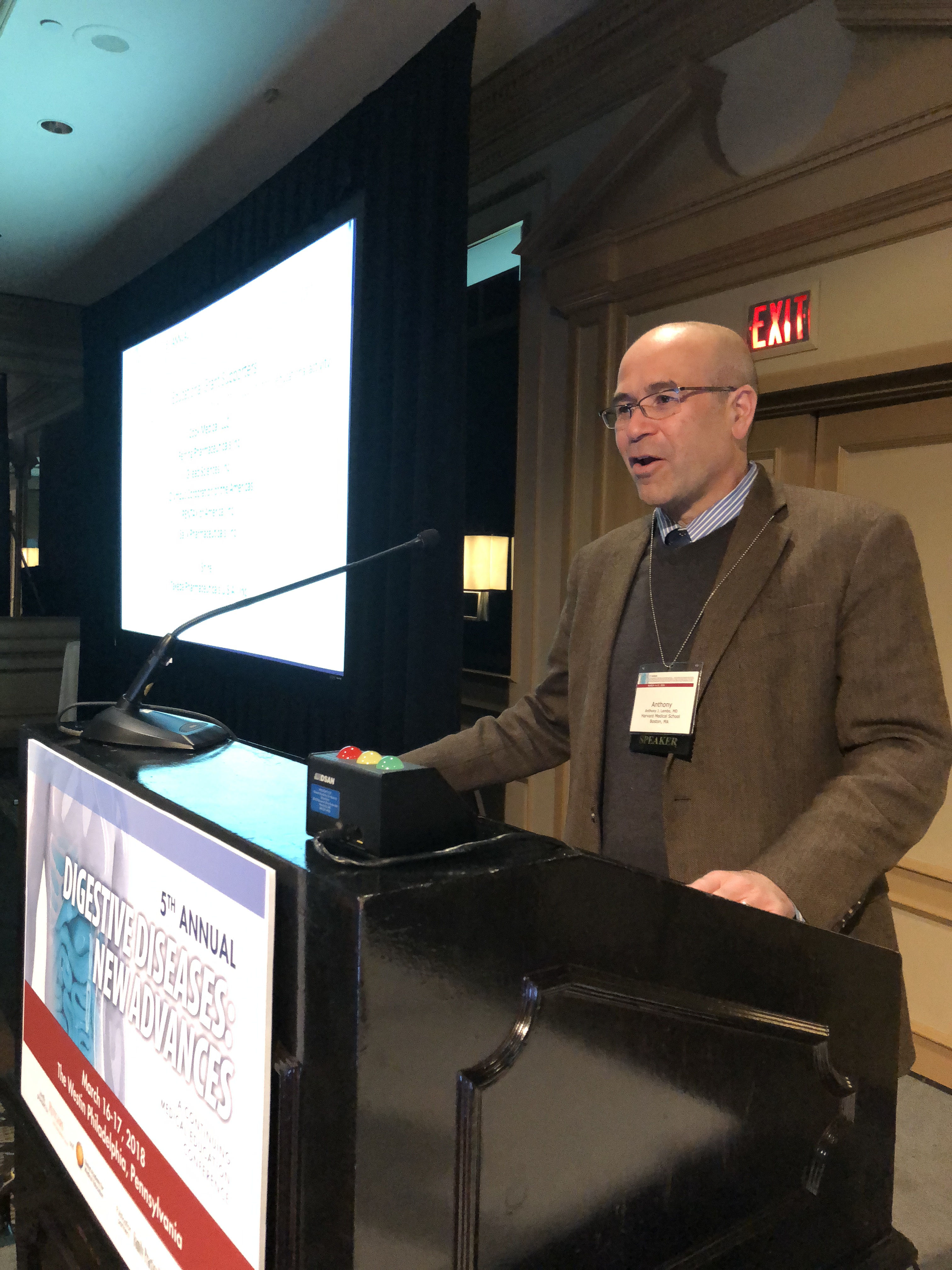
Clinicians in states where cannabis use is increasing might need to be particularly alert for cannabis-related issues. According to the study by Dr. Lembo, the Midwest and West regions registered higher rates of vomiting with cannabis use disorder, compared with the Northeast and South.
Whether cannabinoids also can be a treatment for nausea or vomiting is a frequently asked question, Dr. Lembo said.
While there are no data for smoked marijuana, Dr. Lembo said, .
Dronabinol is indicated for adults for the treatment of chemotherapy-associated nausea and vomiting in patients who don’t respond adequately to conventional antiemetics, according to the agent’s prescribing information.
The cannabinoid medication is an isomer of tetrahydrocannabinol (THC), one of the active compounds in marijuana, according to Dr. Lembo.
“If you smoke marijuana, the levels go up high very quickly,” Dr. Lembo said. “If you take dronabinol, it takes 45 minutes to an hour. It’s a slower rise of it, so people are less likely to abuse dronabinol.”
In his talk, Dr. Lembo reported disclosures related to Allergan, Ironwood Pharmaceuticals, Salix Pharmaceuticals, and Takeda Pharmaceuticals.
Global Academy for Medical Education and this news organization are owned by the same company.
REPORTING FROM DIGESTIVE DISEASES: NEW ADVANCES
Adding biomarkers beats NICE guidelines for detecting preeclampsia
FROM ULTRASOUND IN OBSTETRICS & GYNECOLOGY
Screening for preeclampsia using the United Kingdom’s National Institute for Health and Care Excellence (NICE) guidelines detects only about one-third of cases, according to a study published in Ultrasound in Obstetrics & Gynecology.
The United Kingdom-based prospective multicenter study, involving 16,747 singleton pregnancies, also looked at the effectiveness of a screening method that used Bayes’ theorem to combine maternal risk factors with biomarkers.
Preeclampsia developed in 473 (2.8%) pregnancies, and in 142 cases (0.8%) this led to preterm birth.
The NICE method of screening labels as high-risk women who have one major risk factor – such as a history of hypertensive disease in pregnancy or chronic kidney disease – or two moderate factors, including first pregnancy older than 40 years or a family history of preeclampsia.
This method of screening detected 30.4% of the cases of preeclampsia that developed and 40.8% of the cases that resulted in pre-term birth. The overall screen-positive rate by the NICE method was 10.3% of all participants in the study (1,727 women).
The Bayes’ theorem-based method assessed maternal risk factors in combination with mean arterial pressure and serum pregnancy-associated plasma protein-A. The detection rate for all preeclampsia using this method was 42.5%, representing an improvement of 11.3% over the NICE method, after adjusting for the effects of aspirin use in both groups. Researchers also examined the effect of adding in the biomarkers of uterine artery pulsatility index and serum placental growth factor, and found this detected 82.4% of preterm preeclampsia.
“The performance of screening by a combination of maternal factors with biomarkers was far superior to that of screening by NICE guidelines,” wrote Min Yi Tan, MD, of King’s College Hospital in London, and co-authors.
Overall, 4.5% of women in the study took aspirin from 14 weeks’ gestation until 36 weeks or delivery, but only 23.2% of women who screened positive according to the NICE guidelines took aspirin.
“Such poor compliance may at least in part be attributed to the generally held belief, based on the results of a meta-analysis in 2007, that aspirin reduces the risk of PE by only about 10%,” Dr. Tan and co-authors wrote.
The authors acknowledged that their study did not explore the health economic implications of the combined screening approach, but said there was now accumulating evidence that the performance of first-trimester screening for preterm preeclampsia could be improved substantially by the additional measurement of biomarkers.The study was sponsored by King’s College London, and supported by the National Institute for Health Research Efficacy and Mechanism Evaluation Programme, the Fetal Medicine Foundation and NIHR Collaboration for Leadership in Applied Health Research and Care South London at King’s College Hospital NHS Foundation Trust, with in-kind support from PerkinElmer Life and Analytical Sciences, and Thermo Fisher Scientific. No conflicts of interest were declared.
obnews@frontlinemedcom.com
SOURCE: Tan MY et al. Ultrasound Obstet & Gynecol. 2018 Mar 14. doi: 10.1002/uog.19039.
Key clinical point: Screening for preeclampsia using the United Kingdom’s National Institute for Health and Care Excellence (NICE) guidelines only detects around one-third of all preeclampsia cases, but the addition of biomarkers can improve this significantly.
Major finding: The NICE guidelines detected 30.4% of cases of preeclampsia, while a Bayes’ theorem-based method using maternal risk factors and biomarkers detected 42.5%.
Data source: A prospective multicenter study of 16,747 singleton pregnancies.
Disclosures: The study was sponsored by King’s College London, and supported by the National Institute for Health Research Efficacy and Mechanism Evaluation Programme, the Fetal Medicine Foundation and NIHR Collaboration for Leadership in Applied Health Research and Care South London at King’s College Hospital NHS Foundation Trust, with in-kind support from PerkinElmer Life and Analytical Sciences, and Thermo Fisher Scientific. No conflicts of interest were declared.
Source: Tan MY et al. Ultrasound Obstet & Gynecol. 2018 Mar 14. doi: 10.1002/uog.19039.
mainbar
Screening for preeclampsia using the United Kingdom’s National Institute for Health and Care Excellence (NICE) guidelines detects only about one-third of cases, according to a study published in Ultrasound in Obstetrics & Gynecology.
The United Kingdom-based prospective multicenter study, involving 16,747 singleton pregnancies, also looked at the effectiveness of a screening method that used Bayes’ theorem to combine maternal risk factors with biomarkers.
Preeclampsia developed in 473 (2.8%) pregnancies, and in 142 cases (0.8%) this led to preterm birth.
The NICE method of screening labels as high-risk women who have one major risk factor – such as a history of hypertensive disease in pregnancy or chronic kidney disease – or two moderate factors, including first pregnancy older than 40 years or a family history of preeclampsia.
This method of screening detected 30.4% of the cases of preeclampsia that developed and 40.8% of the cases that resulted in pre-term birth. The overall screen-positive rate by the NICE method was 10.3% of all participants in the study (1,727 women).
The Bayes’ theorem-based method assessed maternal risk factors in combination with mean arterial pressure and serum pregnancy-associated plasma protein-A. The detection rate for all preeclampsia using this method was 42.5%, representing an improvement of 11.3% over the NICE method, after adjusting for the effects of aspirin use in both groups. Researchers also examined the effect of adding in the biomarkers of uterine artery pulsatility index and serum placental growth factor, and found this detected 82.4% of preterm preeclampsia.
“The performance of screening by a combination of maternal factors with biomarkers was far superior to that of screening by NICE guidelines,” wrote Min Yi Tan, MD, of King’s College Hospital in London, and co-authors.
Overall, 4.5% of women in the study took aspirin from 14 weeks’ gestation until 36 weeks or delivery, but only 23.2% of women who screened positive according to the NICE guidelines took aspirin.
“Such poor compliance may at least in part be attributed to the generally held belief, based on the results of a meta-analysis in 2007, that aspirin reduces the risk of PE by only about 10%,” Dr. Tan and co-authors wrote.
The authors acknowledged that their study did not explore the health economic implications of the combined screening approach, but said there was now accumulating evidence that the performance of first-trimester screening for preterm preeclampsia could be improved substantially by the additional measurement of biomarkers.
The study was sponsored by King’s College London, and supported by the National Institute for Health Research Efficacy and Mechanism Evaluation Programme, the Fetal Medicine Foundation and NIHR Collaboration for Leadership in Applied Health Research and Care South London at King’s College Hospital NHS Foundation Trust, with in-kind support from PerkinElmer Life and Analytical Sciences, and Thermo Fisher Scientific. No conflicts of interest were declared.
obnews@frontlinemedcom.com
SOURCE: Tan MY et al. Ultrasound Obstet & Gynecol. 2018 Mar 14. doi: 10.1002/uog.19039.
High-normal TSH linked to unexplained infertility
CHICAGO – Levels of thyroid stimulating hormone were significantly higher in women with unexplained infertility than in a cohort whose partners had severe male factor infertility, though TSH levels still fell within the reference range.
For women with unexplained infertility, thyroid stimulating hormone (TSH) averaged 1.95 mIU/mL (95% confidence interval, 1.54-2.61), compared with 1.66 mIU/mL for the group of women with severe male factor infertility, such as severe oligospermia or azoospermia (95% CI, 1.25-2.17; P = .003).
“We found, very interestingly, that in the unexplained group, TSH was higher in those women, compared with women whose partners had severe male factor infertility,” suggesting that , the study’s first author, Lindsay T. Fourman, MD, said in an interview at the annual meeting of the Endocrine Society.
In terms of TSH levels, “clearly, the cutoff of the upper limit of the normal range is controversial,” said Dr. Fourman. “Some studies have shown that 95% of the population has a TSH of less than 2.5.”
However, the numbers that guide treatment for hypothyroidism are different. “We use a cutoff, generally, of 4 or 5, but maybe that cutoff should be 2.5, and maybe that’s significant for some people,” Dr. Fourman said.
“Current guidelines do not recommend treatment of subclinical hypothyroidism among auto-antibody negative women attempting to conceive naturally,” wrote Dr. Fourman and her collaborators in the poster presenting their finding.
Twice as many women in the group with unexplained infertility had TSH levels in the upper half of the normal range – above 2.5 mIU/mL – than in the male factor infertility group, “again, suggesting this association with TSH and unexplained infertility,” Dr. Fourman said. The thyroid axis is known to play a role in oocyte development. Of women with unexplained infertility, 26.9% had a TSH above 5 mIU/mL, compared with 13.5% of those with severe male factor infertility (P less than .05).
The chart review of records from 187 women with unexplained fertility and 52 women whose partners had severe male factor infertility included women aged 18-39. The unexplained cohort included women for whom all causes of infertility were excluded “in the setting of a very thorough workup – and that would include any ovulatory issues, male factor issues, and by definition, these women had to have a normal FSH, TSH and prolactin,” said Dr. Fourman, an endocrine fellow at Massachusetts General Hospital, Boston.
Control patients were those who had TSH and prolactin levels available and whose partners were being seen for severe male factor infertility, meaning that their partner had severe oligospermia or azoospermia.
Dr. Fourman acknowledged that she and her and her collaborators couldn’t exclude some female factor infertility among the control group. “That is an assumption, but it’s an assumption that would bias us to the null,” strengthening the study’s findings.
Clinical characteristics were similar between study groups, though women with unexplained infertility were slightly older than those with severe male factor infertility (mean 31.5 years versus 30.1 years, P = .01); they also had slightly lower body mass indices (median 23 versus 24.4 kg/m2; P less than .04).
No association was found between prolactin levels, “which suggests that prolactin may not contribute to unexplained infertility in these women,” Dr. Fourman said.
The investigators were able to control for such potentially confounding variables as age, tobacco use, BMI; they excluded from analysis women who had positive thyroid peroxidase antibodies.
“This is very interesting, because it really raises the question of whether we should be treating TSH, even to the lower half of the normal range, to see if that can improve outcomes,” she said. “We are looking for modifiable things that we can treat to try to improve fertility, so if we can identify some cause – like a hormonal cause – we may be able to improve conception outcomes and reduce the need for invasive treatment.”
Based in part on the strength of these findings, Dr. Fourman said she and her collaborators are planning a prospective study to see whether treating women with infertility to achieve a TSH of less than 2.5 can speed time to conception and reduce the need for invasive infertility treatment.
Dr. Fourman reported no conflicts of interest and no external sources of funding.
koakes@frontlinemedcom.com
SOURCE: Fourman, L, et al. ENDO 2018, Abstract SAT-288.
REPORTING FROM ENDO 2018
