User login
Ebola research update: November 2016
The struggle to defeat Ebola virus disease continues globally, although it may not always make the headlines. To catch up on what you may have missed, here are some notable news items and journal articles published over the past few weeks that are worth a second look.
A study in Cell found that specific amino acid substitutions in the Ebola virus glycoprotein have increased tropism for human cells, while reducing tropism for bat cells, and such increased infectivity may have contributed to the wide geographic distribution of some viral lineages.
A recent case study of a West African Ebola survivor demonstrated the persistence of T-cell activation well beyond viral clearance and detected Ebola-specific T cells, providing insights into lymphocyte specificity during the reconvalescent phase of Ebola virus disease.
The Ebola virus glycoprotein mutant GP-A82V arose early and dominated the 2013-2016 West African epidemic, according to a recent study, and it was weakly associated with increased mortality during the epidemic.
A modeling study on a region of Sierra Leone provided numerical estimates for the effectiveness of ring vaccination to control future Ebola virus outbreaks. Investigators showed that outbreaks with moderate transmission potential can be successfully contained, and more extensive vaccination and reinforcement of the health care system would increase the likelihood of containment even if the virus were more transmissible than in the past.
An RT-PCR (reverse transcriptase polymerase chain reaction) diagnostic assay using a glycoprotein target is more sensitive for the detection of Ebola virus in clinical samples than is an RT-PCR using a nucleoprotein target, according to a study in Diagnostic Microbiology & Infectious Disease.
Convalescent whole blood therapy is promising for treating Ebola virus disease in resource-poor settings, especially in the early phases of outbreaks when resource mobilization is done, according to a study in the Journal of Infection.
Rapid chart reviews at Ebola virus disease survivor clinics should be repeated regularly to assess the extent of illness and prioritize service delivery, according to a study in Sierra Leone.
The 2014-2016 outbreak of Ebola virus disease in West Africa harmed the tourism industry in Kenya, according to a study by the University of Nairobi.
Researchers have developed Microreact, a free, real-time epidemic visualization and tracking platform that has been used to monitor outbreaks of Ebola, Zika, and antibiotic-resistant microbes, according to a report in Microbial Genomics.
A study in the Journal of Virology found that Ebola virus variants with improved fitness emerged early during the West African Ebola virus outbreak, impacting virus transmissibility and pathogenicity.
A literature review published in BMC Infectious Diseases found the pooled case fatality rate and seroprevalence for Ebola and Marburg viruses to be lower than usually reported, with species differences despite high heterogeneity between studies.
A study of the scope and scale of the 2014-2015 Ebola virus outbreaks in West Africa found broad socioeconomic impacts, including reduced community cohesion, education loss, reduced child protection, widespread job losses, and food insecurity.
rpizzi@frontlinemedcom.com
On Twitter @richpizzi
Optimal duration of extended AI therapy? Flip a coin
SAN ANTONIO – When does adjuvant therapy with an aromatase inhibitor become too much of a good thing? Or to put it another way, what’s the optimal duration of extended aromatase inhibitor therapy? That’s the question that three clinical trials have tried – but largely failed – to answer.
For example, the randomized, double-blinded NSABP B-42 trial, comparing extended therapy with letrozole (Femara) in postmenopausal women with hormone receptor–positive (HR+) breast cancer who have completed previous adjuvant therapy with an aromatase inhibitor (AI) showed no difference in disease-free survival (DFS) after 7 years of follow-up between women treated with extended letrozole or placebo.
DATA data
In the DATA study, also presented here, investigators from the Netherlands compared 6 years of anastrozole (Arimidex) to 3 years of anastrozole following 2 or 3 years of adjuvant tamoxifen for postmenopausal women with estrogen receptor–positive (ER+), and/or progesterone receptor–positive (PR+) breast cancer.
They found that “adapted” DFS (DFS starting 3 years after randomization) and adapted overall survival (OS) were similar between the two groups.
“The findings of the DATA study do not support extended adjuvant AI use after 5 years of sequential endocrine therapy for all postmenopausal hormone receptor–positive breast cancer patients,” said Vivianne Tjan-Heijnen, MD, of Maastricht University Medical Center in the Netherlands.
Less than ideal
In the optimistically named IDEAL trial, a separate team of investigators, also from the Netherlands, looked at the relative merits of continuing adjuvant therapy with letrozole for 2.5 or 5 years following 5 years of adjuvant therapy with tamoxifen, an AI, or a combination in postmenopausal women with HR+ breast cancer.
They found no differences in either DFS or OS between patients treated for 5 years or those treated for only half that long.
“We conclude that there is no benefit of extending AI-based therapy longer than two-and-a-half years,” said Erik Blok, MD, of Leiden University Medical Center in the Netherlands.
Give what to whom for how long?
Results of the trials raise more questions than they answer, said Michael Gnant, MD, of the Medical University of Vienna, the invited discussant.
“Essentially, these three trials did not reach the necessary statistical significance levels to demonstrate a clear benefit for the respective AI extension,” he said.
“Can other agents we use in luminal breast cancer help? Frankly, I don’t think so. Based on their tolerability profile, and in part also on financial toxicity, I don’t think that the promising agents we explore in many situations for the treatment of hormone receptor–positive breast cancer will realistically be used in the extended adjuvant setting,” he said.
What’s needed, he said, are new strategies for targeting the chronic part of luminal breast cancer recurrence risk. Using endocrine therapies in this setting will likely be ineffective. Instead, agents that could directly target dormant cancer stem cells would “eliminate the source of late metastases for good.”
The best evidence to date clearly points to individualized treatment plans for patients, Dr. Gnant said.
For example, for a patient who has had 2-5 years of tamoxifen, an AI for 2.5-5 additional years can help to prevent recurrences, provided that the patient has risk factors for recurrence and excellent bone health.
“Based on the trial results, it is more complex for a patient who comes off initial or sequential AI. There are factors favoring the extension of AI treatment, and other factors to speak against such extension. I suggest to start with patient features at this time,” he said.
Currently, the main factor driving the choice of extended AI therapy will be how well the patient has tolerated AIs in the first years of therapy and whether she is at increased risk for fractures, suggesting younger age as a factor favoring extended AI use.
Patients with higher clinicopathologic risk factors such as node positivity or more luminal type tumors, as well as higher risk according to genomic studies, might also benefit from extended AI therapy, he said.
Biomarkers needed
“What the data from these and other trials tell us is that endocrine therapy is not for everyone. We need biomarkers that can tell us who should be getting extended endocrine therapy, be it 10 years or even a longer duration of time, versus a subgroup that might do very well with 5 five years of AI,” Aditya Bardia, MBBS, MPH, of the breast cancer division at Massachusetts General Hospital Cancer Center in Boston, said in an interview.
There are several such biomarkers under investigation, but they need validation and testing in large scale clinical trials before they find their way into day-to-practice, he said.
Dr. Bardia was not involved in the studies.
NSABP B-42 was sponsored by PrECOG with financial support from Novartis. Dr. Mamounas reported having no conflicts of interest. The DATA trial was sponsored by the Dutch Breast Cancer Research Group and Novartis. Dr. Tjan-Heijnen reported nothing to disclose. IDEAL was supported by the Dutch Breast Cancer Research Group and Novartis. Dr. Blok reported nothing to disclose.
AT SABCS 2016
Key clinical point: The optimal duration of aromatase inhibitor (AI) therapy following 5 years of endocrine therapy in postmenopausal women is still unknown.
Major finding: There were no significant differences in disease-free or overall survival in three studies investigating extended AI therapy.
Data source: Randomized phase II NSABP B-42 with 3,996 patients; randomized phase III DATA study with 1,912 patients; randomized phase III IDEAL trial with 1,824 patients.
Disclosures: NSABP B-42 was sponsored by PrECOG with financial support from Novartis. Dr. Mamounas reported having no conflicts of interest. The DATA trial was sponsored by the Dutch Breast Cancer Research Group and Novartis. Dr. Tjan-Heijnen reported nothing to disclose. IDEAL was supported by the Dutch Breast Cancer Research Group and Novartis. Dr. Blok reported nothing to disclose.
Hepatitis Outlook: November 2016
If you work on the front lines of medical care treating patients with hepatitis, you may not have time to review all the hepatitis research that enters the medical literature every month. Here’s a quick look at some notable news items and journal articles published over the past month, covering a variety of the major hepatitis viruses.
The introduction of universal mass vaccination against hepatitis A in countries with intermediate endemicity for HAV infection led to a considerable decrease in the incidence of HAV in vaccinated and in nonvaccinated age groups alike.
Mortality was high among chronic hepatitis C patients, with and without cirrhosis, compared with the general population, a Danish cohort study found. Curing CHC was associated with reduced mortality among cirrhotic patients but remained higher than the general population.
A hepatitis C outbreak in a North Dakota skilled nursing facility highlights the importance of prompt reporting and investigation of incident HCV infection, and the need for adherence to basic infection control procedures by health care personnel.
A recent study identified a novel hepatitis B virus subgenotype D10 circulating in Ethiopia, underlining the high genetic variability of HBV strains in Africa.
A study in the Journal of Viral Hepatitis found that baseline hepatitis B core antibody predicts treatment response in chronic hepatitis B patients receiving long-term entecavir.
A novel quantitative microarray antibody capture assay was able to identify extremely high hepatitis delta virus prevalence amongst hepatitis B virus–infected Mongolians.
The albumin-bilirubin (ALBI) score was effective in predicting the long-term prognosis for patients with hepatitis B virus–related cirrhosis and was more accurate than Child-Pugh and Model for End-Stage Liver Disease (MELD) scores.
A study in Hepatology found that proanthocyanidin (PAC) and its analogs present a new class of anti–hepatitis B virus agents that directly target the preS1 region of the HBV large surface protein and could contribute to the development of a potent, well-tolerated, and broadly active inhibitor of HBV infection.
A hepatitis C virus core antigen (HCV-Ag) assay proved to be useful in monitoring treatment of HCV-infected patients with sustained viral response and in patients who experienced treatment failures, according to a study in Diagnostic Microbiology and Infectious Disease.
The introduction of a managed care network for patients infected with hepatitis C virus increased access to care and reduced all-cause mortality, according to a recent study.
A study in South Korea found that hepatitis B infection was associated with an increased incidence of thrombocytopenia in healthy adults without cirrhosis.
A proof-of-concept study demonstrated that peritransplant immunoprophylaxis combined with a single oral direct-acting antiviral in the immediate post-transplant period can prevent HCV recurrence.
A study in the Journal of Viral Hepatitis established the baseline mortality and hepatocellular carcinoma progression rates in decompensated cirrhosis patients against which the impact of new antiviral therapies could be measured.
Genetic distance-based network analyses can be used to identify characteristics associated with hepatitis C virus transmission, informing targeted prevention and treatment strategies, according to a recent study.
According to a new study, primary T-cell immunodeficiency is associated with a lower spontaneous clearance of hepatitis C virus, while female sex and coinfection with hepatitis B virus are associated with a higher spontaneous clearance.
A study in the Journal of Viral Hepatitis found that the induction of humoral and cellular immune response to hepatitis B virus vaccine can be upregulated by CpG oligonucleotides complexed with Dectin-1 ligand.
rpizzi@frontlinemedcom.com
On Twitter @richpizzi
Durvalumab could offer option for difficult-to-treat NSCLC
VIENNA – In a heavily pretreated population of patients with non–small-cell lung cancer (NSCLC), the investigational checkpoint inhibitor durvalumab was associated with good objective response rates (ORR) and ‘impressive’ overall survival (OS) in a phase II study, Marina Garassino, MD, reported at the World Conference on Lung Cancer.
The primary endpoint of ORR in the open-label, single-arm ATLANTIC trial was achieved by 7% of patients with low (less than 25% of tumor cells) expression of the programmed death ligand 1 (PD-L1) that durvalumab targets (n = 93), by 16.4% of patients with PD-L1 expression of 25% or higher (n = 146), and by 30.9% in patients with PD-L1 expression of 90% or more (n = 68).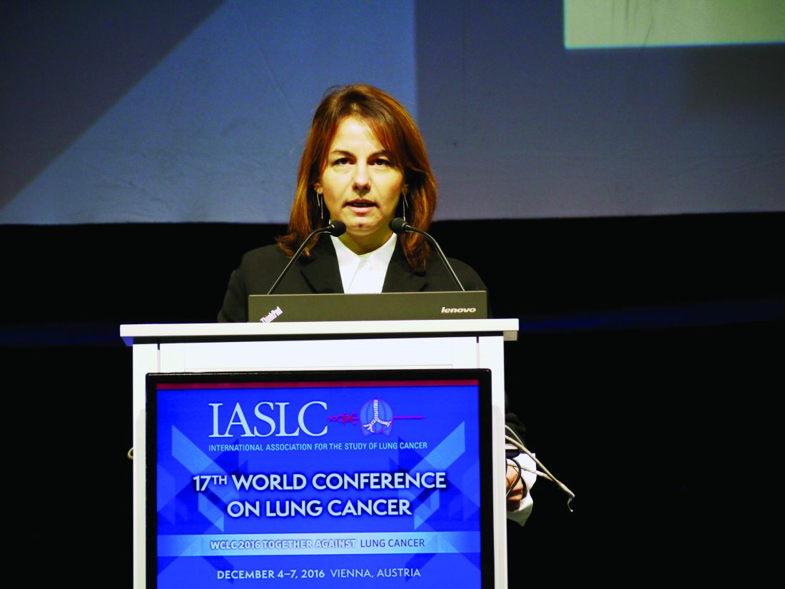
Yet the results of ATLANTIC show not only that durvalumab is active, but also that it can produce long-lasting responses in patients who have been treated with a mean of three prior regimens, she reported.
The median duration of response to date was not reached in patients with PD-L1 expression of 25% or less, was 12.3 months in patients with greater than 25% PD-L1 expression, and had again not yet been reached in those with greater than 90% PD-L1 expression. At the time the data were pulled for analysis, June 2016, 18 of 21 patients in the latter group were progression free.
The disease control rate at 6 months was 20.4%, 28.8%, 38.2% for patients with PD-L1 expression of less than 25%, 25%-90%, and greater than 90%, respectively.
“One-year overall survival was about 48% in patients with PD-L1 greater than 25% and about 51% in those with PD-L1 greater than 90%,” Dr. Garassino said at the meeting, which was sponsored by the International Association for the Study of Lung Cancer.
The respective median OS times for patients with 25% and greater and less than 25% PD-L1 expression were 10.9 (95% CI 8.6-13.6) and 9.3 (95% CI 5.9-10.8) months, and not yet reached for patients with 90% or greater PD-L1 expression.
“Most adverse events were low grade and immune mediated adverse events were easily manageable,” Dr. Garassino said. Overall, 10.2% of patients had grade 3 or 4 treatment-related adverse events and 2.7% had treatment-related adverse events that led to discontinuation.
Any immune-mediated adverse event occurred in 12.3% of patients, of which 6.3% were due to an underactive and 2.4% to a hyperactive thyroid.
“Results are consistent with other anti-PD-1/PD-L1 compounds in metastatic, relapsed NSCLC and we are awaiting the final results of phase III trials to clarify the role of durvalumab [treatment] alone or in combination in NSCLC,” Dr. Garassino said.
Overall, 1,990 patients were screened for inclusion in the ATLANTIC study, which recruited 444 patients with stage IIIB-IV NSCLC who had received at least two prior systemic treatments, one of which had to be platinum based and had a recent (within 3 months) tumor biopsy and archived tissue available for PD-L1 assessment. Dr. Garassino reported results for two of the three cohorts.
Durvalumab was given at an intravenous dose of 10 mg/kg every 2 weeks for up to 1 year. The mean number of prior regimens was 3.2 for patients with less than 25% and greater than or equal to 25% PD-L1 expression combined and 2.6 for those with greater than 90% PD-L1 expression.
“This drug clearly does have activity and we can see that in the response rates, which range from 7% all the way up to 31%,” said Michael Boyer, MBBS, chief clinical officer of Chris O’Brien Lifehouse in Melbourne, who was the invited discussant for the trial. “The higher the PD-L1 expression, the higher the response rate,” he observed.
“There was quite impressive survival, if you bear in mind that this is a third-line, or more than third-line cohort of patients, with a median overall survival in the strongest PD-L1 expressing patients that is clearly going to exceed 1 year” he added.
The big question for the future is what will be the relevance of having an immunotherapy that works well in such a heavily pretreated population when these drugs are more likely to be used second- or even first-line, especially in those who have high PD-L1 expression where the drug seems to be at its most effective, Dr. Boyer said.
AstraZeneca funded the study. Dr. Garassino disclosed relationships with AstraZeneca, Bristol Myers Squibb, Roche, Merck, Sharp & Dohme, Boehringer Ingelheim, and Eli Lilly. Dr. Boyer disclosed that he had received research funding, honoraria, or both that were paid to his institution from Pfizer, Roche, Eli Lilly, Merck, Sharp & Dohme, Bristol Myers Squibb, AstraZeneca, and Clovis.
Key clinical point: Durvalumab had substantial antitumor activity and produced long-lasting responses in patients with previously treated advanced non–small-cell lung cancer (NSCLC).
Major finding: Objective response rates of 16.4% and 30.1% were achieved in patients with the highest PD-L1 expression profiles.
Data source: Phase II, open-label, single-arm ATLANTIC trial of 444 patients with relapsed, locally advanced or metastatic NSCLC treated with two or more prior systemic regimens.
Disclosures: AstraZeneca funded the study. Dr. Garassino disclosed relationships with AstraZeneca, Bristol Myers Squibb, Roche, Merck, Sharp & Dohme, Boehringer Ingelheim, and Eli Lilly. Dr. Boyer disclosed that he had received research funding, honoraria, or both that were paid to his institution from Pfizer, Roche, Eli Lilly, Merck, Sharp & Dohme, Bristol Myers Squibb, AstraZeneca, and Clovis.
Next-generation sequencing highlights evolution of ER+ breast cancer
SAN ANTONIO – The “genomic landscape” of resistant estrogen receptor–positive (ER+) metastatic breast cancer differs significantly from that of ER+ primary breast cancer, according to findings from a study involving whole-exome sequencing and transcriptome sequencing of such cancers.
Multiple genes were recurrently altered in ER+ metastatic breast cancers at a rate significantly higher than in ER+ primary breast cancers (with 3- to 33-fold enrichment in metastatic vs. primary samples), Ofir Cohen, PhD, explained at the San Antonio Breast Cancer Symposium.
This finding, which suggests that recurrent alterations in genes in ER+ metastatic breast cancers are often acquired after treatment and thus may play a role in resistance to ER-directed therapies and/or metastasis, highlights the value of genomic profiling of metastatic biopsies and has implications for drug development, said Dr. Cohen of the Broad Institute of MIT (Massachusetts Institute of Technology) and Harvard in Cambridge.
Although the genomic and molecular landscape of ER+ primary breast cancer is better understood, that of ER+ metastatic breast cancer is underexplored, he noted.
Thus, he and his colleagues performed whole-exome sequencing on 149 metastatic tumor biopsies from patients with ER+ metastatic tumors and resistance to at least one ER-directed therapy and compared the results with those from 44 matched primary samples from the same patient. They also performed transcriptome sequencing (RNA-seq) of 128 metastatic biopsies.
A key observation from the study is that ESR1 genes were mutated in 24% of the cohort, and the mutations were acquired in 14 of the 15 samples with matched primary samples.
“This emphasized the important role that these mutations may play, specifically in the metastatic and drug-resistant settings,” Dr. Cohen said.
Similarly, ERBB2 mutations occurred in 7% of samples, and seemed to be acquired in five of six metastatic samples with matched primary samples, he said.
“For both of these genes, other than being acquired and suggestive of importance, they also have clear clinical implications, as finding those mutations may guide treatment choices for those patients,” he said.
Another example involves RB1 mutations, which were found in 5% of samples, and which appeared to be acquired in three of five of those with matched primaries. More frequent alterations were also found in PIK3CA, PTEN, and AKTI genes, among others, in metastatic vs. primary cancers.
The observation that these tumors evolve and have different mutations in the primary and metastatic settings may be important, because this suggests that knowledge of the alterations and mutations in the primary setting is insufficient to guide treatment in the metastatic setting.
“So the take-home message here is that tumors do evolve, and that the metastatic setting is different than the primary setting,” Dr. Cohen said.
Sequencing both the exome and the transcriptome may “give us a handle to assess that,” he said.
“Our goal is really to understand evolved resistance. That is, to try and understand the mechanisms that drive the evolution of resistance in ER+ metastatic breast cancer, and, once we understand that, [to determine] how we can translate that knowledge in the clinic,” he said, noting that resistance also may develop through other nongenomic mechanisms, such as epigenetic and regulatory mechanisms.
“As long as those mechanisms leave a footprint on the transcriptome … sequencing the transcriptome together with the exome may yield relevant insights into resistance states that do not derive specifically from exome mutations,” he added.
The findings are important because, despite tremendous advances in the treatment of ER+ metastatic breast cancer, therapeutic resistance remains a common problem, and improved understanding of the underlying resistance mechanisms is critical for enabling durable control of this disease, Dr. Cohen said, explaining that resistant tumors remain the most common cause of breast cancer death and that there is an urgent need to develop new therapeutic strategies for patients who no longer respond to existing therapies.
In a written statement, senior investigator Nikhil Wagle, MD, of Dana-Farber Cancer Institute and Harvard Medical School, Boston, said that the ultimate goal of the project is “to integrate the functional and clinical findings into a unified ‘Resistance Atlas’ for ER+ metastatic breast cancer, which should help inform treatment decisions for individual patients as well as propel the development of new combination treatment strategies for ER-positive metastatic breast cancer.”
This study was funded by the National Cancer Institute, the National Human Genome Research Institute, the Department of Defense Breast Cancer Research Program, Susan G. Komen, the V Foundation, the Breast Cancer Alliance, the AACR-Landon Foundation, the Friends of Dana-Farber Cancer Institute, and the Breast Cancer Research Foundation. Dr. Cohen reported having no conflicts of interest. Dr. Wagle is an equity holder in Foundation Medicine, a consultant to Novartis, and a recipient of sponsored research support from Novartis, Genentech, and Merck.
AT SABCS 2016
Key clinical point:
Major finding: ESR1 genes were mutated in 24% of the cohort, and the mutations were acquired in 14 of the 15 samples with matched primary samples.
Data source: Whole-exome sequencing of 149 metastatic breast cancer biopsies and 44 matched primary tumor biopsies, and transcriptome sequencing of 128 metastatic biopsies.
Disclosures: This study was funded by the National Cancer Institute, the National Human Genome Research Institute, the Department of Defense Breast Cancer Research Program, Susan G. Komen, the V Foundation, the Breast Cancer Alliance, the AACR-Landon Foundation, the Friends of Dana-Farber Cancer Institute, and the Breast Cancer Research Foundation. Dr. Cohen reported having no conflicts of interest. Dr. Wagle is an equity holder in Foundation Medicine, a consultant to Novartis, and a recipient of sponsored research support from Novartis, Genentech, and Merck.
VIDEO: Veliparib misses PFS endpoint, advances to phase III trial
SAN ANTONIO – The investigational selective PARP-1 and PARP-2 inhibitor veliparib failed to significantly improve progression-free or overall survival, compared with placebo, when added to carboplatin and paclitaxel in patients with BRCA1 or BRCA2 mutations and locally recurrent or metastatic breast cancer in the randomized phase II BROCADE study.
The potent, orally bioavailable drug did, however, show a trend toward improvement on these measures, as well as a significant improvement in overall response-rate findings that warrant further investigation in a phase III trial, Hyo Han, MD, said at the San Antonio Breast Cancer Symposium.
Patients were randomized to receive placebo (98 patients) or 120 mg veliparib twice daily on days 1-7 (95 patients) in addition to carboplatin and 175 mg/m2 of paclitaxel every 3 weeks.
Median progression-free survival – the primary endpoint of the study – was 12.3 months in the placebo group and 14.1 months in the veliparib group (hazard ratio, 0.789), and overall survival was 25.9 and 28.3 months, respectively (HR, 0.750), said Dr. Han of Moffitt Cancer Center, Tampa, Fla.
The overall response rate was 61.3% vs. 77.8% in the groups, respectively.
Although the study did not meet its primary endpoint, the findings are encouraging as it was powered to detect only dramatic differences between the groups, Dr. Han said.
In a video interview, she discussed veliparib, its safety, the BROCADE findings – including data from a third study arm looking at veliparib in combination with temozolomide, and plans for evaluating veliparib in the ongoing phase III BROCADE3 trial.
The video associated with this article is no longer available on this site. Please view all of our videos on the MDedge YouTube channel
AT SABCS 2016

Fulvestrant/everolimus improves PFS in HR+, HER2– advanced breast cancer
SAN ANTONIO – Adding everolimus to fulvestrant doubled median progression-free survival among postmenopausal women with hormone-receptor positive, human epidermal growth factor receptor 2–negative (HER2-negative) metastatic breast cancer resistant to therapy with an aromatase inhibitor [AI] in the PrECOG 0102 study.
In the randomized phase II trial, the combination of the mammalian target of rapamycin (mTOR) inhibitor everolimus (Afinitor) with the selective estrogen receptor down-regulator [SERD] fulvestrant (Faslodex) was associated with a median progression-free survival of 10.4 months, compared with 5.1 months for fulvestrant plus placebo, reported Noah S. Kornblum, MD, of Montefiore Einstein Center for Cancer Care, New York.
This study provides additional evidence that adding everolimus to anti-estrogen therapy in AI-resistant disease improves clinical outcomes,” he said at the San Antonio Breast Cancer Symposium.
Most women with hormone receptor–positive breast cancer treated with an AI will eventually develop resistance to these agents. Strategies for overcoming resistance include the addition of everolimus to a steroid AI, exemestane (Aromasin), as in the BOLERO-2 trial.
“Another strategy for overcoming AI resistance is by more completely blocking estrogen-receptor signaling through the use of a selective estrogen receptor down-regulator, which may result in more complete blockade of the ER signaling pathway than a steroidal AI such as exemestane,” Dr. Kornblum said.
To test this hypothesis, the investigators enrolled 131 postmenopausal women with inoperable locally advanced or metastatic hormone receptor–positive, HER2-negative breast cancer resistant to AIs.
AI resistance was defined as relapse while receiving adjuvant AI therapy, and/or progression after one or more AIs for metastatic disease. The patients could have had no more than one prior chemotherapy regimen for metastatic disease.
The patients were stratified by Eastern Cooperative Oncology Group performance status, presence of measurable disease, and prior chemotherapy status, and were then randomized to receive either high-dose fulvestrant (500 mg on day 1 and 15 of cycle 1, and then on day 1 of cycles 2-12) plus oral everolimus 10 mg/day, or fulvestrant and placebo.
The trial had an induction phase, in which patients were treated until evidence of progressive disease or unacceptable toxicity for a maximum of 12 28-day cycles, and a continuation phase in which patients who had neither disease progression nor experienced unacceptable toxicities could have their data unblinded and could continue on fulvestrant/everolimus.
The trial did not include the use of corticosteroid-containing mouthwash for prevention of treatment-associated stomatitis, because the trial was designed before the evidence of the benefit of such prophylaxis became public, Dr. Kornblum said.
As noted before, the primary endpoint of PFS by investigator assessment was significantly better with the combination, at 10.4 vs. 5.1 months for the fulvestrant/placebo group. The hazard ratio was 0.60 (P = .02).
There was no difference in overall survival (OS), however. Median OS was 24.8 months among patients treated with everolimus, compared with not yet reached in the placebo arm (not statistically significant).
The combination was associated with more grade 3 adverse events than fulvestrant/placebo (48% vs. 14%, respectively). The most common grade 3 adverse events occurring in more than 5% of patients were stomatitis, pneumonitis, fatigue and hyperglycemia. Overall, the safety profile of the combination was consistent with that seen in BOLERO-2, Dr. Kornblum said.
In all, 10% of patients assigned to the combination and 12% assigned to placebo withdrew from the study because of adverse events; these patients were included in the analysis, which was by intention-to-treat.
Regarding future directions, Dr. Kornblum noted that “our study was completed prior to the availability of the CD4/6 inhibitors, which are effective added to both first-line AI therapy or second-line fulvestrant in AI-resistant disease. On the other hand, mTOR inhibitors are effective as second line therapy in AI-resistant disease, but possibly not as first-line therapy.”
As only a few patients in the study received a prior CDK4/6 inhibitor, “further work is required to define whether prior CDk4/6 inhibitor therapy would impact response to combination mTOR-inhibitor/SERD therapy,” he added.
Potential role for the combination?
Following the presentation, SABCS fixture Steven “Vogl, New York” Vogl, MD, asked what to do when fulvestrant-based therapy fails.*
“I’m interested in what to do after progression. Let’s say the patient gets fulvestrant, gets everolimus, has a nice response – 9 months later, the tumor gets worse. We’re all pretty sure we shouldn’t continue the fulvestrant. I would love PrECOG to do a study in those patients of giving megestrol or megestrol/everolimus, and see if the everolimus extended beyond progression does the patient some good, as does trastuzumab,” he said.
“Sounds like a good idea. I’m game, let’s roll up our sleeves and do it together,” Dr. Kornblum replied.
The study was sponsored by PrECOG with financial support from Novartis. Dr. Kornblum reported having no conflicts of interest.
Correction 12/8/16: An earlier version of this article misstated Dr. Steven Vogl's name.
AT SABCS 2016
Key clinical point: This study provides further evidence of the benefits of adding an mTOR inhibitor to anti-hormonal therapy in postmenopausal women with advanced breast cancer resistant to aromatase inhibitors.
Major finding: Fulvestrant/everolimus was associated with progression-free survival of 10.4 months, vs. 5.1 months for fulvestrant/placebo.
Data source: Randomized phase II trial of 131 women with hormone-receptor positive, HER2-negative locally advanced of metastatic breast cancer resistant to aromatase inhibitors.
Disclosures: The study was sponsored by PrECOG with financial support from Novartis. Dr. Kornblum reported having no conflicts of interest.
Cannabis use, childhood trauma correlated in schizophrenia patients
Childhood trauma was correlated with cannabis use in schizophrenia patients, but no significant interaction between the two factors was found, based on data from 366 individuals, including 295 with schizophrenia and 71 with schizoaffective disorder.
“Childhood trauma and cannabis consumption are among the most studied environmental risk factors for schizophrenia and are also considered to be risk-modifying factors,” wrote Grégoire Baudin of Université François-Rabelais in Tours, France, and colleagues (Schizophr Res. 2016;175:161-7).
Overall, the proportion of patients with a history of childhood trauma (CT) was not significantly different in those with and without cannabis use disorders, the researchers reported. However, CT was a significant predictor of the number of hospitalizations and of high scores on measures of excitement and emotional distress, and poor levels of function and quality of life.
The study was limited by not being representative of the schizophrenia population as a whole, the researchers noted. However, “our results emphasize the need for clinicians to systematically inquire about the traumatic history of patients with psychotic disorders, and consider trauma-focused therapy” for people with schizophrenia and for people at risk, the investigators added.
The researchers had no financial conflicts to disclose. The study was funded in part by the FondaMental Foundation, INSERM, and Investissements d’Avenir programs. Mr. Baudin has received a research grant from the FondaMental Foundation.
Find the full study here: doi: 10.1016/j.schres.2016.04.042.
FROM SCHIZOPHRENIA RESEARCH
Prevalence of Low Vitamin D Levels in Patients With Orthopedic Trauma
The role of vitamin D in general health maintenance is a topic of increasing interest and importance in the medical community. Not only has vitamin D deficiency been linked to a myriad of nonorthopedic maladies, including cancer, diabetes, and cardiovascular disease, but it has demonstrated an adverse effect on musculoskeletal health.1 Authors have found a correlation between vitamin D deficiency and muscle weakness, fragility fractures, and, most recently, fracture nonunion.1 Despite the detrimental effects of vitamin D deficiency on musculoskeletal and general health, evidence exists that vitamin D deficiency is surprisingly prevalent.2 This deficiency is known to be associated with increasing age, but recent studies have also found alarming rates of deficiency in younger populations.3,4
Although there has been some discussion regarding optimal serum levels of 25-hydroxyvitamin D, most experts have defined vitamin D deficiency as a 25-hydroxyvitamin D level of 20 ng/mL or less and insufficiency as 21 to 32 ng/mL.5 Hollis and Wagner5 found increased serum parathyroid hormone and bone resorption and impaired dietary absorption of calcium when 25-hydroxyvitamin D levels were under 32 ng/mL. Given these data, a 25-hydroxyvitamin D level of 21 to 32 ng/mL (52-72 nmol/L) can be considered as indicating a relative insufficiency of vitamin D, and a level of 20 ng/mL or less can be considered as indicating vitamin D deficiency.
Vitamin D plays a vital role in bone metabolism and has been implicated in increased fracture risk and in fracture healing ability. Therefore, documenting the prevalence of vitamin D deficiency in patients with trauma is the first step in raising awareness among orthopedic traumatologists and further developing a screening-and-treatment strategy for vitamin D deficiency in these patients. Steele and colleagues6 retrospectively studied 44 patients with high- and low-energy fractures and found an almost 60% prevalence of vitamin D insufficiency. If vitamin D insufficiency is this prevalent, treatment protocols for patients with fractures may require modifications that include routine screening and treatment for low vitamin D levels.
After noting a regular occurrence of hypovitaminosis D in our patient population (independent of age, sex, or medical comorbidities), we conducted a study to determine the prevalence of vitamin D deficiency in a large orthopedic trauma population.
Patients and Methods
After obtaining Institutional Review Board approval for this study, we retrospectively reviewed the charts of all patients with a fracture treated by 1 of 4 orthopedic traumatologists within a 21-month period (January 1, 2009 to September 30, 2010). Acute fracture and recorded 25-hydroxyvitamin D level were the primary criteria for study inclusion. Given the concern about vitamin D deficiency, it became common protocol to check the serum 25-hydroxyvitamin D levels of patients with acute fractures during the review period. Exclusion criteria were age under 18 years and presence of vitamin D deficiency risk factors, including renal insufficiency (creatinine level, ≥2 mg/dL), malabsorption, gastrectomy, active liver disease, acute myocardial infarction, alcoholism, anorexia nervosa, and steroid dependency.
During the period studied, 1830 patients over age 18 years were treated by 4 fellowship-trained orthopedic traumatologists. Of these patients, 889 (487 female, 402 male) met the inclusion criteria. Mean age was 53.8 years. Demographic data (age, sex, race, independent living status, comorbid medical conditions, medications) were collected from the patients’ medical records. Clinical data collected were mechanism of injury, fracture location and type, injury date, surgery date and surgical procedure performed (when applicable), and serum 25-hydroxyvitamin D levels.
Statistical Methods
Descriptive statistics (mean, median, mode) were calculated. The χ2 test was used when all cell frequencies were more than 5, and the Fisher exact probability test was used when any cell frequency was 5 or less. Prevalence of vitamin D deficiency and insufficiency was calculated in multiple patient populations. Patients were analyzed according to age and sex subgroups.
Definitions
Vitamin D deficiency was defined as a serum 25-hydroxyvitamin D level of 20 ng/mL or less and insufficiency as 21 to 32 ng/mL.2 As the serum test was performed independent of the investigators and with use of standard medical laboratory protocols and techniques, there should be no bias in the results. We had intended to have all patients undergo serum testing during the review period because that was our usual protocol. However, test results were available for only 889 (49%) of the 1830 patients with orthopedic trauma during the review period. Although a false-positive is theoretically possible, this series of orthopedic trauma patients is the largest in the literature and therefore should be more accurate than the previously reported small series.
Results
There were no significant (P < .05) age or sex differences in prevalence of vitamin D deficiency or insufficiency in our patient population. Overall prevalence of deficiency/insufficiency was 77.39%, and prevalence of deficiency alone was 39.03% (Table 1). 

Women in the 18- to 25-year age group had a lower prevalence of deficiency (25%; P = .41) and insufficiency (41.7%; P = .16) than women in the other age groups (Table 3). 

Discussion
We conducted this study to determine the prevalence of vitamin D deficiency in a large population of patients with orthopedic trauma. Results showed that vitamin D deficiency and insufficiency were prevalent in this population, which to our knowledge is the largest studied for vitamin D deficiency. In a 6-month study of 44 fractures, Steele and colleagues6 found an overall 60% rate of deficiency/insufficiency. Although their investigation is important—it was the first of its kind to evaluate patients with various fracture types, including those with high-energy causes—its numbers were small, and the period evaluated (June 1, 2006 to February 1, 2007) was short (8 months). Use of that time frame may have led to an underestimate of the prevalence of vitamin D deficiency, as vitamin D levels are higher in late summer because of increased sun exposure. Our study of 889 patients over 21 months allowed for seasonal variability of vitamin D levels. We did not notice a specific difference in patients who were treated during winter vs summer. Furthermore, our 77% prevalence of vitamin D insufficiency and 39% prevalence of vitamin D deficiency indicate how widespread low vitamin D levels are in a large Midwestern orthopedic trauma population. In the Pacific Northwest, Bee and colleagues7 studied seasonal differences in patients with surgically treated fractures and found an average difference of 3 ng/mL between winter and summer serum levels. However, the real issue, which should not be overlooked, is that the average 25-hydroxyvitamin D level was under 30 ng/mL in both cohorts (26.4 ng/mL in winter vs 29.8 ng/mL in summer). The emphasis should be that both levels were insufficient and that seasonal variance does not really change prevalence.
With use of the current definitions, it has been estimated that 1 billion people worldwide have vitamin D deficiency or insufficiency, with the elderly and certain ethnic populations at higher risk.8-10Vitamin D deficiency is a common diagnosis among elderly patients with hip fractures. According to various reports, 60% to 90% of patients treated for hip fractures are deficient or insufficient in vitamin D.8,9Hypovitaminosis D has also been noted in medical inpatients with and without risks for this deficiency.2 Surprisingly, low vitamin D levels are not isolated to the elderly. In Massachusetts, Gordon and colleagues11 found a 52% prevalence of vitamin D deficiency in Hispanic and black adolescents. Nesby-O’Dell and colleagues10 found that 42% of 15- to 49-year-old black women in the United States had vitamin D deficiency at the end of winter. Bogunovic and colleagues12 noted 5.5 times higher risk of low vitamin D levels in patients with darker skin tones. Although vitamin D deficiency has been linked to specific races, it frequently occurs in lower-risk populations as well. Sullivan and colleagues4 found a 48% prevalence of vitamin D deficiency in white preadolescent girls in Maine. Tangpricha and colleagues3 reported a 32% prevalence of vitamin D deficiency in otherwise fit healthcare providers sampled at a Boston hospital. Bogunovic and colleagues12 also showed that patients between ages 18 years and 50 years, and men, were more likely to have low vitamin D levels.
Establishing the prevalence of hypovitaminosis D in orthopedic trauma patients is needed in order to raise awareness of the disease and modify screening and treatment protocols. Brinker and O’Connor13 found vitamin D deficiency in 68% of patients with fracture nonunions, which suggests that hypovitaminosis D may partly account for difficulty in achieving fracture union. Bogunovic and colleagues12 found vitamin D insufficiency in 43% of 723 patients who underwent orthopedic surgery. Isolating the 121 patients on the trauma service revealed a 66% prevalence of low vitamin D levels. Our 77% prevalence of low vitamin D levels in 889 patients adds to the evidence that low levels are common in patients with orthopedic trauma. Understanding the importance of vitamin D deficiency can be significant in reducing the risk of complications, including delayed unions and nonunions, associated with treating orthopedic trauma cases.
Although our study indicates an alarming prevalence of insufficient vitamin D levels in our patient population, it does not provide a cause-and-effect link between low serum 25-hydroxyvitamin D levels and risk of fracture or nonunion. However, further investigations may yield clinically relevant data linking hypovitaminosis D with fracture risk. Although we did not include patients with nonunion in this study, new prospective investigations will address nonunions and subgroup analysis of race, fracture type, management type (surgical vs nonsurgical), injury date (to determine seasonal effect), and different treatment regimens.
The primary limitation of this study was its retrospective design. In addition, though we collected vitamin D data from 889 patients with acute fracture, our serum collection protocols were not standardized. Most patients who were admitted during initial orthopedic consultation in the emergency department had serum 25-hydroxyvitamin D levels drawn during their hospital stay, and patients initially treated in an ambulatory setting may not have had serum vitamin D levels drawn for up to 2 weeks after injury (the significance of this delay is unknown). Furthermore, the serum result rate for the overall orthopedic trauma population during the review period was only 49%, which could indicate selection bias. There are multiple explanations for the low rate. As with any new protocol or method, it takes time for the order to become standard practice; in the early stages, individuals can forget to ask for the test. In addition, during the review period, the serum test was also relatively new at our facility, and it was a “send-out” test, which could partly account for the lack of consistency. For example, some specimens were lost, and, in a number of other cases, excluded patients mistakenly had their 1,25-hydroxyvitamin D levels measured and were not comparable to included patients. Nevertheless, our sample of 889 patients with acute fractures remains the largest (by several hundred) reported in the literature.
From a practical standpoint, the present results were useful in updating our treatment protocols. Now we typically treat patients only prophylactically, with 50,000 units of vitamin D2 for 8 weeks and daily vitamin D3 and calcium until fracture healing. Patients are encouraged to continue daily vitamin D and calcium supplementation after fracture healing to maintain bone health. Compliance, however, remains a continued challenge and lack thereof can potentially explain the confusing effect of a supplementation protocol on the serum 25-hydroxyvitamin D level.14 The only patients who are not given prophylactic treatment are those who previously had been denied it (patients with chronic kidney disease or elevated blood calcium levels).
Vitamin D deficiency and insufficiency are prevalent in patients with orthopedic trauma. Studies are needed to further elucidate the relationship between low vitamin D levels and risk of complications. Retrospectively, without compliance monitoring, we have not seen a direct correlation with fracture complications.15 Our goal here was to increase orthopedic surgeons’ awareness of the problem and of the need to consider addressing low serum vitamin D levels. The treatment is low cost and low risk. The ultimate goal—if there is a prospective direct correlation between low serum vitamin D levels and complications—is to develop treatment strategies that can effectively lower the prevalence of low vitamin D levels.
Am J Orthop. 2016;45(7):E522-E526. Copyright Frontline Medical Communications Inc. 2016. All rights reserved.
1. Zaidi SA, Singh G, Owojori O, et al. Vitamin D deficiency in medical inpatients: a retrospective study of implications of untreated versus treated deficiency. Nutr Metab Insights. 2016;9:65-69.
2. Thomas MK, Lloyd-Jones DM, Thadhani RI, et al. Hypovitaminosis D in medical inpatients. N Engl J Med. 1998;338(12):777-783.
3. Tangpricha V, Pearce EN, Chen TC, Holick MF. Vitamin D insufficiency among free-living healthy young adults. Am J Med. 2002;112(8):659-662.
4. Sullivan SS, Rosen CJ, Halteman WA, Chen TC, Holick MF. Adolescent girls in Maine are at risk for vitamin D insufficiency. J Am Diet Assoc. 2005;105(6):971-974.
5. Hollis BW, Wagner CL. Normal serum vitamin D levels. N Engl J Med. 2005;352(5):515-516.
6. Steele B, Serota A, Helfet DL, Peterson M, Lyman S, Lane JM. Vitamin D deficiency: a common occurrence in both high- and low-energy fractures. HSS J. 2008;4(2):143-148.
7. Bee CR, Sheerin DV, Wuest TK, Fitzpatrick DC. Serum vitamin D levels in orthopaedic trauma patients living in the northwestern United States. J Orthop Trauma. 2013;27(5):e103-e106.
8. Bischoff-Ferrari HA, Can U, Staehelin HB, et al. Severe vitamin D deficiency in Swiss hip fracture patients. Bone. 2008;42(3):597-602.
9. Pieper CF, Colon-Emeric C, Caminis J, et al. Distribution and correlates of serum 25-hydroxyvitamin D levels in a sample of patients with hip fracture. Am J Geriatr Pharmacother. 2007;5(4):335-340.
10. Nesby-O’Dell S, Scanlon KS, Cogswell ME, et al. Hypovitaminosis D prevalence and determinants among African American and white women of reproductive age: third National Health and Nutrition Examination Survey, 1988–1994. Am J Clin Nutr. 2002;76(1):187-192.
11. Gordon CM, DePeter KC, Feldman HA, Grace E, Emans SJ. Prevalence of vitamin D deficiency among healthy adolescents. Arch Pediatr Adolesc Med. 2004;158(6):531-537.
12. Bogunovic L, Kim AD, Beamer BS, Nguyen J, Lane JM. Hypovitaminosis D in patients scheduled to undergo orthopaedic surgery: a single-center analysis. J Bone Joint Surg Am. 2010;92(13):2300-2304.
13. Brinker MR, O’Connor DP. Outcomes of tibial nonunion in older adults following treatment using the Ilizarov method. J Orthop Trauma. 2007;21(9):634-642.
14. Robertson DS, Jenkins T, Murtha YM, et al. Effectiveness of vitamin D therapy in orthopaedic trauma patients. J Orthop Trauma. 2015;29(11):e451-e453.
15. Bodendorfer BM, Cook JL, Robertson DS, et al. Do 25-hydroxyvitamin D levels correlate with fracture complications: J Orthop Trauma. 2016;30(9):e312-e317.
The role of vitamin D in general health maintenance is a topic of increasing interest and importance in the medical community. Not only has vitamin D deficiency been linked to a myriad of nonorthopedic maladies, including cancer, diabetes, and cardiovascular disease, but it has demonstrated an adverse effect on musculoskeletal health.1 Authors have found a correlation between vitamin D deficiency and muscle weakness, fragility fractures, and, most recently, fracture nonunion.1 Despite the detrimental effects of vitamin D deficiency on musculoskeletal and general health, evidence exists that vitamin D deficiency is surprisingly prevalent.2 This deficiency is known to be associated with increasing age, but recent studies have also found alarming rates of deficiency in younger populations.3,4
Although there has been some discussion regarding optimal serum levels of 25-hydroxyvitamin D, most experts have defined vitamin D deficiency as a 25-hydroxyvitamin D level of 20 ng/mL or less and insufficiency as 21 to 32 ng/mL.5 Hollis and Wagner5 found increased serum parathyroid hormone and bone resorption and impaired dietary absorption of calcium when 25-hydroxyvitamin D levels were under 32 ng/mL. Given these data, a 25-hydroxyvitamin D level of 21 to 32 ng/mL (52-72 nmol/L) can be considered as indicating a relative insufficiency of vitamin D, and a level of 20 ng/mL or less can be considered as indicating vitamin D deficiency.
Vitamin D plays a vital role in bone metabolism and has been implicated in increased fracture risk and in fracture healing ability. Therefore, documenting the prevalence of vitamin D deficiency in patients with trauma is the first step in raising awareness among orthopedic traumatologists and further developing a screening-and-treatment strategy for vitamin D deficiency in these patients. Steele and colleagues6 retrospectively studied 44 patients with high- and low-energy fractures and found an almost 60% prevalence of vitamin D insufficiency. If vitamin D insufficiency is this prevalent, treatment protocols for patients with fractures may require modifications that include routine screening and treatment for low vitamin D levels.
After noting a regular occurrence of hypovitaminosis D in our patient population (independent of age, sex, or medical comorbidities), we conducted a study to determine the prevalence of vitamin D deficiency in a large orthopedic trauma population.
Patients and Methods
After obtaining Institutional Review Board approval for this study, we retrospectively reviewed the charts of all patients with a fracture treated by 1 of 4 orthopedic traumatologists within a 21-month period (January 1, 2009 to September 30, 2010). Acute fracture and recorded 25-hydroxyvitamin D level were the primary criteria for study inclusion. Given the concern about vitamin D deficiency, it became common protocol to check the serum 25-hydroxyvitamin D levels of patients with acute fractures during the review period. Exclusion criteria were age under 18 years and presence of vitamin D deficiency risk factors, including renal insufficiency (creatinine level, ≥2 mg/dL), malabsorption, gastrectomy, active liver disease, acute myocardial infarction, alcoholism, anorexia nervosa, and steroid dependency.
During the period studied, 1830 patients over age 18 years were treated by 4 fellowship-trained orthopedic traumatologists. Of these patients, 889 (487 female, 402 male) met the inclusion criteria. Mean age was 53.8 years. Demographic data (age, sex, race, independent living status, comorbid medical conditions, medications) were collected from the patients’ medical records. Clinical data collected were mechanism of injury, fracture location and type, injury date, surgery date and surgical procedure performed (when applicable), and serum 25-hydroxyvitamin D levels.
Statistical Methods
Descriptive statistics (mean, median, mode) were calculated. The χ2 test was used when all cell frequencies were more than 5, and the Fisher exact probability test was used when any cell frequency was 5 or less. Prevalence of vitamin D deficiency and insufficiency was calculated in multiple patient populations. Patients were analyzed according to age and sex subgroups.
Definitions
Vitamin D deficiency was defined as a serum 25-hydroxyvitamin D level of 20 ng/mL or less and insufficiency as 21 to 32 ng/mL.2 As the serum test was performed independent of the investigators and with use of standard medical laboratory protocols and techniques, there should be no bias in the results. We had intended to have all patients undergo serum testing during the review period because that was our usual protocol. However, test results were available for only 889 (49%) of the 1830 patients with orthopedic trauma during the review period. Although a false-positive is theoretically possible, this series of orthopedic trauma patients is the largest in the literature and therefore should be more accurate than the previously reported small series.
Results
There were no significant (P < .05) age or sex differences in prevalence of vitamin D deficiency or insufficiency in our patient population. Overall prevalence of deficiency/insufficiency was 77.39%, and prevalence of deficiency alone was 39.03% (Table 1).
Women in the 18- to 25-year age group had a lower prevalence of deficiency (25%; P = .41) and insufficiency (41.7%; P = .16) than women in the other age groups (Table 3).
Discussion
We conducted this study to determine the prevalence of vitamin D deficiency in a large population of patients with orthopedic trauma. Results showed that vitamin D deficiency and insufficiency were prevalent in this population, which to our knowledge is the largest studied for vitamin D deficiency. In a 6-month study of 44 fractures, Steele and colleagues6 found an overall 60% rate of deficiency/insufficiency. Although their investigation is important—it was the first of its kind to evaluate patients with various fracture types, including those with high-energy causes—its numbers were small, and the period evaluated (June 1, 2006 to February 1, 2007) was short (8 months). Use of that time frame may have led to an underestimate of the prevalence of vitamin D deficiency, as vitamin D levels are higher in late summer because of increased sun exposure. Our study of 889 patients over 21 months allowed for seasonal variability of vitamin D levels. We did not notice a specific difference in patients who were treated during winter vs summer. Furthermore, our 77% prevalence of vitamin D insufficiency and 39% prevalence of vitamin D deficiency indicate how widespread low vitamin D levels are in a large Midwestern orthopedic trauma population. In the Pacific Northwest, Bee and colleagues7 studied seasonal differences in patients with surgically treated fractures and found an average difference of 3 ng/mL between winter and summer serum levels. However, the real issue, which should not be overlooked, is that the average 25-hydroxyvitamin D level was under 30 ng/mL in both cohorts (26.4 ng/mL in winter vs 29.8 ng/mL in summer). The emphasis should be that both levels were insufficient and that seasonal variance does not really change prevalence.
With use of the current definitions, it has been estimated that 1 billion people worldwide have vitamin D deficiency or insufficiency, with the elderly and certain ethnic populations at higher risk.8-10Vitamin D deficiency is a common diagnosis among elderly patients with hip fractures. According to various reports, 60% to 90% of patients treated for hip fractures are deficient or insufficient in vitamin D.8,9Hypovitaminosis D has also been noted in medical inpatients with and without risks for this deficiency.2 Surprisingly, low vitamin D levels are not isolated to the elderly. In Massachusetts, Gordon and colleagues11 found a 52% prevalence of vitamin D deficiency in Hispanic and black adolescents. Nesby-O’Dell and colleagues10 found that 42% of 15- to 49-year-old black women in the United States had vitamin D deficiency at the end of winter. Bogunovic and colleagues12 noted 5.5 times higher risk of low vitamin D levels in patients with darker skin tones. Although vitamin D deficiency has been linked to specific races, it frequently occurs in lower-risk populations as well. Sullivan and colleagues4 found a 48% prevalence of vitamin D deficiency in white preadolescent girls in Maine. Tangpricha and colleagues3 reported a 32% prevalence of vitamin D deficiency in otherwise fit healthcare providers sampled at a Boston hospital. Bogunovic and colleagues12 also showed that patients between ages 18 years and 50 years, and men, were more likely to have low vitamin D levels.
Establishing the prevalence of hypovitaminosis D in orthopedic trauma patients is needed in order to raise awareness of the disease and modify screening and treatment protocols. Brinker and O’Connor13 found vitamin D deficiency in 68% of patients with fracture nonunions, which suggests that hypovitaminosis D may partly account for difficulty in achieving fracture union. Bogunovic and colleagues12 found vitamin D insufficiency in 43% of 723 patients who underwent orthopedic surgery. Isolating the 121 patients on the trauma service revealed a 66% prevalence of low vitamin D levels. Our 77% prevalence of low vitamin D levels in 889 patients adds to the evidence that low levels are common in patients with orthopedic trauma. Understanding the importance of vitamin D deficiency can be significant in reducing the risk of complications, including delayed unions and nonunions, associated with treating orthopedic trauma cases.
Although our study indicates an alarming prevalence of insufficient vitamin D levels in our patient population, it does not provide a cause-and-effect link between low serum 25-hydroxyvitamin D levels and risk of fracture or nonunion. However, further investigations may yield clinically relevant data linking hypovitaminosis D with fracture risk. Although we did not include patients with nonunion in this study, new prospective investigations will address nonunions and subgroup analysis of race, fracture type, management type (surgical vs nonsurgical), injury date (to determine seasonal effect), and different treatment regimens.
The primary limitation of this study was its retrospective design. In addition, though we collected vitamin D data from 889 patients with acute fracture, our serum collection protocols were not standardized. Most patients who were admitted during initial orthopedic consultation in the emergency department had serum 25-hydroxyvitamin D levels drawn during their hospital stay, and patients initially treated in an ambulatory setting may not have had serum vitamin D levels drawn for up to 2 weeks after injury (the significance of this delay is unknown). Furthermore, the serum result rate for the overall orthopedic trauma population during the review period was only 49%, which could indicate selection bias. There are multiple explanations for the low rate. As with any new protocol or method, it takes time for the order to become standard practice; in the early stages, individuals can forget to ask for the test. In addition, during the review period, the serum test was also relatively new at our facility, and it was a “send-out” test, which could partly account for the lack of consistency. For example, some specimens were lost, and, in a number of other cases, excluded patients mistakenly had their 1,25-hydroxyvitamin D levels measured and were not comparable to included patients. Nevertheless, our sample of 889 patients with acute fractures remains the largest (by several hundred) reported in the literature.
From a practical standpoint, the present results were useful in updating our treatment protocols. Now we typically treat patients only prophylactically, with 50,000 units of vitamin D2 for 8 weeks and daily vitamin D3 and calcium until fracture healing. Patients are encouraged to continue daily vitamin D and calcium supplementation after fracture healing to maintain bone health. Compliance, however, remains a continued challenge and lack thereof can potentially explain the confusing effect of a supplementation protocol on the serum 25-hydroxyvitamin D level.14 The only patients who are not given prophylactic treatment are those who previously had been denied it (patients with chronic kidney disease or elevated blood calcium levels).
Vitamin D deficiency and insufficiency are prevalent in patients with orthopedic trauma. Studies are needed to further elucidate the relationship between low vitamin D levels and risk of complications. Retrospectively, without compliance monitoring, we have not seen a direct correlation with fracture complications.15 Our goal here was to increase orthopedic surgeons’ awareness of the problem and of the need to consider addressing low serum vitamin D levels. The treatment is low cost and low risk. The ultimate goal—if there is a prospective direct correlation between low serum vitamin D levels and complications—is to develop treatment strategies that can effectively lower the prevalence of low vitamin D levels.
Am J Orthop. 2016;45(7):E522-E526. Copyright Frontline Medical Communications Inc. 2016. All rights reserved.
1. Zaidi SA, Singh G, Owojori O, et al. Vitamin D deficiency in medical inpatients: a retrospective study of implications of untreated versus treated deficiency. Nutr Metab Insights. 2016;9:65-69.
2. Thomas MK, Lloyd-Jones DM, Thadhani RI, et al. Hypovitaminosis D in medical inpatients. N Engl J Med. 1998;338(12):777-783.
3. Tangpricha V, Pearce EN, Chen TC, Holick MF. Vitamin D insufficiency among free-living healthy young adults. Am J Med. 2002;112(8):659-662.
4. Sullivan SS, Rosen CJ, Halteman WA, Chen TC, Holick MF. Adolescent girls in Maine are at risk for vitamin D insufficiency. J Am Diet Assoc. 2005;105(6):971-974.
5. Hollis BW, Wagner CL. Normal serum vitamin D levels. N Engl J Med. 2005;352(5):515-516.
6. Steele B, Serota A, Helfet DL, Peterson M, Lyman S, Lane JM. Vitamin D deficiency: a common occurrence in both high- and low-energy fractures. HSS J. 2008;4(2):143-148.
7. Bee CR, Sheerin DV, Wuest TK, Fitzpatrick DC. Serum vitamin D levels in orthopaedic trauma patients living in the northwestern United States. J Orthop Trauma. 2013;27(5):e103-e106.
8. Bischoff-Ferrari HA, Can U, Staehelin HB, et al. Severe vitamin D deficiency in Swiss hip fracture patients. Bone. 2008;42(3):597-602.
9. Pieper CF, Colon-Emeric C, Caminis J, et al. Distribution and correlates of serum 25-hydroxyvitamin D levels in a sample of patients with hip fracture. Am J Geriatr Pharmacother. 2007;5(4):335-340.
10. Nesby-O’Dell S, Scanlon KS, Cogswell ME, et al. Hypovitaminosis D prevalence and determinants among African American and white women of reproductive age: third National Health and Nutrition Examination Survey, 1988–1994. Am J Clin Nutr. 2002;76(1):187-192.
11. Gordon CM, DePeter KC, Feldman HA, Grace E, Emans SJ. Prevalence of vitamin D deficiency among healthy adolescents. Arch Pediatr Adolesc Med. 2004;158(6):531-537.
12. Bogunovic L, Kim AD, Beamer BS, Nguyen J, Lane JM. Hypovitaminosis D in patients scheduled to undergo orthopaedic surgery: a single-center analysis. J Bone Joint Surg Am. 2010;92(13):2300-2304.
13. Brinker MR, O’Connor DP. Outcomes of tibial nonunion in older adults following treatment using the Ilizarov method. J Orthop Trauma. 2007;21(9):634-642.
14. Robertson DS, Jenkins T, Murtha YM, et al. Effectiveness of vitamin D therapy in orthopaedic trauma patients. J Orthop Trauma. 2015;29(11):e451-e453.
15. Bodendorfer BM, Cook JL, Robertson DS, et al. Do 25-hydroxyvitamin D levels correlate with fracture complications: J Orthop Trauma. 2016;30(9):e312-e317.
Purple Curvilinear Papules on the Back
The Diagnosis: Blaschkoid Graft-vs-host Disease
The patient had a history of myelodysplastic syndrome and underwent a bone marrow transplant 1 year prior to presentation. She had acute graft-vs-host disease (GVHD) 6 weeks following the transplant, which resolved with high-dose prednisone followed by UVB phototherapy. Skin biopsy demonstrated lichenoid dermatitis with vacuolar degeneration, dyskeratosis, and prominent pigment incontinence (Figure). Based on these findings and her clinical presentation, a diagnosis of blaschkoid GVHD was made.

Although acute GVHD is the result of immunocompetent donor T cells recognizing host tissues as foreign and initiating an immune response, the pathophysiology of chronic GVHD is not well understood.1,2 Theories for disease pathogenesis in chronic GVHD suggest an underlying autoimmune and/or alloreactive process.2-5 The skin often is the first organ affected in acute GVHD, and patients generally present with a pruritic morbilliform eruption that begins on the trunk and spreads to the rest of the body.1,2 Cutaneous manifestations of chronic GVHD may be protean. Lesions can resemble systemic sclerosis or morphea, lichen planus, psoriasis, ichthyosis, and many other conditions.2
The differential diagnosis of linear dermatoses includes herpes zoster, contact dermatitis, lichen striatus (blaschkitis), nevus unius lateris, inflammatory linear verrucous epidermal nevus, and incontinentia pigmenti.6,7 Lichen planus-like chronic GVHD occurring in a linear distribution has been described.6-14 Distinction between dermatomal and blaschkoid processes is diagnostically important. In the case of GVHD, dermatomal distribution may suggest an association between GVHD and prior herpes simplex virus or varicella-zoster virus infection.6,8 Herpesvirus may alter surface antigens of keratinocytes, rendering them targets of donor lymphocytes, and antibodies to viral particles may cross-react with host keratinocyte HLA antigens. It also is possible that dermatomal GVHD may simply be a type of isomorphic response (Köbner phenomenon).8
When cutaneous GVHD follows Blaschko lines, other mechanisms appear to be at play.9-14 It is plausible that these patients have an underlying genetic mosaicism, perhaps the result of a postzygotic mutation, that results in a daughter cell population that expresses surface antigens different from those of the primary cell population found elsewhere in the skin. Donor lymphocytes may selectively react to this mosaic population, leading to the clinical picture of chronic GVHD oriented along Blaschko lines.10,11,13,14
In conclusion, lichenoid linear GVHD following Blaschko lines is an uncommon presentation of chronic GVHD that highlights the heterogeneity of this disease and should be considered in the appropriate clinical setting.
- Ferrara JL, Levine JE, Reddy P, et al. Graft-versus-host disease. Lancet. 2009;373:1550-1561.
- Hymes SR, Alousi AM, Cowen EW. Graft-versus-host disease: part I. pathogenesis and clinical manifestations of graft-versus-host disease. J Am Acad Dermatol. 2012;66:515.e1-515.e18; quiz 533-534.
- Patriarca F, Skert C, Sperotto A, et al. The development of autoantibodies after allogeneic stem cell transplantation is related with chronic graft-vs-host disease and immune recovery. Exp Hematol. 2006;34:389-396.
- Shimada M, Onizuka M, Machida S, et al. Association of autoimmune disease-related gene polymorphisms with chronic graft-versus-host disease. Br J Haematol. 2007;139:458-463.
- Zhang C, Todorov I, Zhang Z, et al. Donor CD4+ T and B cells in transplants induce chronic graft-versus-host disease with autoimmune manifestations. Blood. 2006;107:2993-3001.
- Freemer CS, Farmer ER, Corio RL, et al. Lichenoid chronic graft-vs-host disease occurring in a dermatomal distribution. Arch Dermatol. 1994;130:70-72.
- Kikuchi A, Okamoto S, Takahashi S, et al. Linear chronic cutaneous graft-versus-host disease. J Am Acad Dermatol. 1997;37:1004-1006.
- Sanli H, Anadolu R, Arat M, et al. Dermatomal lichenoid graft-versus-host disease within herpes zoster scars. Int J Dermatol. 2003;42:562-564.
- Kennedy FE, Hilari H, Ferrer B, et al. Lichenoid chronic graft-vs-host disease following Blaschko lines. ActasDermosifiliogr. 2014;105:89-92.
- Lee SW, Kim YC, Lee E, et al. Linear lichenoid graft versus host disease: an unusual configuration following Blaschko's lines. J Dermatol. 2006;33:583-584.
- Beers B, Kalish RS, Kaye VN, et al. Unilateral linear lichenoid eruption after bone marrow transplantation: an unmasking of tolerance to an abnormal keratinocyte clone? J Am Acad Dermatol. 1993;28(5, pt 2):888-892.
- Wilson B, Lockman D. Linear lichenoid graft-vs-host disease. Arch Dermatol. 1994;130(9):1206-1208.
- Reisfeld PL. Lichenoid chronic graft-vs-host disease. Arch Dermatol. 1994;130:1207-1208.
- Vassallo C, Derlino F, Ripamonti F, et al. Lichenoid cutaneous chronic GvHD following Blaschko lines. Int J Dermatol. 2014;53:473-475.
The Diagnosis: Blaschkoid Graft-vs-host Disease
The patient had a history of myelodysplastic syndrome and underwent a bone marrow transplant 1 year prior to presentation. She had acute graft-vs-host disease (GVHD) 6 weeks following the transplant, which resolved with high-dose prednisone followed by UVB phototherapy. Skin biopsy demonstrated lichenoid dermatitis with vacuolar degeneration, dyskeratosis, and prominent pigment incontinence (Figure). Based on these findings and her clinical presentation, a diagnosis of blaschkoid GVHD was made.
Although acute GVHD is the result of immunocompetent donor T cells recognizing host tissues as foreign and initiating an immune response, the pathophysiology of chronic GVHD is not well understood.1,2 Theories for disease pathogenesis in chronic GVHD suggest an underlying autoimmune and/or alloreactive process.2-5 The skin often is the first organ affected in acute GVHD, and patients generally present with a pruritic morbilliform eruption that begins on the trunk and spreads to the rest of the body.1,2 Cutaneous manifestations of chronic GVHD may be protean. Lesions can resemble systemic sclerosis or morphea, lichen planus, psoriasis, ichthyosis, and many other conditions.2
The differential diagnosis of linear dermatoses includes herpes zoster, contact dermatitis, lichen striatus (blaschkitis), nevus unius lateris, inflammatory linear verrucous epidermal nevus, and incontinentia pigmenti.6,7 Lichen planus-like chronic GVHD occurring in a linear distribution has been described.6-14 Distinction between dermatomal and blaschkoid processes is diagnostically important. In the case of GVHD, dermatomal distribution may suggest an association between GVHD and prior herpes simplex virus or varicella-zoster virus infection.6,8 Herpesvirus may alter surface antigens of keratinocytes, rendering them targets of donor lymphocytes, and antibodies to viral particles may cross-react with host keratinocyte HLA antigens. It also is possible that dermatomal GVHD may simply be a type of isomorphic response (Köbner phenomenon).8
When cutaneous GVHD follows Blaschko lines, other mechanisms appear to be at play.9-14 It is plausible that these patients have an underlying genetic mosaicism, perhaps the result of a postzygotic mutation, that results in a daughter cell population that expresses surface antigens different from those of the primary cell population found elsewhere in the skin. Donor lymphocytes may selectively react to this mosaic population, leading to the clinical picture of chronic GVHD oriented along Blaschko lines.10,11,13,14
In conclusion, lichenoid linear GVHD following Blaschko lines is an uncommon presentation of chronic GVHD that highlights the heterogeneity of this disease and should be considered in the appropriate clinical setting.
- Ferrara JL, Levine JE, Reddy P, et al. Graft-versus-host disease. Lancet. 2009;373:1550-1561.
- Hymes SR, Alousi AM, Cowen EW. Graft-versus-host disease: part I. pathogenesis and clinical manifestations of graft-versus-host disease. J Am Acad Dermatol. 2012;66:515.e1-515.e18; quiz 533-534.
- Patriarca F, Skert C, Sperotto A, et al. The development of autoantibodies after allogeneic stem cell transplantation is related with chronic graft-vs-host disease and immune recovery. Exp Hematol. 2006;34:389-396.
- Shimada M, Onizuka M, Machida S, et al. Association of autoimmune disease-related gene polymorphisms with chronic graft-versus-host disease. Br J Haematol. 2007;139:458-463.
- Zhang C, Todorov I, Zhang Z, et al. Donor CD4+ T and B cells in transplants induce chronic graft-versus-host disease with autoimmune manifestations. Blood. 2006;107:2993-3001.
- Freemer CS, Farmer ER, Corio RL, et al. Lichenoid chronic graft-vs-host disease occurring in a dermatomal distribution. Arch Dermatol. 1994;130:70-72.
- Kikuchi A, Okamoto S, Takahashi S, et al. Linear chronic cutaneous graft-versus-host disease. J Am Acad Dermatol. 1997;37:1004-1006.
- Sanli H, Anadolu R, Arat M, et al. Dermatomal lichenoid graft-versus-host disease within herpes zoster scars. Int J Dermatol. 2003;42:562-564.
- Kennedy FE, Hilari H, Ferrer B, et al. Lichenoid chronic graft-vs-host disease following Blaschko lines. ActasDermosifiliogr. 2014;105:89-92.
- Lee SW, Kim YC, Lee E, et al. Linear lichenoid graft versus host disease: an unusual configuration following Blaschko's lines. J Dermatol. 2006;33:583-584.
- Beers B, Kalish RS, Kaye VN, et al. Unilateral linear lichenoid eruption after bone marrow transplantation: an unmasking of tolerance to an abnormal keratinocyte clone? J Am Acad Dermatol. 1993;28(5, pt 2):888-892.
- Wilson B, Lockman D. Linear lichenoid graft-vs-host disease. Arch Dermatol. 1994;130(9):1206-1208.
- Reisfeld PL. Lichenoid chronic graft-vs-host disease. Arch Dermatol. 1994;130:1207-1208.
- Vassallo C, Derlino F, Ripamonti F, et al. Lichenoid cutaneous chronic GvHD following Blaschko lines. Int J Dermatol. 2014;53:473-475.
A 56-year-old woman with a history of bone marrow transplant presented for evaluation of a nonpruritic rash of 3 months' duration. Physical examination revealed confluent purple-colored and hyperpigmented papules localized to the back and right arm in a curvilinear pattern. Laboratory results were notable for mildly elevated aspartate transaminase and alanine transaminase levels.