User login
Cancer survival tied to reduction in treatment

Photo by Bill Branson
Results of a large study suggest that long-term survivors of childhood cancer are living longer, partly due to a reduction in the use of certain treatments.
The 15-year death rate among the more than 34,000 childhood cancer survivors studied decreased steadily from 1970 onward.
And this decline coincided with changes in pediatric cancer therapy, including reductions in the use and dose of radiation therapy and anthracyclines.
These therapies are known to put cancer survivors at increased risk for developing second malignancies, heart failure, and other serious health problems.
“This study is the first to show that younger survivors from more recent treatment eras are less likely to die from the late effects of cancer treatment and more likely to enjoy longer lives,” said study author Greg Armstrong, MD, of St. Jude Children's Research Hospital in Memphis, Tennessee.
He and his colleagues reported these results in NEJM.
The study included 34,033 subjects who had been diagnosed with cancer and received treatment between 1970 and 1999 when they were age 20 or younger. All patients lived at least 5 years after their cancers were discovered and were considered long-term survivors.
Changes in mortality
At a median follow-up of 21 years (range, 5 to 38), there were 3958 deaths. Forty-one percent of deaths (n=1618) were considered health-related. This included 746 deaths from subsequent neoplasms, 241 from cardiac causes, 137 from pulmonary causes, and 494 from other causes.
The 15-year death rate (death from any cause) fell from 12.4% in the early 1970s to 6% in the 1990s (P<0.001). During the same period, the rate of death from health-related causes fell from 3.5% to 2.1% (P<0.001).
The researchers said there were significant reductions across treatment eras in the rates of death from any health-related cause among patients with acute lymphoblastic leukemia (ALL), Hodgkin lymphoma (HL), Wilms’ tumor, and astrocytoma, but not among patients with other cancers.
The rate of health-related death among ALL patients fell from 3.2% in the early 1970s to 2.1% in the 1990s (P<0.001). The rate fell from 5.3% to 2.6% (P=0.006) for HL patients, from 2.6% to 0.4% (P=0.005) for Wilms’ tumor patients, and from 4.7% to 1.8% (P=0.02) for astrocytoma patients.
The researchers said these reductions in mortality were attributable to decreases in the rates of death from subsequent neoplasm (P<0.001), cardiac causes (P<0.001), and pulmonary causes (P=0.04).
Treatment changes
The overall use of anthracyclines fell from 73% in the 1970s to 42% in the 1990s. And the use of any radiation decreased from 77% to 41%.
The use of cranial radiotherapy for ALL fell from 85% to 19%. The use of abdominal radiotherapy for Wilms’ tumor decreased from 78% to 43%. And the use of chest radiotherapy for HL fell from 87% to 61%.
The researchers noted that temporal reductions in 15-year rates of death from health-related causes followed temporal reductions in therapeutic exposure for patients with ALL, HL, Wilms’ tumor, and astrocytoma.
However, when the team adjusted their analysis for therapy (eg, anthracycline dose), the effect of the treatment era on the relative rate of death from health-related causes was attenuated in ALL (unadjusted relative rate=0.88, adjusted relative rate=1.02) and Wilms’ tumor (0.68 and 0.80, respectively) but not in HL (0.79 in both models) and astrocytoma (0.81 and 0.82, respectively).
Still, the researchers said the results of this study suggest the strategy of lowering therapeutic exposure has contributed to the decline in late mortality among 5-year survivors of childhood cancer. ![]()
No benefit in open massage over closed compressions in trauma cardiac arrest
SAN ANTONIO – Open-chest cardiac massage offers no benefit over closed-chest compressions in patients with traumatic cardiac arrest, according to a prospective observational study from the University of Maryland Shock Trauma Center in Baltimore.
The investigators compared 16 open-chest cardiac massage (OCCM) patients with 17 closed-chest compression (CCC) patients delivered directly to the level 1 trauma center in cardiac arrest. The open-massage group received closed compressions for a mean of 66 seconds before being converted to open massage for reasons that weren’t captured by the data.

End-tidal carbon dioxide (ETCO2) – the gold standard for determining the effectiveness of chest compressions and return of spontaneous circulation – was used as a surrogate for cardiac output and adequacy of resuscitation. Continuous high-resolution ETCO2 measurements were collected every 6 seconds in both groups.
When periods of OCCM were compared to equivalent periods of CCC, there were no differences in the initial, final, peak, or mean ETCO2 values, and there was no difference in return of spontaneous circulation (OCCM, 23.5% versus CCC, 38.9%; P = .53).
“Unless the patient has a thoracic injury that you need to get into the chest to fix, we didn’t see any benefit in opening the chest just to massage the heart. The data suggest that maybe we shouldn’t be so aggressive in doing open cardiac massage. There’s renewed interest in performing endovascular balloon occlusion techniques for the aorta to obtain hemorrhage control; if you do that and you do closed-chest compressions, it’s just as effective as opening up the chest and doing cardiac massage,” said investigator Dr. Matthew Bradley, a trauma surgeon at the Shock Trauma Center, at the annual scientific assembly of the Eastern Association for the Surgery of Trauma.
Most of the patients were men, and there was a higher percentage of penetrating trauma in the OCCM group (81% versus 47%; P = .04).
The results were the same, however, in subgroup analyses limited to blunt and penetrating trauma.
All of the open massage patients died, but there were a few survivors in the CCC group. Dr. Bradley didn’t think the closed versus open approach was the reason for the survival difference.
Resuscitative endovascular balloon occlusion of the aorta patients were excluded from the trial to prevent confounding.
The investigators have no relevant disclosures.
SAN ANTONIO – Open-chest cardiac massage offers no benefit over closed-chest compressions in patients with traumatic cardiac arrest, according to a prospective observational study from the University of Maryland Shock Trauma Center in Baltimore.
The investigators compared 16 open-chest cardiac massage (OCCM) patients with 17 closed-chest compression (CCC) patients delivered directly to the level 1 trauma center in cardiac arrest. The open-massage group received closed compressions for a mean of 66 seconds before being converted to open massage for reasons that weren’t captured by the data.
End-tidal carbon dioxide (ETCO2) – the gold standard for determining the effectiveness of chest compressions and return of spontaneous circulation – was used as a surrogate for cardiac output and adequacy of resuscitation. Continuous high-resolution ETCO2 measurements were collected every 6 seconds in both groups.
When periods of OCCM were compared to equivalent periods of CCC, there were no differences in the initial, final, peak, or mean ETCO2 values, and there was no difference in return of spontaneous circulation (OCCM, 23.5% versus CCC, 38.9%; P = .53).
“Unless the patient has a thoracic injury that you need to get into the chest to fix, we didn’t see any benefit in opening the chest just to massage the heart. The data suggest that maybe we shouldn’t be so aggressive in doing open cardiac massage. There’s renewed interest in performing endovascular balloon occlusion techniques for the aorta to obtain hemorrhage control; if you do that and you do closed-chest compressions, it’s just as effective as opening up the chest and doing cardiac massage,” said investigator Dr. Matthew Bradley, a trauma surgeon at the Shock Trauma Center, at the annual scientific assembly of the Eastern Association for the Surgery of Trauma.
Most of the patients were men, and there was a higher percentage of penetrating trauma in the OCCM group (81% versus 47%; P = .04).
The results were the same, however, in subgroup analyses limited to blunt and penetrating trauma.
All of the open massage patients died, but there were a few survivors in the CCC group. Dr. Bradley didn’t think the closed versus open approach was the reason for the survival difference.
Resuscitative endovascular balloon occlusion of the aorta patients were excluded from the trial to prevent confounding.
The investigators have no relevant disclosures.
AT THE EAST SCIENTIFIC ASSEMBLY
Key clinical point: There’s no benefit in opening the chest just to massage the heart.
Major finding: When periods of OCCM were compared to equivalent periods of CCC, there were no differences in initial, final, peak, or mean ETCO2 values, and there was no difference in return of spontaneous circulation (OCCM, 23.5% versus CCC, 38.9%; P = .53).
Data source: Prospective observational study in 33 patients
Disclosures: The investigators have no relevant disclosures.

Trauma hospitalists reduce mortality, readmissions
SAN ANTONIO – Embedding a hospitalist in the trauma service reduces mortality and readmissions, according to a review from Christiana Care Health System’s Level 1 trauma center in Newark, Del.
Investigators wanted to see how their trauma hospitalist program – launched in 2013 – was working, so they matched 469 patients who were comanaged by a hospitalist in 2014 to 938 patients who were not, based on age, injury severity score, and comorbidities.
“We were pleasantly surprised to see a dramatic reduction in mortality [2.9% to 0.4%] and 30-day trauma-related readmissions [2.3% to 0.6%]” when hospitalists were involved with care, said Dr. Mark Cipolle, chief of trauma surgery at the center. The findings were statistically significant.

More hospitalist patients were upgraded to the ICU [4.3% versus 2.1%], and hospitalist patients stayed in the hospital about a day and half longer. The increased ICU upgrades is probably from the extra vigilance trauma hospitalists bring to the team. As for length of stay, hospitalists probably “kept patients an extra day to tune up their diabetes, hypertension,” and other problems, and ensure they had good follow-up. “We strongly feel that many of these patients go home with their comorbidities better managed than when they came in,” Dr. Cipolle said at the annual scientific assembly of the Eastern Association for the Surgery of Trauma.
Hospitalists also seemed to improve patient satisfaction on surveys.
Christiana currently employs about eight trauma hospitalists in Newark who rotate through week-long shifts. They’ve become so central to trauma care there that Dr. Cipolle suspects it would be difficult to find patients without hospitalist management for a new control group.
His hospitalists tend to work with older patients and manage comorbidities, especially insulin-dependent diabetes, heart failure, other significant heart disease, and chronic renal injury. Trauma surgeons, meanwhile, manage injury-related issues, such as additional diagnostics and pain control.
There can be some disagreements when two attendings care for the same patient, but, overall, “we get along pretty darn well,” Dr. Cipolle said.
Hospitalists in the study made no significant difference in the frequency of cardiology, nephrology, neurology, or endocrinology consultations. There was no difference in the development of venous thromboembolism, pneumonia, stroke, urinary tract infection, bacteremia, or alcohol withdrawal.
The patients were about 72 years old, on average, with a mean injury severity score of 10. Roughly 8% were recovering from a stroke, about 10% had heart failure, a quarter were diabetic, and three-quarters were hypertensive.
Dr. Cipolle said he has no relevant disclosures.
SAN ANTONIO – Embedding a hospitalist in the trauma service reduces mortality and readmissions, according to a review from Christiana Care Health System’s Level 1 trauma center in Newark, Del.
Investigators wanted to see how their trauma hospitalist program – launched in 2013 – was working, so they matched 469 patients who were comanaged by a hospitalist in 2014 to 938 patients who were not, based on age, injury severity score, and comorbidities.
“We were pleasantly surprised to see a dramatic reduction in mortality [2.9% to 0.4%] and 30-day trauma-related readmissions [2.3% to 0.6%]” when hospitalists were involved with care, said Dr. Mark Cipolle, chief of trauma surgery at the center. The findings were statistically significant.
More hospitalist patients were upgraded to the ICU [4.3% versus 2.1%], and hospitalist patients stayed in the hospital about a day and half longer. The increased ICU upgrades is probably from the extra vigilance trauma hospitalists bring to the team. As for length of stay, hospitalists probably “kept patients an extra day to tune up their diabetes, hypertension,” and other problems, and ensure they had good follow-up. “We strongly feel that many of these patients go home with their comorbidities better managed than when they came in,” Dr. Cipolle said at the annual scientific assembly of the Eastern Association for the Surgery of Trauma.
Hospitalists also seemed to improve patient satisfaction on surveys.
Christiana currently employs about eight trauma hospitalists in Newark who rotate through week-long shifts. They’ve become so central to trauma care there that Dr. Cipolle suspects it would be difficult to find patients without hospitalist management for a new control group.
His hospitalists tend to work with older patients and manage comorbidities, especially insulin-dependent diabetes, heart failure, other significant heart disease, and chronic renal injury. Trauma surgeons, meanwhile, manage injury-related issues, such as additional diagnostics and pain control.
There can be some disagreements when two attendings care for the same patient, but, overall, “we get along pretty darn well,” Dr. Cipolle said.
Hospitalists in the study made no significant difference in the frequency of cardiology, nephrology, neurology, or endocrinology consultations. There was no difference in the development of venous thromboembolism, pneumonia, stroke, urinary tract infection, bacteremia, or alcohol withdrawal.
The patients were about 72 years old, on average, with a mean injury severity score of 10. Roughly 8% were recovering from a stroke, about 10% had heart failure, a quarter were diabetic, and three-quarters were hypertensive.
Dr. Cipolle said he has no relevant disclosures.
AT THE EAST SCIENTIFIC ASSEMBLY
Key clinical point: The addition of a hospitalist to the trauma service resulted in statistically significant improvements in mortality and readmissions.
Major finding: When hospitalists were added to the trauma team, mortality fell from 2.9 to 0.4%, and 30-day readmissions fell from 2.3 to 0.6%.
Data source: Retrospective, single-center study of 1,407 trauma patients.
Disclosures: Dr. Cipolle said he has no relevant disclosures.

The Diagnosis Either You Know or You Don’t
A 54-year-old woman self-refers to dermatology for evaluation of asymptomatic lesions on her legs. Over the past several years, they have slowly grown larger, redder, and shinier. She denies any other skin problems. Medical history is significant for type 2 diabetes, which, according to the patient, is under good control.
The patient has sought care from a variety of professionals, including her primary care provider, her endocrinologist, and her gynecologist. Various diagnoses, including “fungal infection,” have been posited; treatment with antifungal cream and oral medications produced no effect.
She also consulted the proprietor of her local health food store, who actually examined her before diagnosing “yeast infection” and advising her to change her diet.
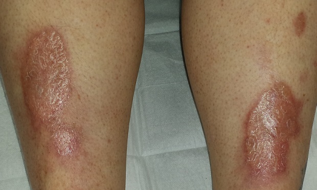
EXAMINATION
There are large, oval, reddish brown shiny plaques, measuring about 8 x 4 cm, on both anterior tibial areas. On closer inspection, the affected skin is yellowish pink and appears quite atrophic. This effect is more pronounced toward the centers of the lesions, which also display distinct telangiectasias. The borders of the plaques are slightly raised.
Examination of the rest of the patient’s skin reveals no other abnormalities.
What is the diagnosis?
DISCUSSION
Necrobiosis lipoidica (NL), though uncommon, is far from rare; it is seen somewhat regularly in dermatology practices. As this case demonstrates, it is one of hundreds of dermatologic diagnoses that you either know about or you don’t. Providers in the latter group invariably call it “fungal infection” because of its rounded borders—and because they simply have nothing else to call it.
True fungal infections are usually very superficial, involving only the outer layer of skin. This means they will manifest with scaling, a feature notably absent in this case. But more to the point, what was needed was a specific diagnosis, rather than another blind attempt at empiric treatment. Correct diagnosis dictates correct treatment.
NL was first described by Oppenhein in 1929, when he saw it in a diabetic patient. But it got its modern name in 1932 from Urbach, who thought it was invariably connected to diabetes. Now we know nondiabetic persons can develop it as well.
A number of theories exist as to the origins of this condition. One pinpoints microangiopathy of the same sort seen in the kidneys and eyes of diabetic patients. Another, proposed to account for the fact that NL is not exclusively seen in diabetes, holds that an autoimmune process might be involved—an opinion bolstered by the finding of increased TNF-alpha in the sera and skin of NL patients.
In any case, these atrophic plaques can grow quite large, eventually breaking down and ulcerating focally. Although the condition is usually asymptomatic, 25% of NL patients with advanced disease report pain.
Definitive diagnosis is made by punch biopsy, though visual diagnosis is quite adequate for those who can recognize the condition. NL lesions are most commonly seen on bilateral anterior tibial areas but can occasionally develop on the arm, face, or even genitals. The lesions can also koebnerize (ie, form along lines of trauma), occasionally being seen in surgical scars or even insulin injection sites.
Treatment, though largely unsatisfactory, includes topical and intralesional steroids. Particular care is taken to avoid or at least limit the formation of ulcers. In advanced cases, patients are referred to a wound care specialist. Oddly enough, neither the severity nor the prognosis of NL seem to have anything to do with how well or poorly the patient’s diabetes is controlled.
This patient was started on topical clobetasol cream, to be applied mostly to the actively advancing peripheral margin. Her prognosis, as with all NL patients, is guarded.
TAKE-HOME LEARNING POINTS
• Necrobiosis lipoidica (NL) is a disease of collagen degeneration that induces a granulomatous response that manifests microscopically with microangiopathy.
• NL usually appears on bilateral anterior tibial areas (but can affect the arms, face, or genitalia), as pinkish brown plaques with atrophic centers that tend to be yellow and telangiectatic.
• Though NL is usually associated with diabetes, it can develop in nondiabetic persons as well.
• The most feared complication of NL is eventual ulceration, which is why treatment is directed at taking care to avoid wounds to the plaques.
A 54-year-old woman self-refers to dermatology for evaluation of asymptomatic lesions on her legs. Over the past several years, they have slowly grown larger, redder, and shinier. She denies any other skin problems. Medical history is significant for type 2 diabetes, which, according to the patient, is under good control.
The patient has sought care from a variety of professionals, including her primary care provider, her endocrinologist, and her gynecologist. Various diagnoses, including “fungal infection,” have been posited; treatment with antifungal cream and oral medications produced no effect.
She also consulted the proprietor of her local health food store, who actually examined her before diagnosing “yeast infection” and advising her to change her diet.
EXAMINATION
There are large, oval, reddish brown shiny plaques, measuring about 8 x 4 cm, on both anterior tibial areas. On closer inspection, the affected skin is yellowish pink and appears quite atrophic. This effect is more pronounced toward the centers of the lesions, which also display distinct telangiectasias. The borders of the plaques are slightly raised.
Examination of the rest of the patient’s skin reveals no other abnormalities.
What is the diagnosis?
DISCUSSION
Necrobiosis lipoidica (NL), though uncommon, is far from rare; it is seen somewhat regularly in dermatology practices. As this case demonstrates, it is one of hundreds of dermatologic diagnoses that you either know about or you don’t. Providers in the latter group invariably call it “fungal infection” because of its rounded borders—and because they simply have nothing else to call it.
True fungal infections are usually very superficial, involving only the outer layer of skin. This means they will manifest with scaling, a feature notably absent in this case. But more to the point, what was needed was a specific diagnosis, rather than another blind attempt at empiric treatment. Correct diagnosis dictates correct treatment.
NL was first described by Oppenhein in 1929, when he saw it in a diabetic patient. But it got its modern name in 1932 from Urbach, who thought it was invariably connected to diabetes. Now we know nondiabetic persons can develop it as well.
A number of theories exist as to the origins of this condition. One pinpoints microangiopathy of the same sort seen in the kidneys and eyes of diabetic patients. Another, proposed to account for the fact that NL is not exclusively seen in diabetes, holds that an autoimmune process might be involved—an opinion bolstered by the finding of increased TNF-alpha in the sera and skin of NL patients.
In any case, these atrophic plaques can grow quite large, eventually breaking down and ulcerating focally. Although the condition is usually asymptomatic, 25% of NL patients with advanced disease report pain.
Definitive diagnosis is made by punch biopsy, though visual diagnosis is quite adequate for those who can recognize the condition. NL lesions are most commonly seen on bilateral anterior tibial areas but can occasionally develop on the arm, face, or even genitals. The lesions can also koebnerize (ie, form along lines of trauma), occasionally being seen in surgical scars or even insulin injection sites.
Treatment, though largely unsatisfactory, includes topical and intralesional steroids. Particular care is taken to avoid or at least limit the formation of ulcers. In advanced cases, patients are referred to a wound care specialist. Oddly enough, neither the severity nor the prognosis of NL seem to have anything to do with how well or poorly the patient’s diabetes is controlled.
This patient was started on topical clobetasol cream, to be applied mostly to the actively advancing peripheral margin. Her prognosis, as with all NL patients, is guarded.
TAKE-HOME LEARNING POINTS
• Necrobiosis lipoidica (NL) is a disease of collagen degeneration that induces a granulomatous response that manifests microscopically with microangiopathy.
• NL usually appears on bilateral anterior tibial areas (but can affect the arms, face, or genitalia), as pinkish brown plaques with atrophic centers that tend to be yellow and telangiectatic.
• Though NL is usually associated with diabetes, it can develop in nondiabetic persons as well.
• The most feared complication of NL is eventual ulceration, which is why treatment is directed at taking care to avoid wounds to the plaques.

Blocking two targets boosted fetal hemoglobin expression
Two distinct proteins appear to control the switch from fetal to adult globin, based on studies performed in a humanized mouse model and human cells. The findings suggest therapies that target both proteins might induce a fetal-type globin state, which could prove therapeutically useful in individuals with human hemoglobinopathies such as sickle cell disease and thalassemia.
The leukemia/lymphoma-related factor (LRF) and B-cell lymphoma/leukemia 11A (BCL11A) are independently involved in the switch, and blocking their production may turn on fetal globin expression, Takeshi Masuda, Ph.D., of Brigham and Women’s Hospital and Harvard Medical School, Boston, and his colleagues report (SCIENCE. 2015 Jan 15;351[6270]:285-9)
Using a humanized mouse model, the researchers knocked out the ZBTB7A gene, which is responsible for producing LRF. This action boosted the expression of genes that control fetal but not adult-type hemoglobin. Knocking out the gene in human cells also resulted in an increase in fetal hemoglobin proteins.
The researchers then examined BCL11A, which is involved with fetal hemoglobin but does not suppress it. When genes were knocked out for both ZBTB7A and BCL11A in the mice, fetal hemoglobin represented a 91%-94% greater percentage of total hemoglobin than when either gene alone was knocked out.
The research was supported by awards and/or grants from the National Institute of Diabetes and Digestive and Kidney Disease, the Doris Duke Charitable Foundation, the National Institutes of Health, and the American Society of Hematology. Dr. Masuda, along with two other study authors, is a contributor to a patent application filed on behalf of Brigham and Women’s Hospital related to therapeutic targeting of the pathways.
Click here to read the study at Science.
FROM SCIENCE
FDA rejects antisense oligonucleotide drug for Duchenne muscular dystrophy
The Food and Drug Administration has decided against approving the exon 51-skipping antisense oligonucleotide drug drisapersen for forms of Duchenne muscular dystrophy amenable to exon skipping.
The agency’s complete response letter to BioMarin Pharmaceutical, the developer of drisapersen (Kyndrisa), said that the standard of substantial evidence of effectiveness had not been met, according to a written statement from the company on Jan. 14.
In a Nov. 24, 2015, meeting of the FDA’s Peripheral and Central Nervous System Drugs Advisory Committee, panel members generally felt that drisapersen’s efficacy data were not persuasive enough for an approval.

Drisapersen targets frame-disrupting mutations found in exon 51 of the dystrophin gene, which produces a nonfunctional protein in individuals with a certain form of Duchenne, by restoring expression of the mutated dystrophin gene. There are currently no FDA-approved drugs to treat Duchenne, which affects approximately 1 in every 3,500 to 1 in 5,000 male children, making it the most common fatal genetic disorder diagnosed in childhood.
BioMarin said that extension studies of drisapersen will continue as the company determines the next steps in its new drug application, as will the ongoing clinical trials for other exon-skipping oligonucleotides it is developing. The drug is still under review by the European Medicines Agency.
The Food and Drug Administration has decided against approving the exon 51-skipping antisense oligonucleotide drug drisapersen for forms of Duchenne muscular dystrophy amenable to exon skipping.
The agency’s complete response letter to BioMarin Pharmaceutical, the developer of drisapersen (Kyndrisa), said that the standard of substantial evidence of effectiveness had not been met, according to a written statement from the company on Jan. 14.
In a Nov. 24, 2015, meeting of the FDA’s Peripheral and Central Nervous System Drugs Advisory Committee, panel members generally felt that drisapersen’s efficacy data were not persuasive enough for an approval.
Drisapersen targets frame-disrupting mutations found in exon 51 of the dystrophin gene, which produces a nonfunctional protein in individuals with a certain form of Duchenne, by restoring expression of the mutated dystrophin gene. There are currently no FDA-approved drugs to treat Duchenne, which affects approximately 1 in every 3,500 to 1 in 5,000 male children, making it the most common fatal genetic disorder diagnosed in childhood.
BioMarin said that extension studies of drisapersen will continue as the company determines the next steps in its new drug application, as will the ongoing clinical trials for other exon-skipping oligonucleotides it is developing. The drug is still under review by the European Medicines Agency.

2015 MS Highlights: The Year in Review
Click here to download the PDF.

Click here to download the PDF.
Click here to download the PDF.

Death from late effects of childhood cancer on decline
The rate of death from treatment-related late effects such as subsequent cancers and cardiopulmonary conditions has decreased among childhood cancer survivors, according to researchers.
At 15 years post diagnosis, the cumulative incidence of death from any cause for survivors diagnosed in the 1970s was 10.7%, 7.9% for those diagnosed in the 1980s, and 5.8% for those diagnosed in the 1990s (P less than .001). The cumulative incidence of death due to health-related causes, which include late effects of cancer therapy, were 3.1%, 2.4%, and 1.9%, respectively (P less than .001).

Results indicate that “the strategy of reducing treatment exposures in order to decrease the frequency of late effects is translating into a significant reduction in observed late mortality and an extension of the life span of children and adolescents who are successfully treated for cancer,” wrote Dr. Gregory Armstrong of St. Jude Children’s Research Hospital, Memphis, Tenn., and colleagues (N Engl J Med. 2016 Jan 13. doi: 10.1056/NEJMoa1510795).
A multivariate model showed that more recent treatment eras were associated with a reduced rate of death. The adjusted relative rate per every 5 years for death due to subsequent neoplasms was 0.83 (95% CI, 0.78-0.88), for cardiac causes, 0.77 (0.68-0.86), and for pulmonary causes, 0.77 (0.66-0.89).
Reductions across treatment eras in the rate of death from health-related causes were observed among survivors of acute lymphoblastic leukemia (3.2% in the early 1970s to 2.1% in the 1990s, P less than .001), Hodgkin lymphoma (5.3% to 2.6%, P = .006), Wilms tumor (2.6% to 0.4%, P = .005), and astrocytoma (4.7% to 1.8%, P = .02).
Temporal reductions in exposure to radiotherapy and anthracyclines occurred in treatment for acute lymphoblastic leukemia, Hodgkin lymphoma, Wilms tumor, and astrocytoma. Health-related mortality reductions were observed concurrently with reduced therapeutic exposures for acute lymphoblastic leukemia and Wilms tumor. For Hodgkin lymphoma and astrocytoma, other factors such as improved screening for late effects of cancer treatment appear to account for reductions in late health-related mortality.
For certain cancers, primarily neuroblastoma, late mortality has increased in recent decades. Increased therapeutic intensity has improved 5-year survival but increased the risk of late effects.
The reduced rate of death from recurrence or progression of primary cancers is the main driver to reductions in all-cause mortality, consistent with results from previous studies.
The retrospective Childhood Cancer Survivor Study evaluated 34,033 patients diagnosed at 31 hospitals in the United States and Canada from 1970 through 1999. In total, 3,958 deaths occurred, 2,002 due to primary cancer and 1,618 due to health-related causes: 746 subsequent neoplasms, 241 cardiac causes, 137 pulmonary causes, and 494 from other causes.
The rate of death from treatment-related late effects such as subsequent cancers and cardiopulmonary conditions has decreased among childhood cancer survivors, according to researchers.
At 15 years post diagnosis, the cumulative incidence of death from any cause for survivors diagnosed in the 1970s was 10.7%, 7.9% for those diagnosed in the 1980s, and 5.8% for those diagnosed in the 1990s (P less than .001). The cumulative incidence of death due to health-related causes, which include late effects of cancer therapy, were 3.1%, 2.4%, and 1.9%, respectively (P less than .001).
Results indicate that “the strategy of reducing treatment exposures in order to decrease the frequency of late effects is translating into a significant reduction in observed late mortality and an extension of the life span of children and adolescents who are successfully treated for cancer,” wrote Dr. Gregory Armstrong of St. Jude Children’s Research Hospital, Memphis, Tenn., and colleagues (N Engl J Med. 2016 Jan 13. doi: 10.1056/NEJMoa1510795).
A multivariate model showed that more recent treatment eras were associated with a reduced rate of death. The adjusted relative rate per every 5 years for death due to subsequent neoplasms was 0.83 (95% CI, 0.78-0.88), for cardiac causes, 0.77 (0.68-0.86), and for pulmonary causes, 0.77 (0.66-0.89).
Reductions across treatment eras in the rate of death from health-related causes were observed among survivors of acute lymphoblastic leukemia (3.2% in the early 1970s to 2.1% in the 1990s, P less than .001), Hodgkin lymphoma (5.3% to 2.6%, P = .006), Wilms tumor (2.6% to 0.4%, P = .005), and astrocytoma (4.7% to 1.8%, P = .02).
Temporal reductions in exposure to radiotherapy and anthracyclines occurred in treatment for acute lymphoblastic leukemia, Hodgkin lymphoma, Wilms tumor, and astrocytoma. Health-related mortality reductions were observed concurrently with reduced therapeutic exposures for acute lymphoblastic leukemia and Wilms tumor. For Hodgkin lymphoma and astrocytoma, other factors such as improved screening for late effects of cancer treatment appear to account for reductions in late health-related mortality.
For certain cancers, primarily neuroblastoma, late mortality has increased in recent decades. Increased therapeutic intensity has improved 5-year survival but increased the risk of late effects.
The reduced rate of death from recurrence or progression of primary cancers is the main driver to reductions in all-cause mortality, consistent with results from previous studies.
The retrospective Childhood Cancer Survivor Study evaluated 34,033 patients diagnosed at 31 hospitals in the United States and Canada from 1970 through 1999. In total, 3,958 deaths occurred, 2,002 due to primary cancer and 1,618 due to health-related causes: 746 subsequent neoplasms, 241 cardiac causes, 137 pulmonary causes, and 494 from other causes.
Key clinical point: Five-year survivors of childhood cancers have increased lifespans due in part to reduced rates of treatment-related late effects.
Major finding: At 15 years post diagnosis, the cumulative incidence of death from any cause for survivors diagnosed in the 1970s was 10.7%; in the 1980s, 7.9%; and in the 1990s, 5.8% (P less than .001). The cumulative incidence of death due to health-related causes, which include late effects of cancer therapy, were 3.1%, 2.4%, and 1.9%, respectively (P less than .001).
Data source: The retrospective Childhood Cancer Survivor Study, evaluating 34,033 patients diagnosed at 31 hospitals in the United States and Canada from 1970 through 1999; 3,958 deaths occurred.
Disclosures: Dr. Armstrong and coauthors reported having no disclosures.

Wound-healing template approved for diabetic foot ulcers
A bilayer matrix used for dermal regeneration and first approved in 1996 as a treatment for third-degree burns is now approved as a treatment for diabetic foot ulcers.
The Integra Dermal Regeneration Template was approved for the new indication based on a study that showed that the matrix device “improved ulcer healing compared to standard diabetic foot ulcer care,” according to a Food and Drug Administration statement announcing the approval on Jan. 7. Specifically, the new indication is for treating “partial and full-thickness neuropathic diabetic foot ulcers that are greater than 6 weeks in duration, with no capsule, tendon or bone exposed, when used in conjunction with standard diabetic ulcer care.”
The product is a dermal-replacement layer that “consists of a porous, three-dimensional matrix, comprised of bovine collagen and chondroitin-6-sulfate,” with a temporary epidermal silicone layer “to provide immediate wound coverage and control moisture loss. … [It] provides an environment for new skin and tissue to regenerate and heal the wound,” according to the agency’s approval summary.
In a multicenter, randomized controlled study, 307 patients were first treated with 0.9% sodium chloride gel, a secondary dressing, and an offloading device for 2 weeks and were then randomized to a treatment or a control group that received continued treatment with the gel. After 16 weeks, 51% of those treated with the device and 32% of those in the control group had healed completely (P = .001). Among those whose wounds healed, the median time to healing was 43 days in the treatment group and 78 days in the control group.
More patients in the control group had severe adverse events (26.8% vs. 15.6%) and moderate adverse events (42.5% vs. 31.8%).The results of the study, funded and sponsored by the manufacturer, were recently published (Wound Repair Regen. 2015;23[6]:891-900).
The product is contraindicated in patients with bovine or chondroitin allergies and in patients with infected wounds.
The manufacturer, Integra LifeSciences, is marketing the device as Integra Omnigraft Dermal Regeneration Matrix for the diabetic foot ulcer indication.
USPSTF Supports Mammography Starting at Age 50
Women aged 50-74 years should undergo biennial screening mammography, and the decision to screen before age 50 should be individualized, according to a final recommendation from the U.S. Preventive Services Task Force.

The recommendation statement, published Jan. 11 in Annals of Internal Medicine, is based on a comprehensive review of data since 2009, when the USPSTF last released breast cancer screening recommendations, and follows a public comment period in early 2015.
“The task force continues to find that the benefit of mammography increases with age, and recommends biennial screening in women ages 50 to 74. Mammography can also be effective for women in their 40s, but the benefits are less and the harms potentially greater. The decision by women to start screening in their 40s should be an individual one, made in partnership with a doctor,” according to a statement from the USPSTF.
The new recommendations will not affect private insurance coverage of mammography without cost sharing as outlined in the Affordable Care Act. As part of a spending bill enacted in December 2015, Congress passed a provision placing a 2-year moratorium on the use of any new USPSTF recommendations related to breast cancer screening to determine coverage, effectively preserving mammography coverage without cost sharing for women aged 40 years and older.
Recommendations by age
The latest USPSTF recommendations by age state that women aged 40-49 years should base their screening decision on personal values, preferences, and health history; women with a family history of breast cancer may benefit more than average-risk women by beginning screening before age 50. This is a C recommendation, indicating “moderate certainty that net benefit is small.”
The recommendation for biennial screening of those aged 50-74 is a B recommendation indicating “high certainty that the net benefit is moderate or there is moderate certainty that the net benefit is moderate to substantial,” Dr. Albert L. Siu, task force chair, reported on behalf of the USPSTF (Ann Intern Med. 2016;164:279-96. doi: 10.7326/M15-2886).
The task force found inadequate evidence to recommend for or against screening those aged 75 and older.
Final evidence documents, including a systematic review of data on the harms associated with breast cancer screening and a modeling study of the benefits and harms associated with different screening strategies, are published along with the recommendation statement.
The USPSTF did not make a recommendations about the use of digital breast tomosynthesis as a primary screening method for breast cancer, noting that the current evidence is insufficient. Evidence also was insufficient to make a recommendation on the benefits and harms of adjunctive screening for breast cancer using breast ultrasonography, magnetic resonance imaging, digital breast tomosynthesis, or other methods in women with dense breasts who had a negative screening mammogram.
Dousing the ‘firestorm’
In an editorial penned by Annals of Internal Medicine Editor-in-Chief and Senior Vice President of the American College of Physicians Christine Laine and her colleagues, they urged a dousing of the “firestorm around breast cancer screening.”
That firestorm was ignited with the 2009 USPSTF breast cancer screening recommendation and was rekindled when the current recommendation was presented in draft form in 2015.
However, “the USPSTF did a difficult job well” and based its recommendations on an important understanding of the updated evidence, as well as potential harms and tradeoffs of different screening strategies, the authors wrote.
“When the USPSTF posted its draft recommendations for comment, it noted, ‘Women deserve to be aware of what the science says so they can make the best choice for themselves, together with their doctor.’ We could not agree more. Let’s douse the flames and clear the smoke so that we can clearly see what the evidence shows and where we need to focus efforts to fill gaps in our knowledge so that women, along with their health care providers, can make the best decision to reduce their risk for breast cancer–related morbidity and mortality,” they wrote (Ann Intern Med. 2016 Jan 11. doi:10.7326/M15-2978).
ACOG supports screening at 40
The American College of Obstetricians and Gynecologists is standing by its recommendation of annual mammograms beginning at age 40 and continues to support use of clinical breast examinations. In a Jan. 11 statement, Dr. Mark S. DeFrancesco, ACOG president, said that “evidence and experience have shown that early detection can lead to improved outcomes in women diagnosed with breast cancer.”
The organization similarly stood by its recommendation in October 2015, when the American Cancer Society released recommendations for annual screening mammography for asymptomatic women at average risk for breast cancer beginning at age 45 years, with a transition to biennial screening mammography beginning at age 55 (JAMA. 2015;314[15]:1599-1614. doi: 10.1001/jama.2015.12783).
ACOG also supports the omnibus legislation passed by Congress in December that provides 2 years of no-copay coverage of breast cancer screening after age 40 via a moratorium on new breast cancer screening recommendations to allow time for additional research, an ACOG spokesperson said in an interview.
“ACOG strongly supports shared decision-making between doctor and patient, and in the case of screening for breast cancer, it is essential,” Dr. DeFrancesco said. “Given the differences among current organizational recommendations on breast cancer screening, we recognize that there may be confusion among women about when they should begin screening for breast cancer. ACOG encourages women to discuss this with their doctor, including concerns such as family history of cancer, risk factors such as overweight, and their own personal experiences with breast cancer. Moreover, it is essential that physicians counsel women about the potential consequences of mammography, including false positives.”

ACOG will convene a consensus conference later in January “with the intent to develop a consistent set of uniform guidelines for breast cancer screening that can be implemented nationwide” in an effort to “avoid the confusion that currently exists among the women we treat,” according to the statement.
The issue of divergence – and convergence – among various guidelines was the topic of another editorial published in conjunction with the USPSTF recommendations. In that article, task force chair Dr. Siu and his colleagues acknowledged that disagreements exist but stressed that “it would be a disservice to women and their clinicians if these disagreements obscured a strong emerging convergence among groups who have recently issued evidence-based guidelines” (Ann Intern Med. 2016 Jan 11. doi:10.7326/M15-3065).
The operations of the USPSTF are supported by the Agency for Healthcare Research and Quality. One of the members of the USPSTF reported receiving past grants and contracts from the National Cancer Institute and the Centers for Disease Control and Prevention.
Women aged 50-74 years should undergo biennial screening mammography, and the decision to screen before age 50 should be individualized, according to a final recommendation from the U.S. Preventive Services Task Force.
The recommendation statement, published Jan. 11 in Annals of Internal Medicine, is based on a comprehensive review of data since 2009, when the USPSTF last released breast cancer screening recommendations, and follows a public comment period in early 2015.
“The task force continues to find that the benefit of mammography increases with age, and recommends biennial screening in women ages 50 to 74. Mammography can also be effective for women in their 40s, but the benefits are less and the harms potentially greater. The decision by women to start screening in their 40s should be an individual one, made in partnership with a doctor,” according to a statement from the USPSTF.
The new recommendations will not affect private insurance coverage of mammography without cost sharing as outlined in the Affordable Care Act. As part of a spending bill enacted in December 2015, Congress passed a provision placing a 2-year moratorium on the use of any new USPSTF recommendations related to breast cancer screening to determine coverage, effectively preserving mammography coverage without cost sharing for women aged 40 years and older.
Recommendations by age
The latest USPSTF recommendations by age state that women aged 40-49 years should base their screening decision on personal values, preferences, and health history; women with a family history of breast cancer may benefit more than average-risk women by beginning screening before age 50. This is a C recommendation, indicating “moderate certainty that net benefit is small.”
The recommendation for biennial screening of those aged 50-74 is a B recommendation indicating “high certainty that the net benefit is moderate or there is moderate certainty that the net benefit is moderate to substantial,” Dr. Albert L. Siu, task force chair, reported on behalf of the USPSTF (Ann Intern Med. 2016;164:279-96. doi: 10.7326/M15-2886).
The task force found inadequate evidence to recommend for or against screening those aged 75 and older.
Final evidence documents, including a systematic review of data on the harms associated with breast cancer screening and a modeling study of the benefits and harms associated with different screening strategies, are published along with the recommendation statement.
The USPSTF did not make a recommendations about the use of digital breast tomosynthesis as a primary screening method for breast cancer, noting that the current evidence is insufficient. Evidence also was insufficient to make a recommendation on the benefits and harms of adjunctive screening for breast cancer using breast ultrasonography, magnetic resonance imaging, digital breast tomosynthesis, or other methods in women with dense breasts who had a negative screening mammogram.
Dousing the ‘firestorm’
In an editorial penned by Annals of Internal Medicine Editor-in-Chief and Senior Vice President of the American College of Physicians Christine Laine and her colleagues, they urged a dousing of the “firestorm around breast cancer screening.”
That firestorm was ignited with the 2009 USPSTF breast cancer screening recommendation and was rekindled when the current recommendation was presented in draft form in 2015.
However, “the USPSTF did a difficult job well” and based its recommendations on an important understanding of the updated evidence, as well as potential harms and tradeoffs of different screening strategies, the authors wrote.
“When the USPSTF posted its draft recommendations for comment, it noted, ‘Women deserve to be aware of what the science says so they can make the best choice for themselves, together with their doctor.’ We could not agree more. Let’s douse the flames and clear the smoke so that we can clearly see what the evidence shows and where we need to focus efforts to fill gaps in our knowledge so that women, along with their health care providers, can make the best decision to reduce their risk for breast cancer–related morbidity and mortality,” they wrote (Ann Intern Med. 2016 Jan 11. doi:10.7326/M15-2978).
ACOG supports screening at 40
The American College of Obstetricians and Gynecologists is standing by its recommendation of annual mammograms beginning at age 40 and continues to support use of clinical breast examinations. In a Jan. 11 statement, Dr. Mark S. DeFrancesco, ACOG president, said that “evidence and experience have shown that early detection can lead to improved outcomes in women diagnosed with breast cancer.”
The organization similarly stood by its recommendation in October 2015, when the American Cancer Society released recommendations for annual screening mammography for asymptomatic women at average risk for breast cancer beginning at age 45 years, with a transition to biennial screening mammography beginning at age 55 (JAMA. 2015;314[15]:1599-1614. doi: 10.1001/jama.2015.12783).
ACOG also supports the omnibus legislation passed by Congress in December that provides 2 years of no-copay coverage of breast cancer screening after age 40 via a moratorium on new breast cancer screening recommendations to allow time for additional research, an ACOG spokesperson said in an interview.
“ACOG strongly supports shared decision-making between doctor and patient, and in the case of screening for breast cancer, it is essential,” Dr. DeFrancesco said. “Given the differences among current organizational recommendations on breast cancer screening, we recognize that there may be confusion among women about when they should begin screening for breast cancer. ACOG encourages women to discuss this with their doctor, including concerns such as family history of cancer, risk factors such as overweight, and their own personal experiences with breast cancer. Moreover, it is essential that physicians counsel women about the potential consequences of mammography, including false positives.”
ACOG will convene a consensus conference later in January “with the intent to develop a consistent set of uniform guidelines for breast cancer screening that can be implemented nationwide” in an effort to “avoid the confusion that currently exists among the women we treat,” according to the statement.
The issue of divergence – and convergence – among various guidelines was the topic of another editorial published in conjunction with the USPSTF recommendations. In that article, task force chair Dr. Siu and his colleagues acknowledged that disagreements exist but stressed that “it would be a disservice to women and their clinicians if these disagreements obscured a strong emerging convergence among groups who have recently issued evidence-based guidelines” (Ann Intern Med. 2016 Jan 11. doi:10.7326/M15-3065).
The operations of the USPSTF are supported by the Agency for Healthcare Research and Quality. One of the members of the USPSTF reported receiving past grants and contracts from the National Cancer Institute and the Centers for Disease Control and Prevention.
FROM ANNALS OF INTERNAL MEDICINE