User login
Geriatric IBD hospitalization carries steep inpatient mortality
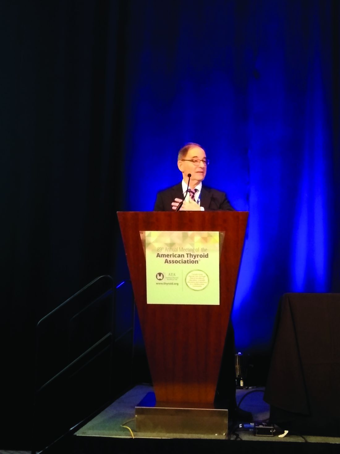
SAN ANTONIO – Jeffrey Schwartz, MD, reported at the annual meeting of the American College of Gastroenterology.

The magnitude of the age-related increased risk highlighted in this large national study was strikingly larger than the differential inpatient mortality between geriatric and nongeriatric patients hospitalized for conditions other than inflammatory bowel disease (IBD). It’s a finding that reveals a major unmet need for improved systems of care for elderly hospitalized IBD patients, according to Dr. Schwartz, an internal medicine resident at Beth Israel Deaconess Medical Center, Boston.
“Given the high prevalence of IBD patients that require inpatient admission, as well as the rapidly aging nature of the U.S. population, it’s our hope that this study will provide some insight to drive efforts to improve standardized guideline-directed therapy and propose interventions to help close what I think is a very important gap in clinical care,” he said.
It’s well established that a second peak of IBD diagnoses occurs in 50- to 70-year-olds. At present, roughly 30% of all individuals carrying the diagnosis of IBD are over age 65, and with the graying of the baby-boomer population, this proportion is climbing.
Dr. Schwartz presented a study of the National Inpatient Sample for 2016, which is a representative sample comprising 20% of all U.S. hospital discharges for that year, the most recent year for which the data are available. The study population included all 71,040 patients hospitalized for acute management of Crohn’s disease or its immediate complications, of whom 10,095 were aged over age 75 years, as well as the 35,950 patients hospitalized for ulcerative colitis, 8,285 of whom were over 75.
Inpatient mortality occurred in 1.5% of the geriatric admissions, compared with 0.2% of nongeriatric admissions for Crohn’s disease. Similarly, the inpatient mortality rate in geriatric patients with ulcerative colitis was 1.0% versus 0.1% in patients under age 75 hospitalized for ulcerative colitis.
There are lots of reasons why the management of geriatric patients with IBD is particularly challenging, Dr. Schwartz noted. They have a higher burden of comorbid conditions, worse nutritional status, and increased risks of infection and cancer. In a regression analysis that attempted to control for such confounders using the Elixhauser mortality index, the nongeriatric Crohn’s disease patients were an adjusted 75% less likely to die in the hospital than those who were older. Nongeriatric ulcerative colitis patients were 81% less likely to die than geriatric patients with the disease. In contrast, nongeriatric patients admitted for reasons other than IBD had only an adjusted 50% lower risk of inpatient mortality than those who were older than 75.
Of note, in this analysis adjusted for confounders, there was no difference between geriatric and nongeriatric IBD patients in terms of resource utilization as reflected in average length of stay and hospital charges, Dr. Schwartz continued.
Asked if he could shed light on any specific complications that drove the age-related disparity in inpatient mortality in the IBD population, the physician replied that he and his coinvestigators were thwarted in their effort to do so because the inpatient mortality of 1.0%-1.5% was so low that further breakdown as to causes of death would have been statistically unreliable. It might be possible to do so successfully by combining several years of National Inpatient Sample data. That being said, it’s reasonable to hypothesize that cardiovascular complications are an important contributor, he added.
Dr. Schwartz reported having no financial conflicts regarding his study, conducted free of commercial support.
SAN ANTONIO – Jeffrey Schwartz, MD, reported at the annual meeting of the American College of Gastroenterology.
The magnitude of the age-related increased risk highlighted in this large national study was strikingly larger than the differential inpatient mortality between geriatric and nongeriatric patients hospitalized for conditions other than inflammatory bowel disease (IBD). It’s a finding that reveals a major unmet need for improved systems of care for elderly hospitalized IBD patients, according to Dr. Schwartz, an internal medicine resident at Beth Israel Deaconess Medical Center, Boston.
“Given the high prevalence of IBD patients that require inpatient admission, as well as the rapidly aging nature of the U.S. population, it’s our hope that this study will provide some insight to drive efforts to improve standardized guideline-directed therapy and propose interventions to help close what I think is a very important gap in clinical care,” he said.
It’s well established that a second peak of IBD diagnoses occurs in 50- to 70-year-olds. At present, roughly 30% of all individuals carrying the diagnosis of IBD are over age 65, and with the graying of the baby-boomer population, this proportion is climbing.
Dr. Schwartz presented a study of the National Inpatient Sample for 2016, which is a representative sample comprising 20% of all U.S. hospital discharges for that year, the most recent year for which the data are available. The study population included all 71,040 patients hospitalized for acute management of Crohn’s disease or its immediate complications, of whom 10,095 were aged over age 75 years, as well as the 35,950 patients hospitalized for ulcerative colitis, 8,285 of whom were over 75.
Inpatient mortality occurred in 1.5% of the geriatric admissions, compared with 0.2% of nongeriatric admissions for Crohn’s disease. Similarly, the inpatient mortality rate in geriatric patients with ulcerative colitis was 1.0% versus 0.1% in patients under age 75 hospitalized for ulcerative colitis.
There are lots of reasons why the management of geriatric patients with IBD is particularly challenging, Dr. Schwartz noted. They have a higher burden of comorbid conditions, worse nutritional status, and increased risks of infection and cancer. In a regression analysis that attempted to control for such confounders using the Elixhauser mortality index, the nongeriatric Crohn’s disease patients were an adjusted 75% less likely to die in the hospital than those who were older. Nongeriatric ulcerative colitis patients were 81% less likely to die than geriatric patients with the disease. In contrast, nongeriatric patients admitted for reasons other than IBD had only an adjusted 50% lower risk of inpatient mortality than those who were older than 75.
Of note, in this analysis adjusted for confounders, there was no difference between geriatric and nongeriatric IBD patients in terms of resource utilization as reflected in average length of stay and hospital charges, Dr. Schwartz continued.
Asked if he could shed light on any specific complications that drove the age-related disparity in inpatient mortality in the IBD population, the physician replied that he and his coinvestigators were thwarted in their effort to do so because the inpatient mortality of 1.0%-1.5% was so low that further breakdown as to causes of death would have been statistically unreliable. It might be possible to do so successfully by combining several years of National Inpatient Sample data. That being said, it’s reasonable to hypothesize that cardiovascular complications are an important contributor, he added.
Dr. Schwartz reported having no financial conflicts regarding his study, conducted free of commercial support.
REPORTING FROM ACG 2019
Key clinical point: A major unmet need exists for better guideline-directed management of geriatric patients hospitalized for inflammatory bowel disease.
Major finding: The inpatient mortality rate among patients aged over age 75 years hospitalized for management of inflammatory bowel disease is four to five times higher than in those who are younger.
Study details: This was a retrospective analysis of all 106,990 hospital admissions for management of inflammatory bowel disease included in the 2016 National Inpatient Sample.
Disclosures: The presenter reported having no financial conflicts regarding his study, conducted free of commercial support.
Source: Schwartz J. ACG 2019, Abstract 42.
Hyperkalemia-related treatment changes linked to death in acute HF
The hyperkalemia that commonly occurs in patients hospitalized for acute heart failure does not affect outcomes, but it can lead to treatment changes that can in turn raise the risk of mortality.
That’s according to an analysis of data from 1,589 patients in the PROTECT trial (Placebo-Controlled Randomized Study of the Selective A1 Adenosine Receptor Antagonist Rolofylline for Patients Hospitalized with Acute Decompensated Heart Failure and Volume Overload to Assess Treatment Effect on Congestion and Renal Function) (N Engl J Med. 2010;363:1419-28).
In PROTECT, patients with acute heart failure and mild or moderate renal impairment (estimated creatinine clearance of 20-80 mL/min) were enrolled and randomized to receive placebo or rolofylline, a selective A1 adenosine receptor antagonist that is no longer in development. Because of the meticulous recording of potassium levels in PROTECT, investigators led by Joost C. Beusekamp of the University of Groningen, the Netherlands, used the data to examine the relations between incident hyperkalemia and changes in treatment, focusing on mineralocorticoid antagonists (MRAs).
They found that of the 35% of the patients who developed hyperkalemia at least once during hospitalization, defined as at least one episode of potassium above 5.0 mEq/L, 53% had been taking MRAs before hospitalization. And of those patients who been taking MRAs before hospitalization, 35% and 44% developed incident hypokalemia and had “a normal potassium” level, respectively. The hyperkalemia patients were also more likely to have their MRAs down-titrated (15%) during their stay than were those with low (8%) and normal (9%) potassium levels.
No significant association was found between in-hospital potassium levels and 180-day mortality or a composite of rehospitalization for cardiovascular or renal causes or all-cause death at 30 days (data not provided). However, there was a significant link between MRA dose reductions and 180-day mortality in a multivariate analysis (HR, 1.73; 95% confidence interval, 1.15-2.60; P = 0.008).
“Incident hyperkalemia was strongly associated with down-titration of MRA therapy which was, in turn, associated with a worse prognosis,” the investigators concluded.
SOURCE: J Am Coll Cardiol HF. 2019 Oct 9. doi: 10.1016/j.jchf.2019.07.010.
FROM JACC: HEART FAILURE
Apps for busy pediatric hospitalists 2.0
PHM19 session
Apps for busy pediatric hospitalists 2.0

Presenters
Tosin Adeyanju, MD, FAAP
Alexander Hogan, MD
Jane Im, MD, FAAP
Kim O’Hara, MD
Michael Tchou, MD, FAAP
Session summary
This presentation at Pediatric Hospital Medicine 2019 started with the sharing of learning tools to help physicians stay current and organized with the ever-expanding body of medical literature.
The instructors shared content aggregators, such as Read by QxMD, that allow the user to follow multiple journals and highlight new articles based on the user’s preferences and chosen keywords. They also shared reference managers, such as Mendeley, which allows users to organize, store, and access their literature library from anywhere and can even be used to simplify citations and bibliographies in articles.
The presenters shared resources and applications that can be used to quickly access information on mobile devices. Applications, such as MDCalc and the CDC STD Tx Guide, can allow users to reference clinical calculators and treatment courses for teaching at the bedside. The presenters also introduced pharmaceutical applications like GoodRx, an application that allows patients and physicians to compare drug prices at various pharmacies. They also introduced the audience to Formulary Search by MMIT that helps users determine which medications are covered by an insurance plan. They also shared some applications that can help users deal with emergencies, like Ped Guide and Pedi Crisis. These apps can help users review emergency algorithms, dose emergency medications, and determine the sizes of emergency equipment.
The presenters closed by sharing teaching applications that allow users to increase interactions with presentation audiences or learners. Teaching tools like Kahoot! and Poll Everywhere allow users to gauge their audiences’ understanding of material. Online software, such as Slack.com and Microsoft.com, allows for collaboration and file sharing across institutions and integrate with many other services.
Key takeaways
• Content aggregators and reference managers help users organize and access literature from anywhere.
• Teaching tools encourage audience participation, immediate assessment of learners.
• Online software tools allow for easy collaboration and file sharing across institutions and easily integrate with many other services.
Dr. Gupta is a pediatric hospitalist at Phoenix Children’s Hospital.
PHM19 session
Apps for busy pediatric hospitalists 2.0
Presenters
Tosin Adeyanju, MD, FAAP
Alexander Hogan, MD
Jane Im, MD, FAAP
Kim O’Hara, MD
Michael Tchou, MD, FAAP
Session summary
This presentation at Pediatric Hospital Medicine 2019 started with the sharing of learning tools to help physicians stay current and organized with the ever-expanding body of medical literature.
The instructors shared content aggregators, such as Read by QxMD, that allow the user to follow multiple journals and highlight new articles based on the user’s preferences and chosen keywords. They also shared reference managers, such as Mendeley, which allows users to organize, store, and access their literature library from anywhere and can even be used to simplify citations and bibliographies in articles.
The presenters shared resources and applications that can be used to quickly access information on mobile devices. Applications, such as MDCalc and the CDC STD Tx Guide, can allow users to reference clinical calculators and treatment courses for teaching at the bedside. The presenters also introduced pharmaceutical applications like GoodRx, an application that allows patients and physicians to compare drug prices at various pharmacies. They also introduced the audience to Formulary Search by MMIT that helps users determine which medications are covered by an insurance plan. They also shared some applications that can help users deal with emergencies, like Ped Guide and Pedi Crisis. These apps can help users review emergency algorithms, dose emergency medications, and determine the sizes of emergency equipment.
The presenters closed by sharing teaching applications that allow users to increase interactions with presentation audiences or learners. Teaching tools like Kahoot! and Poll Everywhere allow users to gauge their audiences’ understanding of material. Online software, such as Slack.com and Microsoft.com, allows for collaboration and file sharing across institutions and integrate with many other services.
Key takeaways
• Content aggregators and reference managers help users organize and access literature from anywhere.
• Teaching tools encourage audience participation, immediate assessment of learners.
• Online software tools allow for easy collaboration and file sharing across institutions and easily integrate with many other services.
Dr. Gupta is a pediatric hospitalist at Phoenix Children’s Hospital.
Findings confirm link between methimazole and risk for acute pancreatitis
CHICAGO –
After 6 months of methimazole use, the odds ratio for acute pancreatitis was 2.02, with a nonsignificant risk elevation for propylthiouracil use after a similar duration, Laszlo Hegedüs, MD, reported at the annual meeting of the American Thyroid Association.
“Ongoing methimazole, but not propylthiouracil, use is associated with an increased risk of acute pancreatitis,” he said.
Dr. Hegedüs, professor of endocrinology and metabolism at the University of Southern Denmark, Odense, said that the European Medicines Agency has noted a few postmarketing reports of acute pancreatitis in patients who received the antithyroid drug methimazole, as well as its prodrug, carbimazole. The agency has accordingly contraindicated antithyroid drug use for patients who previously experienced acute pancreatitis after receiving this drug, advising that methimazole should be “discontinued immediately” should a patient develop acute pancreatitis.
However, investigation of the antithyroid drug–pancreatitis association had been limited to aggregating those case reports, so Dr. Hegedüs and colleagues decided to use Danish medical record and registry data to investigate the association in a nationwide, controlled study that looked at both duration of therapy and total antithyroid drug use.
During the period from 1995-2018, a total of 118,649 patients who used antithyroid drugs were found in the 5.5 million individuals in the Statistics Denmark registry. Dr. Hegedüs and his colleagues also pulled in patient registry and national prescription registry data, as well as civil vital statistics data.
Of those who used antithyroid drugs, 103,825 patients used methimazole, and 14,824 used propylthiouracil. The researchers found 43,580 instances of hospitalization for first-time acute pancreatitis in the pooled antithyroid drug data. Of those, however, just 226 (0.5%) occurred in patients using methimazole, and 19 (0.04%) in those using propylthiouracil at the time of pancreatitis onset.
To ascertain the risk of acute pancreatitis in patients using antithyroid drugs for various durations, Dr. Hegedüs and his colleagues used a case-crossover study design. In the case-crossover technique, patients served as their own controls, because each patient was both exposed and not exposed to antithyroid drugs at some point during the study period. Antithyroid drugs are well suited to this study design, explained Dr. Hegedüs, because they are given for a limited time. A case-crossover design can be used with a small sample size and effectively controls for potentially confounding variables.
The odds ratio for acute pancreatitis in methimazole users after 3 months of exposure was 1.51, with a 95% confidence interval of 1.12-2.02. After 3 months of propylthiouracil exposure, the odds ratio for acute pancreatitis was 1.16 (95% CI 0.46-2.3). At 6 months, the odds ratio of 2.02 for methimazole was similarly statistically significant (95% CI, 1.50-2.78), whereas the odds ratio of 1.40 for propylthiouracil use was not significant (95% CI, 0.58-3.34).
The researchers also wanted to find out whether the cumulative drug dose affected the risk of acute pancreatitis, so they drew from the antithyroid drug population to conduct a case-control study. Here, the investigators matched data from four control patients to each case of acute pancreatitis. The researchers also controlled for sex, age, comorbidities, and prior use of drugs associated with pancreatitis.
Overall, 20% of the 692 methimazole users and their controls were men, as were 16% of the 108 propylthiouracil users, in the case-control study.
Just more than half of patients overall had a total dose exposure of 200 to 1,200 defined daily dose (DDD) – a measure developed by the World Health Organization to denote the assumed average adult dose per day of a medication – with about a quarter of patients receiving a total antithyroid drug dose more than 1,200 DDD and about 20% receiving a dose exposure of less than 200 DDD. The risk of acute pancreatitis did not increase with increased total exposure to antithyroid drugs.
“There is no evidence of a cumulative dose effect of either methimazole or propylthiouracil on the risk of acute pancreatitis,” said Dr. Hegedüs. However, “the warning of the European Medicines Agency seems justified,” he added. “The frequency of acute pancreatitis in acute methimazole users is of a similar magnitude [to that] reported for agranulocytosis,” a known, dire complication of antithyroid drug use. Patients should be advised of the potential complication and informed of signs and symptoms of acute pancreatitis, he said.
Dr. Hegedüs noted that the study had the advantage of using validated epidemiologic methods to look at drug exposure and outcomes at a nationwide scale. However, the registries from which the data were drawn also have limitations. The investigators could not determine the severity of hyperthyroidism, he said, and the relatively rare occurrence of acute pancreatitis meant that there was not sufficient statistical power to look at the subgroup of individuals who had Graves disease and to compare them with those with nodular toxic goiter.
He advised conducting a confirmatory study in an independent cohort, as well as further investigating the yet unknown mechanism of action for the link between the antithyroid drug and acute pancreatitis.
Dr. Hegedüs reported that he had no relevant conflicts of interest and reported no outside sources of funding.
SOURCE: Hegedüs, L. et al. ATA 2019, Short Call Oral Abstract 6 .
REPORTING FROM ATA 2019
Ask about vaping in patients with respiratory symptoms, CDC says
“Do you vape?” may be one of the most important questions health care can providers can ask patients who present with respiratory symptoms this winter.

according to the Centers for Disease Control and Prevention.
Accordingly, providers need to ask patients with respiratory, gastrointestinal, or constitutional symptoms about their use of e-cigarette or vaping products, according to one several new CDC recommendations that appear in the Morbidity and Mortality Weekly Review.
“E-cigarette or vaping product use–associated lung injury (EVALI) remains a diagnosis of exclusion because, at present, no specific test or marker exists for its diagnosis, and evaluation should be guided by clinical judgment,” the CDC report reads.
As of Nov. 13, there have been 2,172 cases of EVALI reported to CDC, of which 42 (1.9%) have been fatal. Most of the patients with EVALI have been white (79%), male (68%), and under the age of 35 years (77%), according to CDC data.
Although vitamin E acetate was recently implicated as a potential cause of EVALI, the agency said evidence is “not sufficient” at this point in their investigation to rule out other chemicals of potential concern.
“Many different substances and product sources are still under investigation, and it might be that there is more than one cause of this outbreak,” CDC said.
Further recommendations
Beyond asking about vape use, providers should evaluate suspected EVALI with pulse oximetry and chest imaging, and should consider outpatient management for patients who are clinically stable, according to the recommendations.
The agency said influenza testing should be “strongly considered,” especially during influenza season, given that EVALI is a diagnosis of exclusion and that it may co-occur with other respiratory illnesses. Antimicrobials (including antivirals) should be given as warranted, they added.
Corticosteroids may be helpful in treating EVALI, but may worsen respiratory infections typically seen in outpatients, and so should be prescribed with caution in the outpatient setting, the CDC recommended.
Behavioral counseling, addiction treatment services, and Food and Drug Administration–approved cessation medications are recommended to help patients quit vaping or e-cigarette products, CDC said.
Health care providers should emphasize the importance of an annual flu shot for all patients 6 months of age or older, including those who use e-cigarette or vaping products, according to the agency.
“It is not known whether patients with EVALI are at higher risk for severe complications of influenza or other respiratory infections,” the report reads.
Blame it on vitamin E? THC? Other?
The report details how, as previously reported, vitamin E acetate was detected in bronchoalveolar lavage fluid samples from 29 patients with EVALI. Although other chemicals could contribute to EVALI, that finding provided “direct evidence” of vitamin E acetate at the primary site of injury, according to CDC.
Most patients with EVALI, 83%, have reported using a tetrahydrocannabinol (THC)-containing e-cigarette or vaping product, according to CDC, while 61% reported using a nicotine-containing product.
Based on that, CDC recommended that people avoid using THC-containing products. However, the agency cautioned that the specific cause or causes of EVALI remain to be elucidated.
“The only way for persons to assure that they are not at risk is to consider refraining from use of all e-cigarette, or vaping, products while this investigation continues,” CDC said in the report.
The need for this additional clinical guidance was assessed in anticipation of the seasonal uptick in influenza and other respiratory infections, according to the CDC, which said the recommendations were based in part on individual clinical perspectives from nine national experts who participated in a previously published clinical guidance on managing patients with EVALI.
SOURCES: Jatlaoui TC et al. MMWR Morb Mortal Wkly Rep. 2019 Nov 19. doi. 10.15585/mmwr.mm6846e2; Chatham-Stephens K et al. MMWR Morb Mortal Wkly Rep. 2019 Nov 19. doi. 10.15585/mmwr.mm6846e1.
“Do you vape?” may be one of the most important questions health care can providers can ask patients who present with respiratory symptoms this winter.
according to the Centers for Disease Control and Prevention.
Accordingly, providers need to ask patients with respiratory, gastrointestinal, or constitutional symptoms about their use of e-cigarette or vaping products, according to one several new CDC recommendations that appear in the Morbidity and Mortality Weekly Review.
“E-cigarette or vaping product use–associated lung injury (EVALI) remains a diagnosis of exclusion because, at present, no specific test or marker exists for its diagnosis, and evaluation should be guided by clinical judgment,” the CDC report reads.
As of Nov. 13, there have been 2,172 cases of EVALI reported to CDC, of which 42 (1.9%) have been fatal. Most of the patients with EVALI have been white (79%), male (68%), and under the age of 35 years (77%), according to CDC data.
Although vitamin E acetate was recently implicated as a potential cause of EVALI, the agency said evidence is “not sufficient” at this point in their investigation to rule out other chemicals of potential concern.
“Many different substances and product sources are still under investigation, and it might be that there is more than one cause of this outbreak,” CDC said.
Further recommendations
Beyond asking about vape use, providers should evaluate suspected EVALI with pulse oximetry and chest imaging, and should consider outpatient management for patients who are clinically stable, according to the recommendations.
The agency said influenza testing should be “strongly considered,” especially during influenza season, given that EVALI is a diagnosis of exclusion and that it may co-occur with other respiratory illnesses. Antimicrobials (including antivirals) should be given as warranted, they added.
Corticosteroids may be helpful in treating EVALI, but may worsen respiratory infections typically seen in outpatients, and so should be prescribed with caution in the outpatient setting, the CDC recommended.
Behavioral counseling, addiction treatment services, and Food and Drug Administration–approved cessation medications are recommended to help patients quit vaping or e-cigarette products, CDC said.
Health care providers should emphasize the importance of an annual flu shot for all patients 6 months of age or older, including those who use e-cigarette or vaping products, according to the agency.
“It is not known whether patients with EVALI are at higher risk for severe complications of influenza or other respiratory infections,” the report reads.
Blame it on vitamin E? THC? Other?
The report details how, as previously reported, vitamin E acetate was detected in bronchoalveolar lavage fluid samples from 29 patients with EVALI. Although other chemicals could contribute to EVALI, that finding provided “direct evidence” of vitamin E acetate at the primary site of injury, according to CDC.
Most patients with EVALI, 83%, have reported using a tetrahydrocannabinol (THC)-containing e-cigarette or vaping product, according to CDC, while 61% reported using a nicotine-containing product.
Based on that, CDC recommended that people avoid using THC-containing products. However, the agency cautioned that the specific cause or causes of EVALI remain to be elucidated.
“The only way for persons to assure that they are not at risk is to consider refraining from use of all e-cigarette, or vaping, products while this investigation continues,” CDC said in the report.
The need for this additional clinical guidance was assessed in anticipation of the seasonal uptick in influenza and other respiratory infections, according to the CDC, which said the recommendations were based in part on individual clinical perspectives from nine national experts who participated in a previously published clinical guidance on managing patients with EVALI.
SOURCES: Jatlaoui TC et al. MMWR Morb Mortal Wkly Rep. 2019 Nov 19. doi. 10.15585/mmwr.mm6846e2; Chatham-Stephens K et al. MMWR Morb Mortal Wkly Rep. 2019 Nov 19. doi. 10.15585/mmwr.mm6846e1.
FROM MMWR
Documentation tips: Acute respiratory failure
It’s always important for everyone to remember why we document things in the chart so that we are on the same page and ultimately do what is best for the patient. We document for insurance companies to prove the need for hospitalization, for legal purposes, and for other clinicians – to clearly communicate the acuity of each patient.

One of the diagnoses that we can often forget to use is acute respiratory failure. Documenting acute respiratory failure matters, regardless if it is, or is not, the primary diagnosis; it increases the estimated Length of Stay (LOS), Severity of Illness (SOI), and Risk of Mortality (ROM). This diagnosis adds an additional degree of specificity to patients with pneumonia, pleural effusions, chronic obstructive pulmonary disease (COPD) exacerbations, etc. While we may be hesitant to document this (perhaps feeling that this applies only to patients who are intubated in the ICU), the reader will hopefully have more confidence using it after reviewing the diagnostic criteria.
Acute respiratory failure can stem from impaired oxygenation or impaired ventilation. The following are some examples that follow these principles:
- Impaired oxygenation. Can be seen in pneumonia, pulmonary edema, and pulmonary embolism, and can present as a low O2 saturation or a low pO2 on an arterial blood gas (ABG) test.
- Impaired ventilation. Can be seen in COPD or asthma where there is increased effort to ventilate the lungs, which can lead to impaired CO2 exchange and subsequent acidosis.
One needs to have two of the following three criteria to make a formal diagnosis of acute respiratory failure:
- pO2 less than 60 mm Hg (hypoxemia).
- pCO2 greater than 50 mm Hg (hypercapnia) with pH less than 7.35.
- Signs and symptoms of acute respiratory distress.
One may think that it would be difficult to meet criteria without an ABG. Although an ABG is the standard, a patient meets criteria 1 without a blood gas if an oxygen saturation less than or equal to 90% is documented. Therefore, in most cases, if you have a documented oxygen saturation less than or equal to 90% on room air with a physical exam showing signs of respiratory distress, your patient will qualify for the diagnosis of acute respiratory failure. This negates the need to always have an ABG.
It is important to document the symptoms and physical exam findings that go along with the diagnosis. Patients should have tachypnea with a respiratory rate (RR) greater than 20 or a decreased rate less than 10. They may have wheezing, difficulty moving air, nasal flaring, and accessory muscle use. All of these findings are extremely helpful to validate the diagnosis and would make it extremely difficult for it to be rejected by a biller or insurance company.
These patients are often given supplemental oxygen (nasal cannula, Venturi mask, non-rebreather) and other treatments including steroids, inhaled bronchodilators, mucolytics, and respiratory therapy. Documenting these interventions in your plans can assist reviewers trying to understand your thought process in the treatment of the patient. If your patient has to be initiated on bilevel positive airway pressure (i.e. – the patient was not on BIPAP at home, but needed to be started because of his/her respiratory status), this almost always means they have acute respiratory failure.
In the two tables accompanying this article, we see some examples of how documenting acute respiratory failure can improve LOS, ROM, SOI, and reimbursement. The number at the top is based off of a specific DRG (Diagnosis Related Group) that is used by coders.
Let’s say we have a 58-year-old male presenting with chest pain, shortness of breath, and concern for unstable angina. Given his symptoms, he is being taken to the cardiac catheterization lab. If we note only that he was hypoxic and required 3L for an O2 saturation of 94%, one can see the ROM, SOI, estimated LOS, and reimbursement in the first column. However, if we write that his oxygen saturation on room air is 87%, he is using intercostal muscles to breathe, and he has marked dyspnea with conversation, we can say that he has acute respiratory failure. Making this distinction increases his expected LOS by almost 4 days and nearly doubles reimbursement.
For the second example, we have an 81-year-old female with diabetes type 2, hypertension, and chronic systolic congestive heart failure who presents with an acute systolic CHF exacerbation. The patient is saturating 85% on room air, has tachypnea (RR 34), and was given large doses of intravenous furosemide in the emergency department. She is stabilized with improvement in her respiratory rate and can go to the floor, but by documenting that this was acute respiratory failure, one can again see the significant improvements in the projected LOS, ROM, and reimbursement as opposed to documenting hypoxia. This has huge implications for our hospitals, and we should continue to strive to document this as clearly as possible.
Key take-home points for hospitalists
- Document accurately, including any comorbid conditions and major comorbid conditions that are applicable.
- Acute respiratory failure comes from impaired oxygenation, impaired ventilation, or both.
- One needs to document two of the three criteria to formally diagnose acute respiratory failure: pO2 less than 60 mm Hg (or room air oxygen saturation less than or equal to 90%), pCO2 greater than 50 mm Hg with pH less than 7.35, and signs/symptoms of respiratory distress.
- Document physical exam findings that correlate with acute respiratory failure (RR greater than 20 or less than 10, wheezing, nasal flaring, accessory muscle use, etc).
- If your patient has to be initiated on BIPAP (i.e. – the patient was not on BIPAP at home, but needed to be started because of his/her respiratory status), they likely have acute respiratory failure.
Dr. DeCaro is a hospitalist and medical director for care coordination at Emory University in Atlanta.
It’s always important for everyone to remember why we document things in the chart so that we are on the same page and ultimately do what is best for the patient. We document for insurance companies to prove the need for hospitalization, for legal purposes, and for other clinicians – to clearly communicate the acuity of each patient.
One of the diagnoses that we can often forget to use is acute respiratory failure. Documenting acute respiratory failure matters, regardless if it is, or is not, the primary diagnosis; it increases the estimated Length of Stay (LOS), Severity of Illness (SOI), and Risk of Mortality (ROM). This diagnosis adds an additional degree of specificity to patients with pneumonia, pleural effusions, chronic obstructive pulmonary disease (COPD) exacerbations, etc. While we may be hesitant to document this (perhaps feeling that this applies only to patients who are intubated in the ICU), the reader will hopefully have more confidence using it after reviewing the diagnostic criteria.
Acute respiratory failure can stem from impaired oxygenation or impaired ventilation. The following are some examples that follow these principles:
- Impaired oxygenation. Can be seen in pneumonia, pulmonary edema, and pulmonary embolism, and can present as a low O2 saturation or a low pO2 on an arterial blood gas (ABG) test.
- Impaired ventilation. Can be seen in COPD or asthma where there is increased effort to ventilate the lungs, which can lead to impaired CO2 exchange and subsequent acidosis.
One needs to have two of the following three criteria to make a formal diagnosis of acute respiratory failure:
- pO2 less than 60 mm Hg (hypoxemia).
- pCO2 greater than 50 mm Hg (hypercapnia) with pH less than 7.35.
- Signs and symptoms of acute respiratory distress.
One may think that it would be difficult to meet criteria without an ABG. Although an ABG is the standard, a patient meets criteria 1 without a blood gas if an oxygen saturation less than or equal to 90% is documented. Therefore, in most cases, if you have a documented oxygen saturation less than or equal to 90% on room air with a physical exam showing signs of respiratory distress, your patient will qualify for the diagnosis of acute respiratory failure. This negates the need to always have an ABG.
It is important to document the symptoms and physical exam findings that go along with the diagnosis. Patients should have tachypnea with a respiratory rate (RR) greater than 20 or a decreased rate less than 10. They may have wheezing, difficulty moving air, nasal flaring, and accessory muscle use. All of these findings are extremely helpful to validate the diagnosis and would make it extremely difficult for it to be rejected by a biller or insurance company.
These patients are often given supplemental oxygen (nasal cannula, Venturi mask, non-rebreather) and other treatments including steroids, inhaled bronchodilators, mucolytics, and respiratory therapy. Documenting these interventions in your plans can assist reviewers trying to understand your thought process in the treatment of the patient. If your patient has to be initiated on bilevel positive airway pressure (i.e. – the patient was not on BIPAP at home, but needed to be started because of his/her respiratory status), this almost always means they have acute respiratory failure.
In the two tables accompanying this article, we see some examples of how documenting acute respiratory failure can improve LOS, ROM, SOI, and reimbursement. The number at the top is based off of a specific DRG (Diagnosis Related Group) that is used by coders.
Let’s say we have a 58-year-old male presenting with chest pain, shortness of breath, and concern for unstable angina. Given his symptoms, he is being taken to the cardiac catheterization lab. If we note only that he was hypoxic and required 3L for an O2 saturation of 94%, one can see the ROM, SOI, estimated LOS, and reimbursement in the first column. However, if we write that his oxygen saturation on room air is 87%, he is using intercostal muscles to breathe, and he has marked dyspnea with conversation, we can say that he has acute respiratory failure. Making this distinction increases his expected LOS by almost 4 days and nearly doubles reimbursement.
For the second example, we have an 81-year-old female with diabetes type 2, hypertension, and chronic systolic congestive heart failure who presents with an acute systolic CHF exacerbation. The patient is saturating 85% on room air, has tachypnea (RR 34), and was given large doses of intravenous furosemide in the emergency department. She is stabilized with improvement in her respiratory rate and can go to the floor, but by documenting that this was acute respiratory failure, one can again see the significant improvements in the projected LOS, ROM, and reimbursement as opposed to documenting hypoxia. This has huge implications for our hospitals, and we should continue to strive to document this as clearly as possible.
Key take-home points for hospitalists
- Document accurately, including any comorbid conditions and major comorbid conditions that are applicable.
- Acute respiratory failure comes from impaired oxygenation, impaired ventilation, or both.
- One needs to document two of the three criteria to formally diagnose acute respiratory failure: pO2 less than 60 mm Hg (or room air oxygen saturation less than or equal to 90%), pCO2 greater than 50 mm Hg with pH less than 7.35, and signs/symptoms of respiratory distress.
- Document physical exam findings that correlate with acute respiratory failure (RR greater than 20 or less than 10, wheezing, nasal flaring, accessory muscle use, etc).
- If your patient has to be initiated on BIPAP (i.e. – the patient was not on BIPAP at home, but needed to be started because of his/her respiratory status), they likely have acute respiratory failure.
Dr. DeCaro is a hospitalist and medical director for care coordination at Emory University in Atlanta.
Pulmonary embolism treatment teams adopted widely for complex disease
NEW YORK – Seven years after the formation of the first pulmonary embolism response team (PERT), more than 100 institutions have joined the PERT Consortium, which was created to guide care and research for this thrombotic complication, according to a status report at a symposium on vascular and endovascular issues sponsored by the Cleveland Clinic Foundation.

“Why are PERTs needed? Pulmonary embolism patients are like snowflakes. No two are the same,” explained Richard Channick, MD, director of the pulmonary vascular disease program, University of California, Los Angeles.
Patient variability is an issue because algorithms for pulmonary embolism (PE) often differ at the point of diagnosis, such as the emergency department or intensive are unit, according to Dr. Channick, who was present when the first PERT was created in 2012 at Massachusetts General Hospital (MGH) in Boston. In addition, treatment algorithms can seem complex at a time when patients are deteriorating quickly.
“The treatment algorithms always say consider this or consider that, and then you get a recommendation with a 2B grade of evidence. So what do you do?” Dr. Channick asked, “This has really been crying for an organized approach.”
PERTs were created to fill this need. In most centers, PERTs are organized to respond to a diagnosis of PE wherever it occurs in the hospital. The goal is rapid activation of a team of experts who can reach a single consensus recommendation.
At MGH and UCLA, a similar relatively simple scheme has been created to guide physicians on how to activate the PERT and which situations make this appropriate.
“A big part of the PERT value has been our ability to conduct a real-time virtual consultation where we leverage online technology to look at images together in order to agree on a strategy,” Dr. Channick explained.
Although frequently asked what specialists are needed for an effective PERT, Dr. Channick said it depends on institutional structures, the types of specialists available, and, in some cases, the specific characteristics of the patient. In many situations, a pulmonary vascular specialist and an interventional radiologist might be sufficient. In others, team members might include some combination of an interventional cardiologist, a cardiac surgeon, and a hematologist.
It is also appropriate to include clinicians likely to participate in care following acute treatment of the PE. “One of the most critical values to PERT is the ability to systematically follow patients” after the PE is treated, Dr. Channick said.
So far, there are no data to confirm patients managed with PERT achieve better outcomes than those who are not. Reductions in mortality, length of stay, and costs are reasonably anticipated and might eventually be demonstrated, but Dr. Channick said that PERTs already have value.
“I think the efficiency of care is important,”he said. He called PERT a “one-stop shopping” approach to ensuring that multiple strategies are considered systematically.
There are many anecdotal examples of the benefits of shared decision-making for PE treatment. In one, a pulmonary specialist in a PERT team narrowly averted a planned thrombolysis in a patient diagnosed with PE who was actually found to have severe pulmonary fibrosis, according to Dr. Channick.
Not least important, the shared decision-making of a PERT could relieve the burden of difficult choices in complex situations. Bad outcomes in PE can be unavoidable even with optimal therapy.
“To me personally, a very important benefit of being part of a PERT is the feeling that we are all in it together,” Dr. Channick said. “Patients can go from being pretty stable to being dead very quickly.”
The PERT Consortium has sponsored an annual meeting on PE since 2015. It also maintains an ongoing registry for PE data from member institutions. These data are expected to have increasing value for comparing the impact of patient characteristics, treatment strategies, and other variables on outcomes.
For clinicians who are uncertain whether the PE incidence at their institution justifies a PERT, Dr. Channick had some advice. “If you build it, they will clot,” he said, meaning that due to the frequency of PE, a PERT will generally have plenty of work once created.
SOURCE: VEITHSYMPOSIUM
NEW YORK – Seven years after the formation of the first pulmonary embolism response team (PERT), more than 100 institutions have joined the PERT Consortium, which was created to guide care and research for this thrombotic complication, according to a status report at a symposium on vascular and endovascular issues sponsored by the Cleveland Clinic Foundation.
“Why are PERTs needed? Pulmonary embolism patients are like snowflakes. No two are the same,” explained Richard Channick, MD, director of the pulmonary vascular disease program, University of California, Los Angeles.
Patient variability is an issue because algorithms for pulmonary embolism (PE) often differ at the point of diagnosis, such as the emergency department or intensive are unit, according to Dr. Channick, who was present when the first PERT was created in 2012 at Massachusetts General Hospital (MGH) in Boston. In addition, treatment algorithms can seem complex at a time when patients are deteriorating quickly.
“The treatment algorithms always say consider this or consider that, and then you get a recommendation with a 2B grade of evidence. So what do you do?” Dr. Channick asked, “This has really been crying for an organized approach.”
PERTs were created to fill this need. In most centers, PERTs are organized to respond to a diagnosis of PE wherever it occurs in the hospital. The goal is rapid activation of a team of experts who can reach a single consensus recommendation.
At MGH and UCLA, a similar relatively simple scheme has been created to guide physicians on how to activate the PERT and which situations make this appropriate.
“A big part of the PERT value has been our ability to conduct a real-time virtual consultation where we leverage online technology to look at images together in order to agree on a strategy,” Dr. Channick explained.
Although frequently asked what specialists are needed for an effective PERT, Dr. Channick said it depends on institutional structures, the types of specialists available, and, in some cases, the specific characteristics of the patient. In many situations, a pulmonary vascular specialist and an interventional radiologist might be sufficient. In others, team members might include some combination of an interventional cardiologist, a cardiac surgeon, and a hematologist.
It is also appropriate to include clinicians likely to participate in care following acute treatment of the PE. “One of the most critical values to PERT is the ability to systematically follow patients” after the PE is treated, Dr. Channick said.
So far, there are no data to confirm patients managed with PERT achieve better outcomes than those who are not. Reductions in mortality, length of stay, and costs are reasonably anticipated and might eventually be demonstrated, but Dr. Channick said that PERTs already have value.
“I think the efficiency of care is important,”he said. He called PERT a “one-stop shopping” approach to ensuring that multiple strategies are considered systematically.
There are many anecdotal examples of the benefits of shared decision-making for PE treatment. In one, a pulmonary specialist in a PERT team narrowly averted a planned thrombolysis in a patient diagnosed with PE who was actually found to have severe pulmonary fibrosis, according to Dr. Channick.
Not least important, the shared decision-making of a PERT could relieve the burden of difficult choices in complex situations. Bad outcomes in PE can be unavoidable even with optimal therapy.
“To me personally, a very important benefit of being part of a PERT is the feeling that we are all in it together,” Dr. Channick said. “Patients can go from being pretty stable to being dead very quickly.”
The PERT Consortium has sponsored an annual meeting on PE since 2015. It also maintains an ongoing registry for PE data from member institutions. These data are expected to have increasing value for comparing the impact of patient characteristics, treatment strategies, and other variables on outcomes.
For clinicians who are uncertain whether the PE incidence at their institution justifies a PERT, Dr. Channick had some advice. “If you build it, they will clot,” he said, meaning that due to the frequency of PE, a PERT will generally have plenty of work once created.
SOURCE: VEITHSYMPOSIUM
REPORTING FROM THE VEITHSYMPOSIUM
Treatment of recurrent C. difficile infection
FMT is an option for some patients
The case

A 67-year-old woman with a past medical history significant for diabetes mellitus type 2 and chronic kidney disease stage 3 was recently hospitalized for a community acquired pneumonia and treated for 5 days with moxifloxacin. In the week following this hospitalization, she began to have watery diarrhea and was found to have Clostridioides difficile diarrhea. She was treated with 10 days of oral vancomycin for her C. difficile infection (CDI). Approximately 3 weeks later, she again developed watery diarrhea with some abdominal cramping and has a leukocyte count of 22.4.
Key clinical questions
When is C. difficile considered recurrent?

C. difficile is considered recurrent when a patient experiences symptom onset and has a positive test in the 2-8 week period following the resolution of symptoms from the previous episode that had been confirmed with a positive test.1
What is the recurrence rate for C. difficile?
Of patients who are initially diagnosed with C. difficile, about 20%-35% develop recurrence of their infection, and of those who experience recurrence, roughly 40%-60% will experience a second recurrence.2
What are the risk factors for recurrent C. difficile?
Risk factors for recurrence of C. difficile include older age (older than 65 years), female sex, Caucasian ethnicity, ongoing antibiotic use, concurrent proton pump inhibitor use, and more severe initial disease.
Also, receiving antineoplastic chemotherapy, being an organ transplant recipient, chronic kidney disease, inflammatory bowel disease, hypogammaglobulinemia, or other immunodeficiency, as well as having exposure to infected adult or infant carrier of C. difficile have all been risk factors for recurrent disease. There is still some degree of ongoing controversy over the role of proton pump inhibitors as a risk factor.2
What are the treatment options for initial C. difficile infection?
The recent Infectious Diseases Society of America (IDSA) guidelines recommend treating for an initial CDI with a 10-day course of oral vancomycin or fidaxomicin instead of metronidazole. This change is based on a combined analysis of two large randomized controlled trials that demonstrated better clinical response rates with vancomycin, compared with metronidazole (81.1% vs. 72.7%; P = .002).1,3
What are the treatment options for first recurrence?
The data is overall limited in treatment of first recurrence of CDI. The IDSA guidelines recommend that a first recurrence of CDI may be treated with oral vancomycin followed by a tapered and pulsed regimen or with a 10-day course of fidaxomicin. If metronidazole was used for the first episode, a 10-day course of vancomycin can be used.1
What are the treatment options for second and subsequent recurrences?
Second or subsequent CDI recurrences may be treated with oral vancomycin as a tapered and pulsed-dose regimen or with fidaxomicin as described above, but this is based on low quality of evidence.
The IDSA guidelines strongly recommend fecal microbiota transplantation (FMT) for patients who have two or more C. difficile recurrences and in whom standard antibiotic treatment has not been successful. FMT has demonstrated high efficacy rates of 80%-90% for clinical remission of recurrent CDI.
FMT can be administered through various routes. The choice of delivery depends in part on local expertise, patient preference, cost, and risk of the procedure.1,4,5,6
What new therapies exist for reducing recurrence?
Bezlotoxumab is a humanized monoclonal antibody directed against C. difficile toxin B that was approved by the Food and Drug Administration in 2016 for prevention of recurrent CDI. Randomized placebo-controlled trials demonstrated that a single infusion of bezlotoxumab, given in combination with usual antibiotics for CDI in adults, was effective in reducing CDI recurrence within 12 weeks (rate of recurrent infection in both trials was 16.5% in the bezlotoxumab groups and 26.6% in the placebo groups).
In a post hoc analysis, the highest benefit was in patients with three or more risk factors: older than 65 years, history of CDI, immunocompromised status, or severe CDI. Although the best strategy for prevention of CDI recurrence remains to be determined, bezlotoxumab remains an option.7,8
Back to the case
The patient had a C. difficile polymerase chain reaction test sent that came back positive for C. difficile. Because she had previously been treated with a 10-day course of oral vancomycin, she was started on a tapered and pulsed-dose regimen of oral vancomycin. Five days later her diarrhea resolved, and her leukocyte count returned to normal.
Dr. Bell is associate clinical professor in the division of hospital medicine at the University of California, San Diego, Medical Center. Dr. Farkhondehpour is a hospitalist and assistant clinical professor at UC San Diego Health.
References
1. McDonald L et al. Clinical practice guidelines for Clostridium difficile infection in adults and children: 2017 update by the Infectious Diseases Society of America (IDSA) and Society for Healthcare Epidemiology of America (SHEA). Clin Infect Dis. 2018 Mar 19;66(7):987-994.
2. Hopkins R and Wilson R. Treatment of recurrent Clostridium difficile colitis: A narrative review. Gastroenterol Rep (Oxf). 2018 Feb;6(1):21-8.
3. Johnson S et al. (2014). Vancomycin, metronidazole, or tolevamer for Clostridium difficile infection: Results from two multinational, randomized, controlled trials. Clin Infect Dis. 2014 Aug 1;59(3):345-54.
4. van Nood E et al. Duodenal infusion of donor feces for recurrent Clostridium difficile. N Engl J Med. 2013 Jan 31;368(5):407-15.
5. Cammarota G et al. Randomised clinical trial: Faecal microbiota transplantation by colonoscopy vs. vancomycin for the treatment of recurrent Clostridium difficile infection. Aliment Pharmacol Ther. 2015 May;41(9):835-43.
6. Kelly C et al. Effect of fecal microbiota transplantation on recurrence in multiple recurrent Clostridium difficile infection. Ann Intern Med. 2016 Nov 1;165(9):609-16.
7. Gerding DN et al. Bezlotoxumab for prevention of recurrent Clostridium difficile infection in patients at increased risk for recurrence. Clin Infect Dis. 2018 Aug 16;67(5):649-56.
8. Wilcox M et al. Bezlotoxumab for prevention of recurrent Clostridium difficile infection. N Engl J Med. 2017 Jan 26;376(4):305-17.
FMT is an option for some patients
FMT is an option for some patients
The case
A 67-year-old woman with a past medical history significant for diabetes mellitus type 2 and chronic kidney disease stage 3 was recently hospitalized for a community acquired pneumonia and treated for 5 days with moxifloxacin. In the week following this hospitalization, she began to have watery diarrhea and was found to have Clostridioides difficile diarrhea. She was treated with 10 days of oral vancomycin for her C. difficile infection (CDI). Approximately 3 weeks later, she again developed watery diarrhea with some abdominal cramping and has a leukocyte count of 22.4.
Key clinical questions
When is C. difficile considered recurrent?
C. difficile is considered recurrent when a patient experiences symptom onset and has a positive test in the 2-8 week period following the resolution of symptoms from the previous episode that had been confirmed with a positive test.1
What is the recurrence rate for C. difficile?
Of patients who are initially diagnosed with C. difficile, about 20%-35% develop recurrence of their infection, and of those who experience recurrence, roughly 40%-60% will experience a second recurrence.2
What are the risk factors for recurrent C. difficile?
Risk factors for recurrence of C. difficile include older age (older than 65 years), female sex, Caucasian ethnicity, ongoing antibiotic use, concurrent proton pump inhibitor use, and more severe initial disease.
Also, receiving antineoplastic chemotherapy, being an organ transplant recipient, chronic kidney disease, inflammatory bowel disease, hypogammaglobulinemia, or other immunodeficiency, as well as having exposure to infected adult or infant carrier of C. difficile have all been risk factors for recurrent disease. There is still some degree of ongoing controversy over the role of proton pump inhibitors as a risk factor.2
What are the treatment options for initial C. difficile infection?
The recent Infectious Diseases Society of America (IDSA) guidelines recommend treating for an initial CDI with a 10-day course of oral vancomycin or fidaxomicin instead of metronidazole. This change is based on a combined analysis of two large randomized controlled trials that demonstrated better clinical response rates with vancomycin, compared with metronidazole (81.1% vs. 72.7%; P = .002).1,3
What are the treatment options for first recurrence?
The data is overall limited in treatment of first recurrence of CDI. The IDSA guidelines recommend that a first recurrence of CDI may be treated with oral vancomycin followed by a tapered and pulsed regimen or with a 10-day course of fidaxomicin. If metronidazole was used for the first episode, a 10-day course of vancomycin can be used.1
What are the treatment options for second and subsequent recurrences?
Second or subsequent CDI recurrences may be treated with oral vancomycin as a tapered and pulsed-dose regimen or with fidaxomicin as described above, but this is based on low quality of evidence.
The IDSA guidelines strongly recommend fecal microbiota transplantation (FMT) for patients who have two or more C. difficile recurrences and in whom standard antibiotic treatment has not been successful. FMT has demonstrated high efficacy rates of 80%-90% for clinical remission of recurrent CDI.
FMT can be administered through various routes. The choice of delivery depends in part on local expertise, patient preference, cost, and risk of the procedure.1,4,5,6
What new therapies exist for reducing recurrence?
Bezlotoxumab is a humanized monoclonal antibody directed against C. difficile toxin B that was approved by the Food and Drug Administration in 2016 for prevention of recurrent CDI. Randomized placebo-controlled trials demonstrated that a single infusion of bezlotoxumab, given in combination with usual antibiotics for CDI in adults, was effective in reducing CDI recurrence within 12 weeks (rate of recurrent infection in both trials was 16.5% in the bezlotoxumab groups and 26.6% in the placebo groups).
In a post hoc analysis, the highest benefit was in patients with three or more risk factors: older than 65 years, history of CDI, immunocompromised status, or severe CDI. Although the best strategy for prevention of CDI recurrence remains to be determined, bezlotoxumab remains an option.7,8
Back to the case
The patient had a C. difficile polymerase chain reaction test sent that came back positive for C. difficile. Because she had previously been treated with a 10-day course of oral vancomycin, she was started on a tapered and pulsed-dose regimen of oral vancomycin. Five days later her diarrhea resolved, and her leukocyte count returned to normal.
Dr. Bell is associate clinical professor in the division of hospital medicine at the University of California, San Diego, Medical Center. Dr. Farkhondehpour is a hospitalist and assistant clinical professor at UC San Diego Health.
References
1. McDonald L et al. Clinical practice guidelines for Clostridium difficile infection in adults and children: 2017 update by the Infectious Diseases Society of America (IDSA) and Society for Healthcare Epidemiology of America (SHEA). Clin Infect Dis. 2018 Mar 19;66(7):987-994.
2. Hopkins R and Wilson R. Treatment of recurrent Clostridium difficile colitis: A narrative review. Gastroenterol Rep (Oxf). 2018 Feb;6(1):21-8.
3. Johnson S et al. (2014). Vancomycin, metronidazole, or tolevamer for Clostridium difficile infection: Results from two multinational, randomized, controlled trials. Clin Infect Dis. 2014 Aug 1;59(3):345-54.
4. van Nood E et al. Duodenal infusion of donor feces for recurrent Clostridium difficile. N Engl J Med. 2013 Jan 31;368(5):407-15.
5. Cammarota G et al. Randomised clinical trial: Faecal microbiota transplantation by colonoscopy vs. vancomycin for the treatment of recurrent Clostridium difficile infection. Aliment Pharmacol Ther. 2015 May;41(9):835-43.
6. Kelly C et al. Effect of fecal microbiota transplantation on recurrence in multiple recurrent Clostridium difficile infection. Ann Intern Med. 2016 Nov 1;165(9):609-16.
7. Gerding DN et al. Bezlotoxumab for prevention of recurrent Clostridium difficile infection in patients at increased risk for recurrence. Clin Infect Dis. 2018 Aug 16;67(5):649-56.
8. Wilcox M et al. Bezlotoxumab for prevention of recurrent Clostridium difficile infection. N Engl J Med. 2017 Jan 26;376(4):305-17.
DAPA-HF: Dapagliflozin’s HFrEF efficacy confirmed in nondiabetics
PHILADELPHIA – The primary outcome results from the practice-changing DAPA-HF trial gave clinicians strong evidence that the diabetes drug dapagliflozin was equally effective at reducing cardiovascular death and acute exacerbations in patients with heart failure with reduced ejection fraction, whether or not they also had type 2 diabetes. More detailed findings from the 2,605 enrolled patients in DAPA-HF who lacked diabetes (55% of the total study population) have now sealed the deal.

“The relative and absolute reductions in cardiovascular death and hospitalizations or urgent visits for heart failure were substantial, clinically important, and consistent in patients with or without type 2 diabetes,” John McMurray, MD, declared at the American Heart Association scientific sessions as he summarized new trial results that confirmed the initial finding he reported previously.
While the initial report of the DAPA-HF (Dapagliflozin and Prevention of Adverse Outcomes in Heart Failure) by the study’s lead investigator, Dr. McMurray, was limited to the finding that the relative risk reduction for the study’s primary endpoint was a highly statistically significant 25% in heart failure patients with diabetes and an equally strongly significant 27% relative cut among patients without diabetes (N Engl J Med. 2019 Sep 19;doi: 10.1056/NEJMoa1911303), the new data showed that same consistency across the range of outcomes studied in the trial as well as across the range of glycosylated hemoglobin levels that patients had at study entry.
In an analysis that divided the entire study population of 4,744 patients with heart failure with reduced ejection fraction (HFrEF) into tertiles based on their entry blood level of hemoglobin A1c, patients with a normal level at or below 5.6% had a 26% relative reduction in the study’s primary endpoint, essentially the same response as the 29% relative cut in adverse events in the tertile of patients with a glycosylated hemoglobin level of 5.7%-5.9% and the relative 28% relative reduction in events in patients diagnosed with type 2 diabetes and having a hemoglobin A1c of 6.0% or greater, reported Dr. McMurray, professor of cardiology at the University of Glasgow. The results also showed a very benign safety profile in the patients without diabetes, similar to patients with diabetes and to placebo, and with no episodes of major hypoglycemia or diabetic ketoacidosis.
“It’s quite impressive that the result was consistent regardless of the level of hemoglobin A1c,” commented Larry A. Allen, MD, professor of medicine at the University of Colorado in Aurora and designated discussant for the report. Even though the patients without diabetes constituted just over half of the full DAPA-HF enrollment, the comparison of the effect of dapagliflozin in patients with or without diabetes was prespecified in a trial that enrolled a relatively large number of patients into each of the two subgroups by diabetes status. “I think there a good chance dapagliflozin will get an indication” for treating HFrEF patients without diabetes, Dr. Allen suggested in a video interview.
If the DAPA-HF results persuade the U.S. Food and Drug Administration to grant a supplemental indication to dapagliflozin for use in cutting cardiovascular deaths and acute heart failure exacerbations in patients without diabetes, it would pave the way for health insurers to pay for the drug. Right now, even though Dr. Allen and other heart failure physicians have been impressed by the DAPA-HF findings and are eager to add the drug to the list of agents that HFrEF patients routinely receive, he’s been stymied so far by patients’ out-of pocket cost for using dapagliflozin off-label, roughly $500 a month.
“The DAPA-HF results suggest there is strong reason to consider dapagliflozin for patients without diabetes, and for payers to pay for it. I’m not prescribing dapagliflozin to HFrEF patients without diabetes right now; not because of the data, but because of noncoverage. Payers have not yet caught up with the data,” he said, and they likely will continue to not pay for the drug when used by patients without diabetes until a new labeled indication appears for those patients.
The immediate availability of dapagliflozin (Farxiga) and the two other approved members of the sodium-glucose co-transporter 2 inhibitor class of drugs, empagliflozin (Jardiance) and canagliflozin (Invokana), to treat patients with HFrEF, and the prospect of soon having dapagliflozin and possibly the other drugs in this class to treat patients with HFrEF but without diabetes also raises issues of drug sequencing in these patients and the overall number of drugs that HFrEF patients must now take to be on optimized medical therapy, Dr. Allen noted.
The already-existing lineup of medications for HFrEF patients includes starting on an ACE inhibitor or angiotensin receptor blocker and adding a beta-blocker, a mineralocorticoid receptor antagonist, then swapping out the initial renin-angiotensin system inhibitor for sacubitril/valsartan, and then, on top of all this, adding dapagliflozin or another drug in the same class. It raises questions of what is objectively the best way to introduce all these drugs into patients, and how to do it without subjecting patients to “financial toxicity,” Dr. Allen said during his discussion of the trial’s results.
DAPA-HF was sponsored by AstraZeneca, which markets dapagliflozin (Farxiga). The University of Glasgow received payment from AstraZeneca to compensate for the time Dr. McMurray spent running the study. Dr. Allen has been a consultant to ACI Clinical, Boston Scientific, and Janssen.
SOURCE: McMurray JJV. AHA 19, Late-Breaking Science 1.
A labeling change for dapagliflozin that says the drug is approved for use in patients with heart failure with reduced ejection fraction (HFrEF) and without diabetes is critical so that payers will get on board with this new and important treatment. The evidence for efficacy and safety in patients without diabetes was so strong in the DAPA-HF trial that I don’t think a second trial will be needed for the Food and Drug Administration to add this indication to dapagliflozin’s label.
For patients with type 2 diabetes as well as HFrEF, it’s already full steam ahead to use dapagliflozin or another drug from the class of sodium glucose co-transporter 2 (SGLT2) inhibitors, empagliflozin and canagliflozin. However, so far these drugs are not being widely prescribed by clinicians to patients with HFrEF but without diabetes. We need to build up the familiarity of clinicians with the SGLT2 inhibitor drugs so that primary care physicians will feel comfortable starting HFrEF patients on them. It’s relatively easy to start patients on the drugs in this class because of their good safety and no signal of problems when using them with other HFrEF medications.
The growing list of key drugs to use on patients with HFrEF means that we need to become smarter on how we start patients on these agents. Currently it’s done without evidence for which order of introduction works best. We also need to confirm that all five types of drugs that now appear indicated for HFrEF patients are all truly additive: an angiotensin receptor blocker coupled with the angiotensin receptor neprilysin inhibitor sacubitril, a beta-blocker, a mineralocorticoid receptor antagonist, and now an SGLT2 inhibitor. I propose that researchers run studies that systematically stop one of these drugs to see whether the overall benefit to HFrEF patients remains unchanged, thereby identifying an agent that could be dropped from what is a growing list of drug classes, with possibly more classes to follow depending on results from studies now underway.
Christopher M. O’Connor, MD, is a heart failure physician and president of the Inova Heart and Vascular Institute in Falls Church, Va. He has been a consultant to Arena, Bayer, Bristol-Meyers Squibb, Merck, and Windtree Therapeutics. He made these comments in an interview.
PHILADELPHIA – The primary outcome results from the practice-changing DAPA-HF trial gave clinicians strong evidence that the diabetes drug dapagliflozin was equally effective at reducing cardiovascular death and acute exacerbations in patients with heart failure with reduced ejection fraction, whether or not they also had type 2 diabetes. More detailed findings from the 2,605 enrolled patients in DAPA-HF who lacked diabetes (55% of the total study population) have now sealed the deal.
“The relative and absolute reductions in cardiovascular death and hospitalizations or urgent visits for heart failure were substantial, clinically important, and consistent in patients with or without type 2 diabetes,” John McMurray, MD, declared at the American Heart Association scientific sessions as he summarized new trial results that confirmed the initial finding he reported previously.
While the initial report of the DAPA-HF (Dapagliflozin and Prevention of Adverse Outcomes in Heart Failure) by the study’s lead investigator, Dr. McMurray, was limited to the finding that the relative risk reduction for the study’s primary endpoint was a highly statistically significant 25% in heart failure patients with diabetes and an equally strongly significant 27% relative cut among patients without diabetes (N Engl J Med. 2019 Sep 19;doi: 10.1056/NEJMoa1911303), the new data showed that same consistency across the range of outcomes studied in the trial as well as across the range of glycosylated hemoglobin levels that patients had at study entry.
In an analysis that divided the entire study population of 4,744 patients with heart failure with reduced ejection fraction (HFrEF) into tertiles based on their entry blood level of hemoglobin A1c, patients with a normal level at or below 5.6% had a 26% relative reduction in the study’s primary endpoint, essentially the same response as the 29% relative cut in adverse events in the tertile of patients with a glycosylated hemoglobin level of 5.7%-5.9% and the relative 28% relative reduction in events in patients diagnosed with type 2 diabetes and having a hemoglobin A1c of 6.0% or greater, reported Dr. McMurray, professor of cardiology at the University of Glasgow. The results also showed a very benign safety profile in the patients without diabetes, similar to patients with diabetes and to placebo, and with no episodes of major hypoglycemia or diabetic ketoacidosis.
“It’s quite impressive that the result was consistent regardless of the level of hemoglobin A1c,” commented Larry A. Allen, MD, professor of medicine at the University of Colorado in Aurora and designated discussant for the report. Even though the patients without diabetes constituted just over half of the full DAPA-HF enrollment, the comparison of the effect of dapagliflozin in patients with or without diabetes was prespecified in a trial that enrolled a relatively large number of patients into each of the two subgroups by diabetes status. “I think there a good chance dapagliflozin will get an indication” for treating HFrEF patients without diabetes, Dr. Allen suggested in a video interview.
If the DAPA-HF results persuade the U.S. Food and Drug Administration to grant a supplemental indication to dapagliflozin for use in cutting cardiovascular deaths and acute heart failure exacerbations in patients without diabetes, it would pave the way for health insurers to pay for the drug. Right now, even though Dr. Allen and other heart failure physicians have been impressed by the DAPA-HF findings and are eager to add the drug to the list of agents that HFrEF patients routinely receive, he’s been stymied so far by patients’ out-of pocket cost for using dapagliflozin off-label, roughly $500 a month.
“The DAPA-HF results suggest there is strong reason to consider dapagliflozin for patients without diabetes, and for payers to pay for it. I’m not prescribing dapagliflozin to HFrEF patients without diabetes right now; not because of the data, but because of noncoverage. Payers have not yet caught up with the data,” he said, and they likely will continue to not pay for the drug when used by patients without diabetes until a new labeled indication appears for those patients.
The immediate availability of dapagliflozin (Farxiga) and the two other approved members of the sodium-glucose co-transporter 2 inhibitor class of drugs, empagliflozin (Jardiance) and canagliflozin (Invokana), to treat patients with HFrEF, and the prospect of soon having dapagliflozin and possibly the other drugs in this class to treat patients with HFrEF but without diabetes also raises issues of drug sequencing in these patients and the overall number of drugs that HFrEF patients must now take to be on optimized medical therapy, Dr. Allen noted.
The already-existing lineup of medications for HFrEF patients includes starting on an ACE inhibitor or angiotensin receptor blocker and adding a beta-blocker, a mineralocorticoid receptor antagonist, then swapping out the initial renin-angiotensin system inhibitor for sacubitril/valsartan, and then, on top of all this, adding dapagliflozin or another drug in the same class. It raises questions of what is objectively the best way to introduce all these drugs into patients, and how to do it without subjecting patients to “financial toxicity,” Dr. Allen said during his discussion of the trial’s results.
DAPA-HF was sponsored by AstraZeneca, which markets dapagliflozin (Farxiga). The University of Glasgow received payment from AstraZeneca to compensate for the time Dr. McMurray spent running the study. Dr. Allen has been a consultant to ACI Clinical, Boston Scientific, and Janssen.
SOURCE: McMurray JJV. AHA 19, Late-Breaking Science 1.
REPORTING FROM AHA 2019
Key clinical point: Dapaglifozin produced as much benefit in HFrEF patients without diabetes as it did in those with type 2 diabetes.
Major finding: The relative risk reduction with dapagliflozin was 26% in patients with a hemoglobin A1c of 5.6% or less.
Study details: DAPA-HF is a multicenter, randomized trial involving 4,744 patients with heart failure with reduced ejection fraction.
Disclosures: DAPA-HF was sponsored by AstraZeneca, which markets dapagliflozin (Farxiga). The University of Glasgow received payment from AstraZeneca to compensate for the time Dr. McMurray spent running the study. Dr. Allen has been a consultant to ACI Clinical, Boston Scientific, and Janssen.
Source: McMurray JJV et al. AHA 19, Late-Breaking Science 1.
FDA approves cefiderocol for multidrug-resistant, complicated urinary tract infections
The Food and Drug Administration announced that it has approved cefiderocol (Fetroja), an IV antibacterial drug to treat complicated urinary tract infections (cUTIs), including kidney infections, caused by multidrug-resistant gram-negative microorganisms in patients 18 years of age or older.

The safety and effectiveness of cefiderocol was demonstrated in a pivotal study of 448 patients with cUTIs. Published results indicated that 73% of patients had resolution of symptoms and eradication of the bacteria approximately 7 days after completing treatment, compared with 55% in patients who received an alternative antibiotic.
observed in comparison to patients treated with other antibiotics in a trial of critically ill patients having multidrug-resistant gram-negative bacterial infections (clinical trials. gov NCT02714595).
The cause of the increase in mortality has not been determined, according to the FDA. Some of the deaths in the study were attributable to worsening or complications of infection, or underlying comorbidities, in patients treated for hospital-acquired/ventilator-associated pneumonia (i.e., nosocomial pneumonia), bloodstream infections, or sepsis. Thus, safety and efficacy of cefiderocol has not been established for the treating these types of infections, according to the announcement.
Adverse reactions observed in patients treated with cefiderocol included diarrhea, constipation, nausea, vomiting, elevations in liver tests, rash, infusion-site reactions, and candidiasis. The FDA added that cefiderocol should not be used in persons known to have a severe hypersensitivity to beta-lactam antibacterial drugs.
“A key global challenge the FDA faces as a public health agency is addressing the threat of antimicrobial-resistant infections, like cUTIs. This approval represents another step forward in the FDA’s overall efforts to ensure safe and effective antimicrobial drugs are available to patients for treating infections,” John Farley, MD, acting director of the Office of Infectious Diseases in the FDA’s Center for Drug Evaluation and Research said in the FDA press statement.
Fetroja is a product of Shionogi.
The Food and Drug Administration announced that it has approved cefiderocol (Fetroja), an IV antibacterial drug to treat complicated urinary tract infections (cUTIs), including kidney infections, caused by multidrug-resistant gram-negative microorganisms in patients 18 years of age or older.
The safety and effectiveness of cefiderocol was demonstrated in a pivotal study of 448 patients with cUTIs. Published results indicated that 73% of patients had resolution of symptoms and eradication of the bacteria approximately 7 days after completing treatment, compared with 55% in patients who received an alternative antibiotic.
observed in comparison to patients treated with other antibiotics in a trial of critically ill patients having multidrug-resistant gram-negative bacterial infections (clinical trials. gov NCT02714595).
The cause of the increase in mortality has not been determined, according to the FDA. Some of the deaths in the study were attributable to worsening or complications of infection, or underlying comorbidities, in patients treated for hospital-acquired/ventilator-associated pneumonia (i.e., nosocomial pneumonia), bloodstream infections, or sepsis. Thus, safety and efficacy of cefiderocol has not been established for the treating these types of infections, according to the announcement.
Adverse reactions observed in patients treated with cefiderocol included diarrhea, constipation, nausea, vomiting, elevations in liver tests, rash, infusion-site reactions, and candidiasis. The FDA added that cefiderocol should not be used in persons known to have a severe hypersensitivity to beta-lactam antibacterial drugs.
“A key global challenge the FDA faces as a public health agency is addressing the threat of antimicrobial-resistant infections, like cUTIs. This approval represents another step forward in the FDA’s overall efforts to ensure safe and effective antimicrobial drugs are available to patients for treating infections,” John Farley, MD, acting director of the Office of Infectious Diseases in the FDA’s Center for Drug Evaluation and Research said in the FDA press statement.
Fetroja is a product of Shionogi.
FROM THE FDA