User login
Formerly Skin & Allergy News
ass lick
assault rifle
balls
ballsac
black jack
bleach
Boko Haram
bondage
causas
cheap
child abuse
cocaine
compulsive behaviors
cost of miracles
cunt
Daech
display network stats
drug paraphernalia
explosion
fart
fda and death
fda AND warn
fda AND warning
fda AND warns
feom
fuck
gambling
gfc
gun
human trafficking
humira AND expensive
illegal
ISIL
ISIS
Islamic caliphate
Islamic state
madvocate
masturbation
mixed martial arts
MMA
molestation
national rifle association
NRA
nsfw
nuccitelli
pedophile
pedophilia
poker
porn
porn
pornography
psychedelic drug
recreational drug
sex slave rings
shit
slot machine
snort
substance abuse
terrorism
terrorist
texarkana
Texas hold 'em
UFC
section[contains(@class, 'nav-hidden')]
section[contains(@class, 'nav-hidden active')]
The leading independent newspaper covering dermatology news and commentary.
No Routine Cancer Screening Option? New MCED Tests May Help
Analyses presented during a session at the American Association for Cancer Research annual meeting, revealed that three new MCED tests — CanScan, MERCURY, and OncoSeek — could detect a range of cancers and recognize the tissue of origin with high accuracy. One — OncoSeek — could also provide an affordable cancer screening option for individuals living in lower-income countries.
The need for these noninvasive liquid biopsy tests that can accurately identify multiple cancer types with a single blood draw, especially cancers without routine screening strategies, is pressing. “We know that the current cancer standard of care screening will identify less than 50% of all cancers, while more than 50% of all cancer deaths occur in types of cancer with no recommended screening,” said co-moderator Marie E. Wood, MD, of the University of Colorado Anschutz Medical Campus, in Aurora, Colorado.
That being said, “the clinical utility of multicancer detection tests has not been established and we’re concerned about issues of overdiagnosis and overtreatment,” she noted.
The Early Data
One new MCED test called CanScan, developed by Geneseeq Technology, uses plasma cell-free DNA fragment patterns to detect cancer signals as well as identify the tissue of origin across 13 cancer types.
Overall, the CanScan test covers cancer types that contribute to two thirds of new cancer cases and 74% of morality globally, said presenter Shanshan Yang, of Geneseeq Research Institute, in Nanjing, China.
However, only five of these cancer types have screening recommendations issued by the US Preventive Services Task Force (USPSTF), Dr. Yang added.
The interim data comes from an ongoing large-scale prospective study evaluating the MCED test in a cohort of asymptomatic individuals between ages 45 and 75 years with an average risk for cancer and no cancer-related symptoms on enrollment.
Patients at baseline had their blood collected for the CanScan test and subsequently received annual routine physical exams once a year for 3 consecutive years, with an additional 2 years of follow-up.
The analysis included 3724 participants with analyzable samples at the data cutoff in September 2023. Among the 3724 participants, 29 had confirmed cancer diagnoses. Among these cases, 14 patients had their cancer confirmed through USPSTF recommended screening and 15 were detected through outside of standard USPSTF screening, such as a thyroid ultrasound, Dr. Yang explained.
Almost 90% of the cancers (26 of 29) were detected in the stage I or II, and eight (27.5%) were not one of the test’s 13 targeted cancer types.
The CanScan test had a sensitivity of 55.2%, identifying 16 of 29 of the patients with cancer, including 10 of 21 individuals with stage I (47.6%), and two of three with stage II (66.7%).
The test had a high specificity of 97.9%, meaning out of 100 people screened, only two had false negative findings.
Among the 15 patients who had their cancer detected outside of USPSTF screening recommendations, eight (53.3%) were found using a CanScan test, including patients with liver and endometrial cancers.
Compared with a positive predictive value of (PPV) of 1.6% with screening or physical exam methods alone, the CanScan test had a PPV of 17.4%, Dr. Yang reported.
“The MCED test holds significant potential for early cancer screening in asymptomatic populations,” Dr. Yang and colleagues concluded.
Another new MCED test called MERCURY, also developed by Geneseeq Technology and presented during the session, used a similar method to detect cancer signals and predict the tissue of origin across 13 cancer types.
The researchers initially validated the test using 3076 patients with cancer and 3477 healthy controls with a target specificity of 99%. In this group, researchers reported a sensitivity of 0.865 and a specificity of 0.989.
The team then performed an independent validation analysis with 1465 participants, 732 with cancer and 733 with no cancer, and confirmed a high sensitivity and specificity of 0.874 and 0.978, respectively. The sensitivity increased incrementally by cancer stage — 0.768 for stage I, 0.840 for stage II, 0.923 for stage III, and 0.971 for stage IV.
The test identified the tissue of origin with high accuracy, the researchers noted, but cautioned that the test needs “to be further validated in a prospective cohort study.”
MCED in Low-Income Settings
The session also featured findings on a new affordable MCED test called OncoSeek, which could provide greater access to cancer testing in low- and middle-income countries.
The OncoSeek algorithm identifies the presence of cancer using seven protein tumor markers alongside clinical information, such as gender and age. Like other tests, the test also predicts the possible tissue of origin.
The test can be run on clinical protein assay instruments that are already widely available, such as Roche cobas analyzer, Mao Mao, MD, PhD, the founder and CEO of SeekIn, of Shenzhen, China, told this news organization.
This “feature makes the test accessible worldwide, even in low- and middle-income countries,” he said. “These instruments are fully-automated and part of today’s clinical practice. Therefore, the test does not require additional infrastructure building and lab personal training.”
Another notable advantage: the OncoSeek test only costs about $20, compared with other MCED tests, which can cost anywhere from $200 to $1000.
To validate the technology in a large, diverse cohort, Dr. Mao and colleagues enrolled approximately 10,000 participants, including 2003 cancer cases and 7888 non-cancer cases.
Peripheral blood was collected from each participant and analyzed using a panel of the seven protein tumor markers — AFP, CA125, CA15-3, CA19-9, CA72-4, CEA, and CYFRA 21-1.
To reduce the risk for false positive findings, the team designed the OncoSeek algorithm to achieve a specificity of 93%. Dr. Mao and colleagues found a sensitivity of 51.7%, resulting in an overall accuracy of 84.6%.
The performance was consistent in additional validation cohorts in Brazil, China, and the United States, with sensitivities ranging from 39.0% to 77.6% for detecting nine common cancer types, including breast, colorectal, liver, lung, lymphoma, esophagus, ovary, pancreas, and stomach. The sensitivity for pancreatic cancer was at the high end of 77.6%.
The test could predict the tissue of origin in about two thirds of cases.
Given its low cost, OncoSeek represents an affordable and accessible option for cancer screening, the authors concluded.
Overall, “I think MCEDs have the potential to enhance cancer screening,” Dr. Wood told this news organization.
Still, questions remain about the optimal use of these tests, such as whether they are best for average-risk or higher risk populations, and how to integrate them into standard screening, she said.
Dr. Wood also cautioned that the studies presented in the session represent early data, and it is likely that the numbers, such as sensitivity and specificity, will change with further prospective analyses.
And ultimately, these tests should complement, not replace, standard screening. “A negative testing should not be taken as a sign to avoid standard screening,” Dr. Wood said.
Dr. Yang is an employee of Geneseeq Technology, Inc., and Dr. Mao is an employee of SeekIn. Dr. Wood had no disclosures to report.
A version of this article appeared on Medscape.com.
Oncologists Voice Ethical Concerns Over AI in Cancer Care
TOPLINE:
Most respondents, for instance, said patients should not be expected to understand how AI tools work, but many also felt patients could make treatment decisions based on AI-generated recommendations. Most oncologists also felt responsible for protecting patients from biased AI, but few were confident that they could do so.
METHODOLOGY:
- The US Food and Drug Administration (FDA) has for use in various medical specialties over the past few decades, and increasingly, AI tools are being integrated into cancer care.
- However, the uptake of these tools in oncology has raised ethical questions and concerns, including challenges with AI bias, error, or misuse, as well as issues explaining how an AI model reached a result.
- In the current study, researchers asked 204 oncologists from 37 states for their views on the ethical implications of using AI for cancer care.
- Among the survey respondents, 64% were men and 63% were non-Hispanic White; 29% were from academic practices, 47% had received some education on AI use in healthcare, and 45% were familiar with clinical decision models.
- The researchers assessed respondents’ answers to various questions, including whether to provide informed consent for AI use and how oncologists would approach a scenario where the AI model and the oncologist recommended a different treatment regimen.
TAKEAWAY:
- Overall, 81% of oncologists supported having patient consent to use an AI model during treatment decisions, and 85% felt that oncologists needed to be able to explain an AI-based clinical decision model to use it in the clinic; however, only 23% felt that patients also needed to be able to explain an AI model.
- When an AI decision model recommended a different treatment regimen than the treating oncologist, the most common response (36.8%) was to present both options to the patient and let the patient decide. Oncologists from academic settings were about 2.5 times more likely than those from other settings to let the patient decide. About 34% of respondents said they would present both options but recommend the oncologist’s regimen, whereas about 22% said they would present both but recommend the AI’s regimen. A small percentage would only present the oncologist’s regimen (5%) or the AI’s regimen (about 2.5%).
- About three of four respondents (76.5%) agreed that oncologists should protect patients from biased AI tools; however, only about one of four (27.9%) felt confident they could identify biased AI models.
- Most oncologists (91%) felt that AI developers were responsible for the medico-legal problems associated with AI use; less than half (47%) said oncologists or hospitals (43%) shared this responsibility.
IN PRACTICE:
“Together, these data characterize barriers that may impede the ethical adoption of AI into cancer care. The findings suggest that the implementation of AI in oncology must include rigorous assessments of its effect on care decisions, as well as decisional responsibility when problems related to AI use arise,” the authors concluded.
SOURCE:
The study, with first author Andrew Hantel, MD, from Dana-Farber Cancer Institute, Boston, was published last month in JAMA Network Open.
LIMITATIONS:
The study had a moderate sample size and response rate, although demographics of participating oncologists appear to be nationally representative. The cross-sectional study design limited the generalizability of the findings over time as AI is integrated into cancer care.
DISCLOSURES:
The study was funded by the National Cancer Institute, the Dana-Farber McGraw/Patterson Research Fund, and the Mark Foundation Emerging Leader Award. Dr. Hantel reported receiving personal fees from AbbVie, AstraZeneca, the American Journal of Managed Care, Genentech, and GSK.
A version of this article appeared on Medscape.com.
EHR Copy and Paste Can Get Physicians Into Trouble
Physicians who misuse the “copy-and-paste” feature in patients’ electronic health records (EHRs) can face serious consequences, including lost hospital privileges, fines, and malpractice lawsuits.
In California, a locum tenens physician lost her hospital privileges after repeatedly violating the copy-and-paste policy developed at Santa Rosa Memorial Hospital, Santa Rosa, California.
“Her use of copy and paste impaired continuity of care,” said Alvin Gore, MD, who was involved in the case as the hospital’s director of utilization management.
Dr. Gore said the hospital warned the doctor, but she did not change her behavior. He did not identify the physician, citing confidentiality. The case occurred more than 5 years ago. Since then, several physicians have been called onto the carpet for violations of the policy, but no one else has lost privileges, Dr. Gore said.
“EHRs are imperfect, time consuming, and somewhat rigid,” said Robert A. Dowling, MD, a practice management consultant for large medical groups. “If physicians can’t easily figure out a complex system, they’re likely to use a workaround like copy and paste.”
Copy-and-paste abuse has also led to fines. A six-member cardiology group in Somerville, New Jersey, paid a $422,000 fine to the federal government to settle copy-and-paste charges, following an investigation by the Office of the Inspector General of the Department of Health and Human Services, according to the Report on Medicare Compliance.
This big settlement, announced in 2016, is a rare case in which physicians were charged with copy-and-paste fraud — intentionally using it to enhance reimbursement.
More commonly, Medicare contractors identify physicians who unintentionally received overpayments through sloppy copy-and-paste practices, according to a coding and documentation auditor who worked for 10 years at a Medicare contractor in Pennsylvania.
Such cases are frequent and are handled confidentially, said the auditor, who asked not to be identified. Practices must return the overpayment, and the physicians involved are “contacted and educated,” she said.
Copy and paste can also show up in malpractice lawsuits. In a 2012 survey, 53% of professional liability carriers said they had handled an EHR-related malpractice claim, and 71% of those claims included copy-and-paste use.
One such case, described by CRICO, a malpractice carrier based in Massachusetts, took place in 2012-2013. “A patient developed amiodarone toxicity because the patient›s history and medications were copied from a previous note that did not document that the patient was already on the medication,» CRICO stated.
“If you do face a malpractice claim, copying and pasting the same note repeatedly makes you look clinically inattentive, even if the copy/pasted material is unrelated to the adverse event,” CRICO officials noted in a report.
The Push to Use Copy and Paste
Copy and paste is a great time-saver. One study linked its use to lower burnout rates. However, it can easily introduce errors into the medical record. “This can be a huge problem,” Dr. Dowling said. “If, for example, you copy forward a previous note that said the patient had blood in their urine ‘6 days ago,’ it is immediately inaccurate.”
Practices can control use of copy and paste through coding clerks who read the medical records and then educate doctors when problems crop up.
The Pennsylvania auditor, who now works for a large group practice, said the group has very few copy-and-paste problems because of her role. “Not charting responsibly rarely happens because I work very closely with the doctors,” she said.
Dr. Dowling, however, reports that many physicians continue to overuse copy and paste. He points to a 2022 study which found that, on average, half the clinical note at one health system had been copied and pasted.
One solution might be to sanction physicians for overusing copy and paste, just as they’re sometimes penalized for not completing their notes on time with a reduction in income or possible termination.
Practices could periodically audit medical records for excessive copy-paste use. EHR systems like Epic’s can indicate how much of a doctor’s note has been copied. But Dr. Dowling doesn’t know of any practices that do this.
“There is little appetite to introduce a new enforcement activity for physicians,” he said. “Physicians would see it just as a way to make their lives more difficult than they already are.”
Monitoring in Hospitals and Health Systems
Some hospitals and health systems have gone as far as disabling copy-and-paste function in their EHR systems. However, enterprising physicians have found ways around these blocks.
Some institutions have also introduced formal policies, directing doctors on how they can copy and paste, including Banner Health in Arizona, Northwell Health in New York, UConn Health in Connecticut, University of Maryland Medical System, and University of Toledo in Ohio.
Definitions of what is not acceptable vary, but most of these policies oppose copying someone else’s notes and direct physicians to indicate the origin of pasted material.
Santa Rosa Memorial’s policy is quite specific. It still allows some copy and paste but stipulates that it cannot be used for the chief complaint, the review of systems, the physical examination, and the assessment and plan in the medical record, except when the information can’t be obtained directly from the patient. Also, physicians must summarize test results and provide references to other providers’ notes.
Dr. Gore said he and a physician educator who works with physicians on clinical documentation proposed the policy about a decade ago. When physicians on staff were asked to comment, some said they would be opposed to a complete ban, but they generally agreed that copy and paste was a serious problem that needed to be addressed, he said.
The hospital could have simply adopted guidelines, as opposed to rules with consequences, but “we wanted our policy to have teeth,” Dr. Gore said.
When violators are identified, Dr. Gore says he meets with them confidentially and educates them on proper use of copy and paste. Sometimes, the department head is brought in. Some physicians go on to violate the policy again and have to attend another meeting, he said, but aside from the one case, no one else has been disciplined.
It’s unclear how many physicians have faced consequences for misusing copy-paste features — such data aren’t tracked, and sanctions are likely to be handled confidentially, as a personnel matter.
Geisinger Health in Pennsylvania regularly monitors copy-and-paste usage and makes it part of physicians’ professional evaluations, according to a 2022 presentation by a Geisinger official.
Meanwhile, even when systems don’t have specific policies, they may still discipline physicians when copy and paste leads to errors. Scott MacDonald, MD, chief medical information officer at UC Davis Health in Sacramento, California, told this news organization that copy-and-paste abuse has come up a few times over the years in investigations of clinical errors.
Holding Physicians Accountable
Physicians can be held accountable for copy and paste by Medicare contractors and in malpractice lawsuits, but the most obvious way is at their place of work: A practice, hospital, or health system.
One physician has lost staff privileges, but more typically, coding clerks or colleagues talk to offending physicians and try to educate them on proper use of copy and paste.
Educational outreach, however, is often ineffective, said Robert Hirschtick, MD, a retired teaching physician at Northwestern University Feinberg School of Medicine, Chicago, Illinois. “The physician may be directed to take an online course,” he said. “When they take the course, the goal is to get it done with, rather than to learn something new.”
Dr. Hirschtick’s articles on copy and paste, including one titled, “Sloppy and Paste,” have put him at the front lines of the debate. “This is an ethical issue,” he said in an interview. He agrees that some forms of copy and paste are permissible, but in many cases, “it is intellectually dishonest and potentially even plagiarism,” he said.
Dr. Hirschtick argues that copy-and-paste policies need more teeth. “Tying violations to compensation would be quite effective,” he said. “Even if physicians were rarely penalized, just knowing that it could happen to you might be enough. But I haven’t heard of anyone doing this.”
A version of this article appeared on Medscape.com.
Consider Skin Cancer, Infection Risks in Solid Organ Transplant Recipients
SAN DIEGO — because of their suppressed immune systems.
“There are over 450,000 people with a solid organ transplant living in the United States. If you do the math, that works out to about 40 organ transplant recipients for every dermatologist, so there’s a lot of them out there for us to take care of,” Sean Christensen, MD, PhD, associate professor of dermatology, Yale University, New Haven, Connecticut, said at the annual meeting of the American Academy of Dermatology (AAD). “If we expand that umbrella to include all types of immunosuppression, that’s over 4 million adults in the US.”
Dr. Christensen encouraged dermatologists to be aware of the varying risks for immunosuppressive drugs and best screening practices for these patients, and to take advantage of a validated skin cancer risk assessment tool for transplant patients.
During his presentation, he highlighted five classes of immunosuppressive drugs and their associated skin cancer risks:
- Calcineurin inhibitors (tacrolimus or cyclosporine), which cause severe immune suppression and pose a severe skin cancer risk. They may also cause gingival hyperplasia and sebaceous hyperplasia.
- Antimetabolites (mycophenolate mofetil or azathioprine), which cause moderate to severe immune suppression and pose a severe skin cancer risk.
- Mammalian target of rapamycin inhibitors (sirolimus or everolimus), which cause severe immune suppression and pose a moderate skin cancer risk. They also impair wound healing.
- Corticosteroids (prednisone), which cause mild to severe immune suppression and pose a minimal skin cancer risk.
- A decoy receptor protein (belatacept), which causes severe immune suppression and poses a mild skin cancer risk.
“Most of our solid-organ transplant recipients will be on both a calcineurin inhibitor and an antimetabolite,” Dr. Christensen said. “In addition to the skin cancer risk associated with immunosuppression, there is an additive risk” that is a direct effect of these medications on the skin. “That means our transplant recipients have a severely and disproportionate increase in skin cancer,” he noted.
Up to half of solid-organ transplant recipients will develop skin cancer, Dr. Christensen said. These patients have a sixfold to 10-fold increased risk for basal cell carcinoma (BCC), a 35- to 65-fold increased risk for squamous cell carcinoma (SCC), a twofold to sevenfold increased risk for melanoma, and a 16- to 100-fold increased risk for Merkel cell carcinoma.
Transplant recipients with SCC, he said, have a twofold to threefold higher risk for metastasis (4%-8% nodal metastasis) and twofold to fivefold higher risk for death (2%-7% mortality) from SCC.
As for other kinds of immunosuppression, HIV positivity, treatment with 6-mercaptopurine or azathioprine (for inflammatory bowel disease and rheumatoid arthritis), and antitumor necrosis factor agents (for psoriasis, inflammatory bowel disease, and rheumatoid arthritis) have been linked in studies to a higher risk for nonmelanoma skin cancer.
Dr. Christensen also highlighted graft-versus-host disease (GVHD). “It does look like there is a disproportionate and increased risk of SCC of the oropharynx and of the skin in patients who have chronic GVHD. This is probably due to a combination of both the immunosuppressive medications that are required but also from chronic and ongoing inflammation in the skin.”
Chronic GVHD has been linked to a 5.3-fold increase in the risk for SCC and a twofold increase in the risk for BCC, he added.
Moreover, new medications for treating GVHD have been linked to an increased risk for SCC, including a 3.2-fold increased risk for SCC associated with ruxolitinib, a Janus kinase (JAK) 1 and JAK2 inhibitor, in a study of patients with polycythemia vera and myelofibrosis; and a case report of SCC in a patient treated with belumosudil, a rho-associated coiled-coil-containing protein kinase-2 kinase inhibitor, for chronic GVHD. Risk for SCC appears to increase based on duration of use with voriconazole, an antifungal, which, he said, is a potent photosynthesizer.
Dr. Christensen also noted the higher risk for infections in immunocompromised patients and added that these patients can develop inflammatory disease despite immunosuppression:
Staphylococcus, Streptococcus, and Dermatophytes are the most common skin pathogens in these patients. There’s a significantly increased risk for reactivation of herpes simplex, varicella-zoster viruses, and cytomegalovirus. Opportunistic and disseminated fungal infections, such as mycobacteria, Candida, histoplasma, cryptococcus, aspergillus, and mucormycosis, can also appear.
More than 80% of transplant recipients develop molluscum and verruca vulgaris/human papillomavirus infection. They may also develop noninfectious inflammatory dermatoses.
Risk Calculator
What can dermatologists do to help transplant patients? Dr. Christensen highlighted the Skin and UV Neoplasia Transplant Risk Assessment Calculator, which predicts skin cancer risk based on points given for race, gender, skin cancer history, age at transplant, and site of transplant.
The tool, validated in a 2023 study of transplant recipients in Europe, is available online and as an app. It makes recommendations to users about when patients should have initial skin screening exams. Those with the most risk — 45% at 5 years — should be screened within 6 months. “We can use [the tool] to triage these cases when we first meet them and get them plugged into the appropriate care,” Dr. Christensen said.
He recommended seeing high-risk patients at least annually. Patients with a prior SCC and a heavy burden of actinic keratosis should be followed more frequently, he said.
In regard to SCC, he highlighted a 2024 study of solid organ transplant recipients that found the risk for a second SCC after a first SCC was 74%, the risk for a third SCC after a second SCC was 83%, and the risk for another SCC after five SCCs was 92%.
Dr. Christensen disclosed relationships with Canfield Scientific Inc. (consulting), Inhibitor Therapeutics (advisory board), and Sol-Gel Technologies Ltd. (grants/research funding).
A version of this article first appeared on Medscape.com.
FROM AAD 2024
Alopecia Areata: Late Responses Complicate Definition of JAK Inhibitor Failure
SAN DIEGO — , according to late breaker data presented at the annual meeting of the American Academy of Dermatology.
Although the majority respond within months, response curves have so far climbed for as long as patients are followed, allowing many with disappointing early results to catch up, according to Rodney D. Sinclair, MD, professor of dermatology at the University of Melbourne, Australia.
His remarks were derived specifically from new long-term follow-up with baricitinib, the first JAK inhibitor approved for AA, but the pattern appears to be similar with ritlecitinib, the only other JAK inhibitor approved for AA, and for several if not all JAK inhibitors in phase 3 AA trials.
“We have had patients on baricitinib where not much was happening at 18 months, but now, at 4 years, they have a SALT score of zero,” Dr. Sinclair reported
A Severity of Alopecia Tool (SALT) score of 0 signifies complete hair regrowth. On a scale with a maximum score of 100 (complete hair loss), a SALT score of 20 or less, signaling clinical success, has been a primary endpoint in many JAK inhibitor trials, including those conducted with baricitinib.
Providing the most recent analysis in patients with severe AA participating in the phase 3 BRAVE-AA1 and BRAVE-AA2 trials of baricitinib, which were published together in 2022, Dr. Sinclair broke the data down into responders, mixed responders, and nonresponders at 52 weeks. The proportion of patients who responded with even longer follow-up were then tallied.
In the as-observed responses over time, the trajectory of response continued to climb through 76 weeks of follow-up in all three groups.
Relative to the 44.5% rate of overall response (SALT ≤ 20 ) at 52 weeks, there was some further growth in every group maintained on JAK inhibitor therapy over longer follow-up. In Dr. Sinclair’s late breaking analysis, this did not include nonresponders, who stopped therapy by week 52, but 78.4% of the combined responders and mixed responders who remained on treatment had reached treatment success at 76 weeks.
Response Curves Climb More Slowly With Severe Alopecia
While improvement in SALT scores was even seen in nonresponders over time as long as they remained on therapy, Dr. Sinclair reported that response curves tended to climb more slowly in those with more severe alopecia at baseline. Yet, they still climbed. For example, 28.1% of those with a baseline SALT score of 95 to 100 had reached treatment success at week 52, but the proportion had climbed to 35.4% by week 76.
The response curves climbed more quickly among those with a SALT score between 50 and 95 at baseline than among those with more severe alopecia, but the differences in SALT scores at 52 weeks and 76 weeks among patients in this range of baseline SALT scores were small.
Basically, “those with a SALT score of 94 did just as well as those with a SALT score of 51 when followed long-term,” he said, noting that this was among several findings that confounded expectations.
Duration of AA was found to be an important prognostic factor, with 4 years emerging as a general threshold separating those with a diminished likelihood of benefit relative to those with a shorter AA duration.
“When the duration of AA is more than 4 years, the response to any JAK inhibitor seems to fall off a cliff,” Dr. Sinclair said.
To clarify this observation, Dr. Sinclair made an analogy between acute and chronic urticaria. Chronicity appears to change the pathophysiology of both urticaria and AA, making durable remissions more difficult to achieve if the inflammatory response was persistently upregulated, he said.
The delayed responses in some patients “suggests that it is not enough to control inflammation for the hair to regrow. You actually have to activate the hair to grow as well as treat the inflammation,” Dr. Sinclair said.
This heterogeneity that has been observed in the speed of AA response to JAK inhibitors might be explained at least in part by the individual differences in hair growth activation. For ritlecitinib, the only other JAK inhibitor approved for AA to date, 62% were categorized as responders in the registration ALLEGRO trials, but only 44% were early responders, meaning SALT scores of ≤ 20 by week 24, according to a summary published last year. Of the remaining 16%, 11% were middle responders, meaning a SALT score of ≤ 20 reached at week 48, and 6% were late responders, meaning a SALT score of ≤ 20 reached at week 96.
In the context of late breaking 68-week data with deuruxolitinib, an oral JAK inhibitor currently under FDA review for treating moderate to severe AA, presented in the same AAD session as Dr. Sinclair’s baricitinib data, Brett King, MD, PhD, associate professor of dermatology, Yale University, New Haven, Connecticut, described similar long-term response curves. At 24 weeks, the SALT ≤ 20 response was achieved in 34.9% of patients, but climbed to 62.8% with continuous therapy over 68 weeks.

The difference between AA and most other inflammatory conditions treated with a JAK inhibitor is that “it takes time to treat,” Dr. King said.
Time Factor Is Important for Response
“What we are learning is that patients keep getting better over time,” Dr. Sinclair said. Asked specifically how long he would treat a patient before giving up, he acknowledged that he used to consider 6 months adequate, but that he has now changed his mind.
“It might be that even 2 years is too short,” he said, although he conceded that a trial of therapy for this long “might be an issue for third-part payers.”
Asked to comment, Melissa Piliang, MD, chair of the department of dermatology at the Cleveland Clinic, agreed with the principle that early responses are not necessarily predictive of complete response.

“In my clinical experience, 6 months is not long enough to assess response,” she told this news organization. “Some patients have hair growth after 18 months to 2 years” of treatment. Additional studies to identify the characteristics and predictors of late response, she said, “would be very helpful, as would trials allowing multiple therapies to simulate real-world practice.”
Like Dr. Sinclair, Dr. Piliang is interested in the possibility of combining a JAK inhibitor with another therapy aimed specially at promoting hair regrowth.
“Using a secondary therapy to stimulate regrowth as an addition to an anti-inflammatory medicine like a JAK inhibitor might speed up response in some patients,” she speculated. Dr. Sinclair reports financial relationships with more than 30 pharmaceutical companies, including Eli Lilly, the manufacturer of baricitinib. Dr. King reports financial relationships with multiple companies, including Concert Pharmaceuticals (consultant and investigator), the manufacturer of deuruxolitinib. Dr. Piliang reports financial relationships with Eli Lilly, Pfizer, and Proctor & Gamble.
SAN DIEGO — , according to late breaker data presented at the annual meeting of the American Academy of Dermatology.
Although the majority respond within months, response curves have so far climbed for as long as patients are followed, allowing many with disappointing early results to catch up, according to Rodney D. Sinclair, MD, professor of dermatology at the University of Melbourne, Australia.
His remarks were derived specifically from new long-term follow-up with baricitinib, the first JAK inhibitor approved for AA, but the pattern appears to be similar with ritlecitinib, the only other JAK inhibitor approved for AA, and for several if not all JAK inhibitors in phase 3 AA trials.
“We have had patients on baricitinib where not much was happening at 18 months, but now, at 4 years, they have a SALT score of zero,” Dr. Sinclair reported
A Severity of Alopecia Tool (SALT) score of 0 signifies complete hair regrowth. On a scale with a maximum score of 100 (complete hair loss), a SALT score of 20 or less, signaling clinical success, has been a primary endpoint in many JAK inhibitor trials, including those conducted with baricitinib.
Providing the most recent analysis in patients with severe AA participating in the phase 3 BRAVE-AA1 and BRAVE-AA2 trials of baricitinib, which were published together in 2022, Dr. Sinclair broke the data down into responders, mixed responders, and nonresponders at 52 weeks. The proportion of patients who responded with even longer follow-up were then tallied.
In the as-observed responses over time, the trajectory of response continued to climb through 76 weeks of follow-up in all three groups.
Relative to the 44.5% rate of overall response (SALT ≤ 20 ) at 52 weeks, there was some further growth in every group maintained on JAK inhibitor therapy over longer follow-up. In Dr. Sinclair’s late breaking analysis, this did not include nonresponders, who stopped therapy by week 52, but 78.4% of the combined responders and mixed responders who remained on treatment had reached treatment success at 76 weeks.
Response Curves Climb More Slowly With Severe Alopecia
While improvement in SALT scores was even seen in nonresponders over time as long as they remained on therapy, Dr. Sinclair reported that response curves tended to climb more slowly in those with more severe alopecia at baseline. Yet, they still climbed. For example, 28.1% of those with a baseline SALT score of 95 to 100 had reached treatment success at week 52, but the proportion had climbed to 35.4% by week 76.
The response curves climbed more quickly among those with a SALT score between 50 and 95 at baseline than among those with more severe alopecia, but the differences in SALT scores at 52 weeks and 76 weeks among patients in this range of baseline SALT scores were small.
Basically, “those with a SALT score of 94 did just as well as those with a SALT score of 51 when followed long-term,” he said, noting that this was among several findings that confounded expectations.
Duration of AA was found to be an important prognostic factor, with 4 years emerging as a general threshold separating those with a diminished likelihood of benefit relative to those with a shorter AA duration.
“When the duration of AA is more than 4 years, the response to any JAK inhibitor seems to fall off a cliff,” Dr. Sinclair said.
To clarify this observation, Dr. Sinclair made an analogy between acute and chronic urticaria. Chronicity appears to change the pathophysiology of both urticaria and AA, making durable remissions more difficult to achieve if the inflammatory response was persistently upregulated, he said.
The delayed responses in some patients “suggests that it is not enough to control inflammation for the hair to regrow. You actually have to activate the hair to grow as well as treat the inflammation,” Dr. Sinclair said.
This heterogeneity that has been observed in the speed of AA response to JAK inhibitors might be explained at least in part by the individual differences in hair growth activation. For ritlecitinib, the only other JAK inhibitor approved for AA to date, 62% were categorized as responders in the registration ALLEGRO trials, but only 44% were early responders, meaning SALT scores of ≤ 20 by week 24, according to a summary published last year. Of the remaining 16%, 11% were middle responders, meaning a SALT score of ≤ 20 reached at week 48, and 6% were late responders, meaning a SALT score of ≤ 20 reached at week 96.
In the context of late breaking 68-week data with deuruxolitinib, an oral JAK inhibitor currently under FDA review for treating moderate to severe AA, presented in the same AAD session as Dr. Sinclair’s baricitinib data, Brett King, MD, PhD, associate professor of dermatology, Yale University, New Haven, Connecticut, described similar long-term response curves. At 24 weeks, the SALT ≤ 20 response was achieved in 34.9% of patients, but climbed to 62.8% with continuous therapy over 68 weeks.
The difference between AA and most other inflammatory conditions treated with a JAK inhibitor is that “it takes time to treat,” Dr. King said.
Time Factor Is Important for Response
“What we are learning is that patients keep getting better over time,” Dr. Sinclair said. Asked specifically how long he would treat a patient before giving up, he acknowledged that he used to consider 6 months adequate, but that he has now changed his mind.
“It might be that even 2 years is too short,” he said, although he conceded that a trial of therapy for this long “might be an issue for third-part payers.”
Asked to comment, Melissa Piliang, MD, chair of the department of dermatology at the Cleveland Clinic, agreed with the principle that early responses are not necessarily predictive of complete response.
“In my clinical experience, 6 months is not long enough to assess response,” she told this news organization. “Some patients have hair growth after 18 months to 2 years” of treatment. Additional studies to identify the characteristics and predictors of late response, she said, “would be very helpful, as would trials allowing multiple therapies to simulate real-world practice.”
Like Dr. Sinclair, Dr. Piliang is interested in the possibility of combining a JAK inhibitor with another therapy aimed specially at promoting hair regrowth.
“Using a secondary therapy to stimulate regrowth as an addition to an anti-inflammatory medicine like a JAK inhibitor might speed up response in some patients,” she speculated. Dr. Sinclair reports financial relationships with more than 30 pharmaceutical companies, including Eli Lilly, the manufacturer of baricitinib. Dr. King reports financial relationships with multiple companies, including Concert Pharmaceuticals (consultant and investigator), the manufacturer of deuruxolitinib. Dr. Piliang reports financial relationships with Eli Lilly, Pfizer, and Proctor & Gamble.
FROM AAD 2024
A 16-Year-Old Female Presents With Multiple Areas of Hair Loss on the Scalp
KOH analysis of the scales from the scalp areas revealed no fungal elements. Given the observed erythema and scaling, a punch biopsy was conducted. Histopathological examination of the biopsy sample displayed interface inflammation affecting both the infundibular and lower portions of hair follicles. The presence of folliculosebaceous units transitioning from intermediate to terminal size follicles was noted. A perifollicular, peri eccrine, superficial, and deep perivascular lymphoplasmacytic infiltrate was identified, alongside increased dermal mucin, findings consistent with a diagnosis of discoid lupus erythematosus.
Subsequent laboratory investigations were largely unremarkable, except for an elevated ANA titer (1:320, with a speckled pattern). The patient was initiated on a treatment regimen comprising intralesional triamcinolone and oral hydroxychloroquine (Plaquenil).
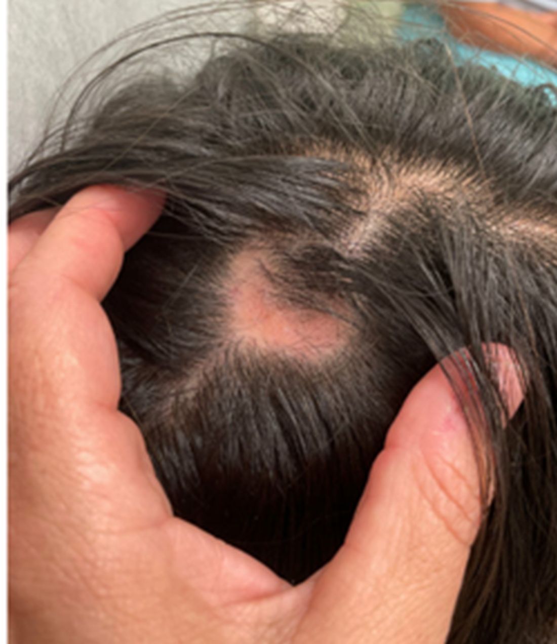
Discussion
It predominantly affects adults, yet pediatric cases account for 5%-7% of DLE diagnoses, with a significant predominance in females. Pediatric scalp DLE is particularly concerning due to its potential for causing scarring and permanent hair loss, which can significantly impact the psychological wellbeing of affected children.
The pathogenesis of DLE is multifactorial, involving genetic predispositions, environmental factors like UV light exposure, and immunological mechanisms leading to skin damage.
In children, DLE typically presents as well-demarcated, erythematous plaques with scale and follicular plugging, primarily affecting the scalp. Lesions may also exhibit changes in pigmentation, atrophy, and telangiectasia. The scalp involvement often leads to scarring alopecia, which can be distressing for pediatric patients. Unlike systemic lupus erythematosus (SLE), DLE is usually limited to the skin without systemic involvement. The progression of DLE to systemic lupus erythematosus in children has been previously described to be 22.2%. In a recent report of 201 pediatric cases of DLE, 12% of the cases progressed to systemic lupus erythematosus (SLE) and 14.5% had concurrent SLE. The onset of symptoms before the age of 10 years was the only statistically significant predictor for progression to SLE. Pruritus is a common symptom and may be correlated with disease activity.
The differential diagnosis for this patient encompassed a variety of conditions, including tinea capitis, alopecia areata, trichotillomania, and lichen planopilaris, each considered based on clinical presentation but ultimately excluded through clinical, microscopic, and biopsy findings.

Management strategies for pediatric scalp DLE aim at preventing disease progression, minimizing scarring, and addressing aesthetic concerns. These include the use of topical and intralesional corticosteroids, calcineurin inhibitors, and antimalarial agents like hydroxychloroquine, alongside stringent photoprotection to mitigate UV-triggered exacerbations.
Conclusion
The prognosis for pediatric scalp DLE can be favorable with timely and appropriate management, underscoring the importance of early diagnosis and intervention to prevent scarring and hair loss. However, ongoing surveillance is crucial for monitoring potential progression to systemic lupus erythematosus, albeit a low-risk transformation.
Dr. Matiz is a pediatric dermatologist at Southern California Permanente Medical Group, San Diego.
References
1. George PM and Tunnessen WW. Childhood discoid lupus erythematosus. Arch Dermatol. 1993;129(5):613-617.
2. Hawat T et al. Pediatric discoid lupus erythematosus: Short report. Dermatol Ther. 2022 Jan;35(1):e15170. doi: 10.1111/dth.15170.
3. Arkin LM et al. Practice-based differences in paediatric discoid lupus erythematosus. Br J Dermatol. 2019 Oct;181(4):805-810. doi: 10.1111/bjd.17780.
KOH analysis of the scales from the scalp areas revealed no fungal elements. Given the observed erythema and scaling, a punch biopsy was conducted. Histopathological examination of the biopsy sample displayed interface inflammation affecting both the infundibular and lower portions of hair follicles. The presence of folliculosebaceous units transitioning from intermediate to terminal size follicles was noted. A perifollicular, peri eccrine, superficial, and deep perivascular lymphoplasmacytic infiltrate was identified, alongside increased dermal mucin, findings consistent with a diagnosis of discoid lupus erythematosus.
Subsequent laboratory investigations were largely unremarkable, except for an elevated ANA titer (1:320, with a speckled pattern). The patient was initiated on a treatment regimen comprising intralesional triamcinolone and oral hydroxychloroquine (Plaquenil).
Discussion
It predominantly affects adults, yet pediatric cases account for 5%-7% of DLE diagnoses, with a significant predominance in females. Pediatric scalp DLE is particularly concerning due to its potential for causing scarring and permanent hair loss, which can significantly impact the psychological wellbeing of affected children.
The pathogenesis of DLE is multifactorial, involving genetic predispositions, environmental factors like UV light exposure, and immunological mechanisms leading to skin damage.
In children, DLE typically presents as well-demarcated, erythematous plaques with scale and follicular plugging, primarily affecting the scalp. Lesions may also exhibit changes in pigmentation, atrophy, and telangiectasia. The scalp involvement often leads to scarring alopecia, which can be distressing for pediatric patients. Unlike systemic lupus erythematosus (SLE), DLE is usually limited to the skin without systemic involvement. The progression of DLE to systemic lupus erythematosus in children has been previously described to be 22.2%. In a recent report of 201 pediatric cases of DLE, 12% of the cases progressed to systemic lupus erythematosus (SLE) and 14.5% had concurrent SLE. The onset of symptoms before the age of 10 years was the only statistically significant predictor for progression to SLE. Pruritus is a common symptom and may be correlated with disease activity.
The differential diagnosis for this patient encompassed a variety of conditions, including tinea capitis, alopecia areata, trichotillomania, and lichen planopilaris, each considered based on clinical presentation but ultimately excluded through clinical, microscopic, and biopsy findings.
Management strategies for pediatric scalp DLE aim at preventing disease progression, minimizing scarring, and addressing aesthetic concerns. These include the use of topical and intralesional corticosteroids, calcineurin inhibitors, and antimalarial agents like hydroxychloroquine, alongside stringent photoprotection to mitigate UV-triggered exacerbations.
Conclusion
The prognosis for pediatric scalp DLE can be favorable with timely and appropriate management, underscoring the importance of early diagnosis and intervention to prevent scarring and hair loss. However, ongoing surveillance is crucial for monitoring potential progression to systemic lupus erythematosus, albeit a low-risk transformation.
Dr. Matiz is a pediatric dermatologist at Southern California Permanente Medical Group, San Diego.
References
1. George PM and Tunnessen WW. Childhood discoid lupus erythematosus. Arch Dermatol. 1993;129(5):613-617.
2. Hawat T et al. Pediatric discoid lupus erythematosus: Short report. Dermatol Ther. 2022 Jan;35(1):e15170. doi: 10.1111/dth.15170.
3. Arkin LM et al. Practice-based differences in paediatric discoid lupus erythematosus. Br J Dermatol. 2019 Oct;181(4):805-810. doi: 10.1111/bjd.17780.

Upon physical examination, the patient exhibited several patches of alopecia with accompanying perifollicular scaling, crusting, and erythema on the affected areas. Examination of her face revealed comedones and papules.
Circulating Tumor DNA Predicts Early Treatment Response in Patients With HER2-Positive Cancers
This was the main finding of new data presented by study author Razelle Kurzrock, MD, at the American Association for Cancer Research annual meeting.
“We found that on-treatment ctDNA can detect progression before standard-of-care response assessments. These data suggest that monitoring ctDNA can provide clinicians with important prognostic information that may guide treatment decisions,” Dr. Kurzrock, professor at the Medical College of Wisconsin, Milwaukee, said during her presentation.
Commenting on the clinical implications of these findings during an interview, she said the results suggest that ctDNA dynamics provide an early window into predicting response to targeted therapies in patients with HER2-altered cancers, confirming previous findings of the predictive value of ctDNA in other cancer types.
“Such monitoring may be useful in clinical trials and eventually in practice,” she added.
Need for new methods to predict early tumor response
Limitations of standard radiographic tumor assessments present challenges in determining clinical response, particularly for patients receiving targeted therapies.
During her talk, Dr. Kurzrock explained that although targeted therapies are effective for patients with specific molecular alterations, standard imaging assessments fail to uncover molecular-level changes within tumors, limiting the ability of clinicians to accurately assess a patient’s response to targeted therapies.
“In addition to limitations with imaging, patients and physicians want to know as soon as possible whether or not the agents are effective, especially if there are side effects,” Dr. Kurzrock during an interview. She added that monitoring early response may be especially important across tumor types, as HER2 therapies are increasingly being considered in the pan-cancer setting.
Commenting on the potential use of this method in other cancer types with HER2 alterations, Pashtoon Murtaza Kasi, MD, MS, noted that since the study relied on a tumor-informed assay, it would be applicable across diverse tumor types.
“It is less about tissue type but more about that particular patient’s tumor at that instant in time for which a unique barcode is created,” said Dr. Kasi, a medical oncologist at Weill Cornell Medicine, New York, who was not involved in the study.
In an interview, he added that the shedding and biology would affect the assay’s performance for some tissue types.
Design of patient-specific ctDNA assays
In this retrospective study, the researchers examined ctDNA dynamics in 58 patients with various HER2-positive tumor types, including breast, colorectal, and other solid malignancies harboring HER2 alterations. All the patients received combination HER2-targeted therapy with trastuzumab and pertuzumab in the phase 2 basket trial My Pathway (NCT02091141).
By leveraging comprehensive genomic profiling of each patient’s tumor, the researchers designed personalized ctDNA assays, tracking 2-16 tumor-specific genetic variants in the patients’ blood samples. FoundationOne Tracker was used to detect and quantify ctDNA at baseline and the third cycle of therapy (cycle 3 day 1, or C3D1).
During an interview, Dr. Kurzrock explained that FoundationOne Tracker is a personalized ctDNA monitoring assay that allows for the detection of ctDNA in plasma, enabling ongoing liquid-based monitoring and highly sensitive quantification of ctDNA levels as mean tumor molecules per milliliter of plasma.
Among the 52 patients for whom personalized ctDNA assays were successfully designed, 48 (92.3%) had ctDNA data available at baseline, with a median of 100.7 tumor molecules per milliliter of plasma. Most patients (89.6%) were deemed ctDNA-positive, with a median of 119.5 tumor molecules per milliliter of plasma.
Changes in ctDNA levels predict patient survival
The researchers found that patients who experienced a greater than 90% decline in ctDNA levels by the third treatment cycle had significantly longer overall survival (OS) than those with less than 90% ctDNA decline or any increase. According to data presented by Dr. Kurzrock, the median OS was not reached in the group with greater than 90% decline in on-treatment ctDNA levels, versus 9.4 months in the group with less than 90% decline or ctDNA increase (P = .007). These findings held true when the analysis was limited to the 14 patients with colorectal cancer, in which median OS was not reached in the group with greater than 90% decline in on-treatment ctDNA levels, versus 10.2 months in the group with less than 90% decline or ctDNA increase (P = 0.04).
Notably, the prognostic significance of ctDNA changes remained even among patients exhibiting radiographic stable disease, underscoring the limitations of relying solely on anatomic tumor measurements and highlighting the potential for ctDNA monitoring to complement standard clinical assessments. In the subset of patients with radiographic stable disease, those with a greater than 90% ctDNA decline had significantly longer OS than those with less ctDNA reduction (not reached versus 9.4 months; P = .01).
“When used as a complement to imaging, tissue-informed ctDNA monitoring with FoundationOne Tracker can provide more accuracy than imaging alone,” Dr. Kurzrock noted in an interview.
Dr. Kasi echoed Dr. Kurzrock’s enthusiasm regarding the clinical usefulness of these findings, saying, “Not only can you see very early on in whom the ctDNA is going down and clearing, but you can also tell apart within the group who has ‘stable disease’ as to who is deriving more benefit.”
The researchers also observed that increases in on-treatment ctDNA levels often preceded radiographic evidence of disease progression by a median of 1.3 months. These findings highlight the potential for ctDNA monitoring to complement standard clinical assessments, allowing us to detect treatment response and disease progression earlier than what is possible with imaging alone, Dr. Kurzrock explained during her talk. “This early warning signal could allow clinicians to intervene and modify treatment strategies before overt clinical deterioration,” she said.
In an interview, Dr. Kasi highlighted that this high sensitivity and specificity and the short half-life of the tumor-informed ctDNA assay make this liquid biopsy of great clinical value. “The short half-life of a few hours means that if you do an intervention to treat cancer with HER2-directed therapy, you can very quickly assess response to therapy way earlier than traditional radiographic methods.”
Dr. Kasi cautioned, however, that this assay would not capture whether new mutations or HER2 loss occurred at the time of resistance. “A repeat tissue biopsy or a next-generation sequencing-based plasma-only assay would be required for that,” he said.
Implementation of ctDNA monitoring in clinical trials
Dr. Kurzrock acknowledged that further research is needed to validate these results in larger, prospective cohorts before FoundationOne Tracker is adopted in the clinic. She noted, however, that this retrospective analysis, along with results from previous studies, provides a rationale for the use of ctDNA monitoring in clinical trials.
“In some centers like ours, ctDNA monitoring is already part of our standard of care since not only does it help from a physician standpoint to have a more accurate and early assessment of response, but patients also appreciate the information gained from ctDNA dynamics,” Dr. Kasi said in an interview. He explained that when radiographic findings are equivocal, ctDNA monitoring is an additional tool in their toolbox to help guide care.
He noted, however, that the cost is a challenge for implementing ctDNA monitoring as a complementary tool for real-time treatment response monitoring. “For serial monitoring, helping to reduce costs would be important in the long run,” he said in an interview. He added that obtaining sufficient tissue for testing using a tumor-informed assay can present a logistical challenge, at least for the first test. “You need sufficient tissue to make the barcode that you then follow along,” he explained.
“Developing guidelines through systematic studies about testing cadence would also be important. This would help establish whether ctDNA monitoring is helpful,” Dr. Kasi said in an interview. He explained that in some situations, biological variables affect the shedding and detection of ctDNA beyond the assay — in those cases, ctDNA monitoring may not be helpful. “Like any test, it is not meant for every patient or clinical question,” Dr. Kasi concluded.
Dr. Kurzrock and Dr. Kasi reported no relationships with entities whose primary business is producing, marketing, selling, reselling, or distributing healthcare products used by or on patients.
FROM AACR 2024
A 30-Year-Old White Female Presented With a 4-Month History of Scaly, Erythematous Patches and Plaques on Her Trunk and Extremities
Tumor necrosis factor (TNF)-alpha inhibitors are used to treat a variety of autoimmune conditions including psoriasis, psoriatic arthritis, rheumatoid arthritis (RA), spondyloarthritis, and inflammatory bowel disease (IBD). Interestingly, they have also been observed to cause paradoxical psoriasis with an incidence between 0.6%-5.3%, most commonly occurring in patients with underlying Crohn’s disease and rheumatoid arthritis (RA). Infliximab is the most common TNF inhibitor associated with this condition (52.6%-62.6% of cases) followed by etanercept (12%-29%). .
Psoriasis is traditionally divided into two types. Patients with type I psoriasis have a family history, develop symptoms before the age of 40 and are often positive for HLA-Cw6. Type II psoriasis is not related to HLA-Cw6, lacks a family history, and typically manifests after age 40. Psoriatic lesions are well-defined, erythematous plaques with silvery scales most commonly appearing on extensor surfaces and the scalp. Variants include nail psoriasis, pustular psoriasis, inverse psoriasis, and guttate psoriasis.
Although psoriasis is typically a clinical diagnosis, histologic examination may be used to differentiate from other dermatoses if necessary. The lesions of TNF inhibitor-induced psoriasis characteristically display patterns similar to primary psoriasis, including parakeratosis, microabscesses, and rete ridges. Eosinophilic hypersensitivity reactions and features overlapping with eczematous hypersensitivity (psoriasiform dermatitis) may also be present.
The pathogenesis of this condition is not well understood, but theories include a variety of immune processes including interferon overproduction, interleukin and T-cell activation, and the presence of an infectious nidus. Classical psoriasis is related to type 1 interferon release, so theoretically, immunosuppression caused by TNF inhibitor treatment may permit uncontrolled production of interferons, resulting in psoriatic lesions. Another theory is that interleukin (IL)-23, a pro-inflammatory cytokine, promotes activation of T-helper 17 (Th17) cells. Th17 cells are part of the pathogenesis of primary psoriasis and other inflammatory conditions, such as RA and inflammatory bowel disease. Of note, individuals with gastrointestinal inflammatory diseases are already known to be at a greater risk for developing psoriasis. Immunosuppression caused by a TNF inhibitor may leave patients more susceptible to other infections, which may induce psoriatic plaques.
There are multiple approaches to treatment depending on the severity of the disease. If the psoriatic eruption is mild, the medication may be continued. This “treat-through” method is often considered when stopping the current immunotherapy would cause the patient significant issues. Moderate to severe cases of TNF inhibitor-induced psoriasis may warrant switching TNF inhibitor therapy or completely changing the drug class used in the treatment of the underlying autoimmune condition. Additional treatments include topical and oral steroids, UV therapy, methotrexate, cyclosporine, and acitretin.
This case and the photo were submitted by Lucas Shapiro, BS, of Nova Southeastern University College of Osteopathic Medicine, Fort Lauderdale, Florida, and Leon S. Maratchi, MD, Gastro Health, Hollywood, Florida. The column was edited by Donna Bilu Martin, MD.
Dr. Bilu Martin is a board-certified dermatologist in private practice at Premier Dermatology, MD, in Aventura, Florida. More diagnostic cases are available at mdedge.com/dermatology. To submit a case for possible publication, send an email to dermnews@mdedge.com.
References
1. Li SJ et al. J Psoriasis Psoriatic Arthritis. 2019 Apr;4(2):70-80. doi: 10.1177/2475530318810851.
2. Lu J and Lu Y. J Transl Autoimmun. 2023 Sep 6:7:100211. doi: 10.1016/j.jtauto.2023.100211.
3. Nair PA and Badri T. Psoriasis. [Updated 2023 Apr 3]. In: StatPearls [Internet]. Treasure Island (FL): StatPearls Publishing; 2024 Jan-. Available from: www.ncbi.nlm.nih.gov/books/NBK448194/
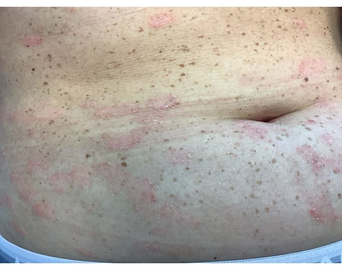
Mild Hidradenitis Suppurativa: Positive Results Reported for Topical Therapy
SAN DIEGO — cream, in a phase 2 trial.
“HS is a chronic, recurring inflammatory skin disease that is associated with painful inflammatory modules and abscesses,” said presenting author Martina J. Porter, MD, a dermatologist at Beth Israel Deaconess Medical Center and Harvard Medical School, both in Boston, Massachusetts. Dr. Porter presented the data during a late-breaking session at the annual meeting of the American Academy of Dermatology.
“Over time, these patients may progress to having tunnels, ulcerations, malodorous discharge, and permanent scarring,” she said. “Currently, there are no approved therapies for milder HS, and the standard treatments that we apply in clinical practice are often inadequate.”
Ruxolitinib is a selective Janus kinase (JAK) 1/JAK2 inhibitor that has demonstrated efficacy in other inflammatory and autoimmune skin diseases. Ruxolitinib cream, 1.5%, is approved for treating mild to moderate atopic dermatitis and nonsegmental vitiligo in patients ages 12 years and older.
The phase 2 double-blind, vehicle-controlled trial evaluated the efficacy and safety of ruxolitinib cream for mild HS. Researchers assigned 69 adults with Hurley stage I or II HS to receive 1.5% ruxolitinib cream or vehicle cream twice daily for 16 weeks. The primary endpoint was the change from baseline in AN count at week 16. To be eligible, patients had to have an AN count between 3 and 10.
“This is much more mild than what we have seen in any systemic therapy trials,” Dr. Porter said. “And, if patients had 3 lesions, they all needed to be in one anatomic area, but if they had 4-10 lesions, they had to have two anatomic areas involved. Also, no patients with active draining tunnels were allowed in the study.”
Of the 69 patients, 34 received ruxolitinib cream and 35 received vehicle. About 51% of patients in the vehicle arm were Black and 34% were White, while about 32% of patients in the ruxolitinib arm were Black and 56% were White.
The mean age of patients overall was 29 years, and about half the patients in both study arms had Hurley stage I disease, while the other half had Hurley stage II disease. Their average AN count ranged between 5.3 and 5.6 — mostly inflammatory nodules and few abscesses. Patients were not allowed to receive any type of intervention or rescue therapy during the study.
Dr. Porter reported that the least square mean change in AN count from baseline to week 16 was -2.42 in the vehicle arm vs -3.61 in the ruxolitinib cream arm (P <.05). The proportion of patients who achieved a 50% decrease in AN count was 79.2% in the ruxolitinib cream arm, compared with 56.5% of patients in the vehicle arm, respectively. More patients in the ruxolitinib cream arm achieved a 75% decrease in AN count (54.2% vs 25%), a 90% decrease in AN count (20.8 vs 12.5%), and a 100% decrease in AN count (20.8% vs 12.5%).
In other findings, 79.2% of patients in the ruxolitinib cream arm achieved a Hidradenitis Suppurativa Clinical Response score from baseline through week 16, compared with 50% of those in the vehicle group. The International Hidradenitis Suppurativa Severity Score System results favored the ruxolitinib cream arm (-4.46 vs -2.66 in the vehicle arm). Skin Pain and Itch numeric rating scale scores were moderate at baseline and improved similarly in both groups during the study.
Ruxolitinib cream was generally well tolerated over 16 weeks. No serious treatment-emergent adverse events were reported. The most common adverse event reported in the ruxolitinib cream group was COVID-19 and nasopharyngitis (two cases each) and one case of an application site reaction.
“Twice-daily 1.5% ruxolitinib cream was effective in patients with milder HS,” Dr. Porter concluded. “Modifications to our traditionally accepted clinical endpoints may be needed in studies of patients with milder HS.”
Jennifer L. Hsiao, MD, a dermatologist who directs the HS clinic at the University of Southern California, Los Angeles, who was asked to comment on the results, characterized the study as exciting for several reasons.
“First, with the global push in recent years to increase HS awareness, I am already seeing more patients earlier in their disease course with milder disease, and there is currently a gap in approved therapies for this patient population,” she told this news organization.
“Second, patients are very interested in topical therapies for HS and are thrilled whenever they learn that topical options are under investigation. This study had small patient numbers, but it was encouraging to see the positive results for ruxolitinib cream and that the treatment appeared well-tolerated.”
The trial was sponsored by the Incyte Corporation. Dr. Porter disclosed that she has received consulting fees from AbbVie, Alumis, Eli Lilly, Incyte, Janssen, Novartis, Pfizer, Prometheus Laboratories, Sanofi, Sonoma Biotherapeutics, Trifecta Clinical, and UCB. Dr. Hsiao disclosed that she is a member of the board of directors for the HS Foundation. She has also served as a consultant for AbbVie, Aclaris, Boehringer Ingelheim, Incyte, Novartis, and UCB; as a speaker for AbbVie, Novartis, and UCB; and as an investigator for Amgen, Boehringer Ingelheim, and Incyte.
A version of this article appeared on Medscape.com .
FROM AAD 2024
Study Identifies Several Factors That Influence Longterm Antibiotic Prescribing for Acne
to follow them, according to the authors of a recently published study.
“This study explored why dermatologists still prescribe a good number of long-term antibiotics for people with acne,” the study’s senior author Howa Yeung, MD, MSc, assistant professor of dermatology at Emory University, Atlanta, said in an interview. “And we found a lot of reasons.” The study was published online in JAMA Dermatology.
Using online surveys and semi-structured video interviews of 30 dermatologists, infectious disease physicians with expertise in antimicrobial stewardship, dermatology residents, and nonphysician clinicians, the investigators assessed respondents’ knowledge and attitudes regarding long-term antibiotics in acne. Salient themes impacting long-term antibiotic prescriptions included the following:
- A perceived dearth of evidence to justify changes in practice.
- Difficulties with iPLEDGE, the Risk Evaluation and Mitigation Strategy (REMS) for managing the teratogenic risks associated with isotretinoin, and with discussing oral contraceptives.
- “Navigating” discussions with about tapering-off of antibiotics.
- Challenging patient demands.
- A lack of effective tools for monitoring progress in antibiotic stewardship.
“It’s surprising there are so many barriers that make it difficult for dermatologists to stick with the guidelines even if they want to,” said Dr. Yeung, a coauthor of the recently released updated American Academy of Dermatology (AAD) acne management guidelines.
A dermatologist who wants to stop systemic antibiotics within 3 months may not know how to do so, he explained, or high demand for appointments may prevent timely follow-ups.
A major reason why dermatologists struggle to limit long-term antibiotic use is that there are very few substitutes that are perceived to work as well, said David J. Margolis, MD, PhD, who was not involved with the study and was asked to comment on the results. He is professor of epidemiology and dermatology at the University of Pennsylvania, Philadelphia.
“Part of the reason antibiotics are being used to treat acne is that they’re effective, and effective for severe disease,” he said. The alternatives, which are mostly topicals, said Dr. Margolis, do not work as well for moderate to severe disease or, with isotretinoin, involve time-consuming hurdles. Dr. Margolis said that he often hears such concerns from individual dermatologists. “But it’s helpful to see these in a well-organized, well-reported qualitative study.”
Infectious disease specialists surveyed considered limiting long-term antibiotic use as extremely important, while several dermatologists “argued that other specialties ‘underestimate the impact acne has on people’s lives,’ ” the authors wrote. Other respondents prioritized making the right choice for the patient at hand.
Although guidelines were never meant to be black and white, Dr. Yeung said, it is crucial to target the goal of tapering off after about 3-4 months — a cutoff with which guidelines from groups including the AAD, the Japanese Dermatological Association in guidelines from 2016, and 2017, respectively, and others concur.
He added, “Some folks believe that if the oral antibiotic is working, why stop? We need to develop evidence to show that reducing oral antibiotic use is important to our patients, not just to a theoretical problem of antibiotic resistance in society.” For example, in a study published in The Lancet in 2004, patients who used strictly topical regimens achieved efficacy similar to that of those who used only oral antibiotics.
In addition, some clinicians worried that limiting antibiotics could reduce patient satisfaction, spurring switches to other providers. However, he and the other authors of the JAMA Dermatology study noted that in a survey of patients with acne published in the Journal of Clinical and Aesthetic Dermatology in 2019, 76.9% said they would be “very or extremely likely” to use effective antibiotic-free treatments if offered.
Because most respondents were highly aware of the importance of antibiotic stewardship, Dr. Yeung said, additional passive education is not necessarily the answer. “It will take a concerted effort by our national societies to come up with resources and solutions for individual dermatologists to overcome some of these larger barriers.” Such solutions could range from training in communication and shared decision-making to implementing systems that provide individualized feedback to support antibiotic stewardship.
Many ongoing studies are examining antibiotic stewardship, Dr. Margolis said in the interview. However, he added, dermatologists’ idea of long-term use is 3 months, versus 1 month or less in other specialties. “Moreover, dermatology patients tend to be much healthier individuals and are rarely hospitalized, so there may be some issues comparing the ongoing studies to individuals with acne.” Future research will need to account for such differences, he said.
The study was funded by an American Acne & Rosacea Society Clinical Research Award. Dr. Yeung is associate editor of JAMA Dermatology. Dr. Margolis has received a National Institutes of Health grant to study doxycycline versus spironolactone in acne.
FROM JAMA DERMATOLOGY