User login
Protocol helped identify hospitalized children at risk for VTE
SAN DIEGO – Following simple institutional care guidelines helped clinicians identify pediatric patients at moderate-to-severe risk of venous thromboembolism (VTE), results from a single-center study showed.
“Hospital-acquired VTE is on the rise in the pediatric population,” lead study author Emily Southard, MD, said at the biennial summit of the Thrombosis & Hemostasis Societies of North America. “This consists of a DVT or [pulmonary embolism] 48 hours or more after admission, or any time at the site of a central venous catheter.”
One published study found a 70% increased incidence in the pediatric population from 2001-2007 (Pediatrics 2009;124[4]:1001-8). More than half of the children in that study (63%) had at least one coexisting complex medical condition, with malignancy being the most common.
Hospital-acquired VTE cases tend to harbor a number of complications, said Dr. Southard, who is a pediatric hematology/oncology fellow at Children’s Hospital Colorado, Aurora. For example, 15%-20% of patients with a DVT will have a pulmonary embolism (PE) as well, 26% of patients with upper or lower extremity DVT develop post-thrombotic syndrome, and 3% of patients with PE develop chronic pulmonary hypertension.
“Medical costs are also impacted,” she said. “The cost for a hospital-acquired VTE in pediatrics increased the length of stay by about 8 days and increased the cost of hospital admission by more than $27,000.”
Known risk factors for VTE in this patient population include ICU admission (Odds Ratio, 2.14), presence of a central venous catheter (OR, 2.12), mechanical ventilation (OR, 1.56), and prolonged admission (OR, 1.03 for each day).
Risk factors in pediatric trauma patients include ICU admission (OR, 6.25), transfusion of blood products (OR, 2.1), lower extremity fracture (OR, 1.8), and neurosurgery (OR, 2.13). She and her associates hypothesized that understanding the relative contributions of clinical, biological, and genetic risk factors for pediatric VTE would help appropriately risk-stratify patients and allow better prophylactic approaches.
In 2012, Children’s Hospital Colorado implemented a VTE risk assessment tool as part of a hospital-wide patient safety initiative. The assessment is triggered via an Epic Best Practice Advisory to complete in certain higher-risk patients, including ICU patients, hematology/oncology floor patients, any patients with a central line catheter, and those who are over age 12 and obese.
Clinicians also assess for risk factors such as significant infection, recent surgery, and personal or family history of thrombophilia. Next, they classify each patient’s risk of hospital-acquired VTE as high, moderate, or low risk.
In a pilot study, Dr. Southard and her associates set out to validate the accuracy of the institution’s VTE risk assessment tool since it was implemented in 2012. She presented findings from 215 hospital-acquired VTE cases in patients younger than age 18, compared with age-matched inpatient controls. Data from patients under 6 months of age is available after October 2016, coinciding with a change in definition of pediatric hospital-acquired VTE.
Most hospital-acquired VTE patients (77.2%) ranged in age from 1-17 years. The number of patients admitted for a trauma diagnosis was similar between VTE cases and controls (7.4% vs. 7.9%, respectively). However, compared with controls, a significantly greater number of VTE cases were immobile (41.8% vs. 10.3%, respectively), required ICU admission (86.4% vs. 26.5%), had a central venous catheter (80.4% vs. 10.9%), had a positive blood culture (16.7% vs. 1.9%), required surgery or a medical procedure (57.7% vs. 36.7%), and had a longer procedure time (a mean of 151 vs. 133 minutes).
The researchers also found that upon initial admission, 7.9% of VTE cases were identified as high risk and another 21.9% were identified as moderate risk, compared with 1.2% and 3.7% in the controls, respectively.
“Patients identified as moderate or high risk for VTE were generally more medically complex patients,” Dr. Southard said.
Future directions of this project include expanding the patient population that has a risk assessment performed.
Dr. Southard reported having no financial disclosures.
SOURCE: Southard E et al. THSNA 2018.
REPORTING FROM THSNA 2018
Key clinical point:
Major finding: A significantly greater number of VTE patients were immobile (41.8% vs. 10.3%, respectively), required ICU admission (86.4% vs. 26.5%), and had a central venous catheter (80.4% vs. 10.9%), compared with controls.
Study details: A retrospective analysis of 215 hospital-acquired VTE cases in patients younger than age 18.
Disclosures: Dr. Southard reported having no financial disclosures.
Source: Southard E et al. THSNA 2018.
Certifications, training to increase addiction medicine specialists
Two new workforce developments aim to increase the number of addiction medicine specialists and provide new training opportunities in the subspecialty.
The American Board of Medical Specialties (ABMS) recently certified its first formal wave of addiction medicine physicians, adding 1,200 specialists to the field. Addiction medicine was first recognized as a subspecialty by ABMS in 2015, followed by the first certification exam in 2017.
The two developments “will change the landscape in substance use prevention, early intervention, and in addiction treatment and management,” said Lon R. Hays, MD, president of The Addiction Medicine Foundation, in Chevy Chase, Md., and director of the addiction medicine fellowship program at the University of Kentucky, Lexington.
“Many more trained physicians will be available to address the opioid crisis and other addictions,” Dr. Hays said in a statement. “They will also be able to help prevent and intervene early with unhealthy substance use in all its forms. For the first time, when aspiring physicians consider a career path, they will now have as an available choice an addiction medicine specialty that meets the highest standards of medicine.”
“When the American Board of Medical Specialties welcomed addiction medicine as its newest subspecialty, it in a lot of ways, legitimized our discipline,” Dr. Brennan said in an interview. “The American Board of Medical Specialties really represents the ‘House of Medicine.’ Being able to enter into that, it gives us a measure of credibility in the eyes of the public, and it basically codifies that these physicians who have passed this board exam have achieved a level of competency and knowledge that makes them trustworthy and safe to provide care to folks suffering from addiction.”
While the 1,200 additional addiction medicine specialists are an improvement, many more are needed, Dr. Brennan said, adding that he is optimistic that the new addiction medicine training opportunities provided by ACGME will help achieve higher numbers.
“For addiction medicine, we’ve had fellowships for about 10 years, but the funding for those fellowships was really challenging,” Dr. Brennan said. “Once you get ACGME-accredited, it gives you the ability to partake of [Centers for Medicare & Medicaid Services] funding that funds most of the graduate medical education residency fellowship spots in the United States. ACGME is the gold standard. I think that makes us much more potentially attractive for graduating physicians who are finishing their residencies.”
The certification of new addiction specialists is welcome news, particularly in the midst of the current epidemic, added Clif Knight, MD, senior vice president for education for the American Academy of Family Physicians.
“This is really good news [especially considering], the difficulty that the country is having with so much addiction – of course opioids are in the forefront – but there are so many different types of addiction,” he said in an interview. “This is good news that the certification is available and that physicians are pursuing obtaining additional expertise and recognition in their ability to treat addictions.”
Dr. Knight expressed appreciation that the ACGME training opportunities in addiction medicine are open to doctors of various specialties, he said.
“A lot of times subspecialty fellowships are only available to one specialty,” said Dr. Knight. “In this case, it looks like they’ve been very deliberate to make this available to multiple specialties. I think that really sends an important message that this is not just one specialty that is focused on [addiction medicine], but really should be something that all specialties are engaged and involved in.”
*This article was updated on 4/2/2018.
Single screening for Lynch syndrome beats sequential tests in CRC
Physicians could more accurately test patients with colon cancer for Lynch syndrome by using a single tumor sequencing test instead of the current protocol of up to six sequential tests, a new study suggests. The process may also be faster in some cases.
“We found that up-front tumor testing is actually more sensitive and more specific for detecting Lynch syndrome than the old, multiple-test model,” study coauthor Rachel Pearlman, MS, a genetic counselor at Ohio State University Wexner Medical Center, said in an interview. “Tumor sequencing resulted in a 10% improvement in Lynch syndrome detection rates while also providing important information about treatment options for the patients.”
According to Ms. Pearlman, screening for Lynch syndrome is recommended for all patients with colon cancer and can require multiple sequential tests. It affects an estimated 3% of these patients, putting them at higher risk of several kinds of cancers including endometrial, ovarian, and gastric.
“Identifying the condition at the time of diagnosis can potentially impact treatment options and also help to facilitate intensive surveillance for other types of cancer,” Ms. Pearlman said. “In addition, we’ll know that the patients’ family members are at risk and will benefit from genetic counseling and testing.”
However, “traditional sequential testing is complex and confusing to patients and clinicians and occurs over a prolonged period, incurring risk for loss to follow-up,” the investigators wrote in JAMA Oncology.
For the new study, the researchers sought to confirm whether tumor sequencing, a form of genetic testing, would be faster and more accurate than the current sequential testing approach.
In a multicenter study, they prospectively tested tumor DNA in 2015 and 2016. They also tested another 46 patients who had been previously confirmed to have Lynch syndrome.
The average age of the patients was 60 years, 52% were women, 89% were white. Hispanics and Asians made up just 1% each of the total. Most of the cancers were stage II (26%) or stage III (40%).
Tumor sequencing identified all of the 46 confirmed cases of Lynch syndrome and turned up 12 more in the larger group, the researchers found.
Sensitivity of tumor sequencing was better (100%; 95% confidence interval, 93.8%-100%) than immunohistochemical testing plus BRAF (89.7%; 95% CI, 78.8%-96.1%; P = .04) and microsatellite instability testing plus BRAF (91.4%; 95% CI, 81.0%-97.1%; P = .07), and its specificity was equal to the other approaches, Ms. Pearlman and her associates reported.
Researchers also reported that tumor sequencing identified nearly 300 cases of tumors with genetic mutations that could impact therapy.
Eesults from tumor sequencing are available in a median of 2 weeks, which may be longer than some other tests, but “it requires less time overall by eliminating multiple follow-up tests in a subset of cases,” the study authors wrote.
“While this new test is currently more expensive than traditional step-wise testing, it will eliminate many other tests for a subset of patients so that it may be more cost-effective overall. If it is not now, it will certainly be in the future as the costs of tumor sequencing continue to decline,” Ms. Pearlman said. “However, formal cost-analysis studies will be necessary to determine if this is a cost-effective approach.”
The study was funded by a grant from Pelotonia, an annual cycling event that supports cancer research, and the National Cancer Institute. Myriad Genetics donated the sequence testing used for some patients.
SOURCE: Hampel H et al. JAMA Oncol. 2018 Mar 29. doi: 10.1001/jamaoncol.2018.0104.
FROM JAMA ONCOLOGY
Key clinical point: Tumor sequencing provides more accurate Lynch syndrome testing in colon cancer.
Major finding: Sensitivity of tumor sequencing was better (100%) than immunohistochemical testing plus BRAF (89.7%) and microsatellite instability testing plus BRAF (91.4%). Specificity was the same.
Study details: Prospective testing of 419 consecutive patients with colon cancer plus analysis of samples from 46 patients with confirmed Lynch syndrome.
Disclosures: The study was funded by a grant from Pelotonia, an annual cycling event that supports cancer research, and the National Cancer Institute. Myriad Genetics provided the sequence testing used for some of the patients.
Source: Hampel H et al. JAMA Oncol. 2018 Mar 29. doi: 10.1001/jamaoncol.2018.0104.

Warfarin dose capping avoided supratherapeutic INRs in hospitalized elderly
SAN DIEGO – A simple intervention of initial warfarin dose capping in hospitalized patients aged 85 years and older led to significant reductions in supratherapeutic INRs without a significant change in length of stay, a single-center study showed.
“We’re excited to see if these results are reproducible,” Jonathan Falsetta, PharmD, said at the biennial summit of the Thrombosis & Hemostasis Societies of North America. “We do feel that it may represent an important step forward in warfarin safety in a vulnerable patient population.”
“From this we gathered that we had an issue with dosing,” said Dr. Falsetta, who is assistant director of pharmacy clinical educational services at Plainview Hospital. “These patients were spiking dangerously high INRs, and we needed to do something about it.”
A review of current medical literature revealed a lack of well-validated recommendations regarding warfarin initiation in older patients, so Dr. Falsetta and his associates set out to create their own dose-capping recommendation. This involved limiting the initial dose of warfarin to 2.5 mg or less for hospitalized patients aged 85 years and older.
“We wanted this to be applicable to patients regardless if they were warfarin naïve or if they had been on warfarin prior to admission,” he said.
Before the roll out, clinical pharmacists and pharmacy residents conducted provider education on the initial dose capping protocol. Providers could order initial doses that exceeded 2.5 mg with valid clinical reasoning. Outcomes of interest were dosing protocol compliance and post-intervention analysis of INRs in this patient population. The pre-intervention period spanned from Nov. 1, 2014 through Oct. 31, 2015, while the post-intervention period spanned from Nov. 1, 2015 through Oct. 31, 2017. Dr. Falsetta reported data from 768 patients.
Between the pre-intervention and post-intervention periods, compliance with dose capping rose from 38.5% to 64.2% (P less than .001), the supratherapeutic INR rate dropped from 20.9% to 13.3% (P= .004), and the length of hospital stay in hours rose from a mean of 145.8 to a mean of 155.8, which was not statistically significant (P= .13).
Following the post-intervention period, the number of peak INRs in the 1 to 2 range rose by 15% and the number of peak INRs in the 2 to 3 range rose by 6%. At the same time, the number of peak INRs in the 3 to 4 range fell by 6%, the number of peak INRs in the 4 to 5 range fell by 36%, and the number of peak INRs in the 5 and greater range fell by 53%. These INR percentages represent relative increases and/or decreases.
“This was a relatively simple intervention that resulted in significant reductions in supratherapeutic INRs,” Dr. Falsetta said.
The researchers also observed that there was less IV vitamin K use after the dose capping intervention. “We can’t say for sure that this was tied to the intervention, but it was interesting, and it is something for us to take a look at, as well as roll this out in future iterations,” he said. “We are on the cusp of rolling this out at some of the tertiary sites within our health care system, and some of the community sites as well.”
He acknowledged certain limitations of the study, including its single-center design, relatively small sample size, and lack of clinical endpoints. “I’d like to be able to tie this to something like reduced bleeding events,” Dr. Falsetta said. “I think that’s something we need to explore in the future.”
Dr. Falsetta reported having no financial disclosures.
SOURCE: Falsetta J et al. THSNA 2018.
REPORTING FROM THSNA 2018
Key clinical point:
Major finding: The supratherapeutic INR rate dropped from 20.9% to 13.3% (P= .004) between the pre- and post-intervention period.
Study details: A single-center study of 768 hospitalized patients aged 85 years and older.
Disclosures: Dr. Falsetta reported having no financial disclosures.
Source: Falsetta J et al., THSNA 2018.
Think about breast cancer surveillance for transgender patients
CHICAGO – , said Christel de Blok, MD, sharing results of a Dutch national study.
The study included 3,078 transgender people (2,064 transgender women) who began hormone therapy (HT) at age 18 years or older. The mean age at which transgender women began HT was 33 years; for transgender men, the mean age was 25 years. In all, transgender women in the study had a total of 30,699 person-years of exposure to HT; for transgender men, the figure was 13,155 person-years.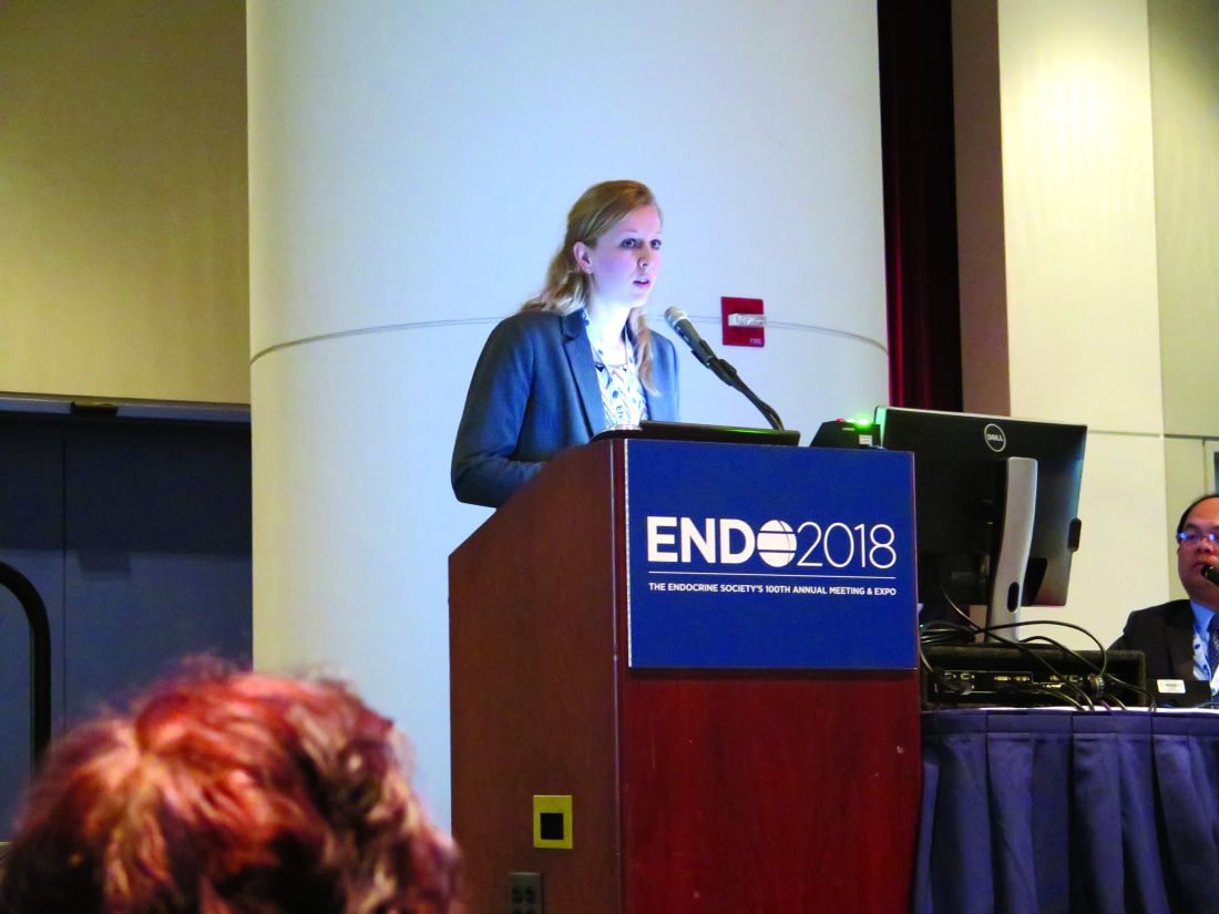
Overall, there were 16 observed cases of breast cancer in transgender women and four in transgender men. After gender-affirming surgery, the transgender women were followed for a median of 146 months, and experienced a median of 193 months of HT. Transgender men who had mastectomies were followed for a median 93 months, and those who had a hysterectomy-oophorectomy were followed for a median 144 months. Transgender men received a median 176 months of HT.
“Breast cancer can still occur after mastectomy in [transgender] men,” Dr. de Blok said at the annual meeting of the Endocrine Society. “What is interesting is that three out of the four cases of breast cancer in [transgender] men happened after mastectomy.”
In the Netherlands, one in eight women and one in 1,000 men will develop cancer at some point during their lives. In patients who have had a subtotal mastectomy and who are BRCA-1/2 carriers, there is still an approximate 5% residual risk of breast cancer, said Dr. de Blok.
A literature review conducted by Dr. de Blok and her colleagues revealed 19 cases of breast cancer in transgender women and 13 in transgender men. However, a more general study of incidence and characteristics of breast cancer in transgender people receiving hormone treatment had not been done, said Dr. de Blok, of the VU University Medical Center, Amsterdam.
The investigators examined data for adult transgender people seen at their center from 1991 to 2017 and started on hormone treatment. This clinic, said Dr. de Blok, sees about 95% of the transgender individuals in the Netherlands.
The study was able to capitalize on comprehensive information from national databases and registries. Investigators drew from a national histopathology and cytopathology registry as well as from a national vital statistics database. A comprehensive cancer database was used to establish both reference incidence values for males and females and the number of expected cases within the study group.
In both transgender men and women, exactly 50% of cases were ductal carcinoma, compared to 85% in the group of reference women.
An additional 31% of the breast cancers in transgender women were lobular, 6% were ductal carcinoma in situ (DCIS), and the remainder were of other types. Of the cancers in transgender women, 82% were estrogen receptor positive, 64% were progesterone receptor positive, and 9% were Her2/neu positive.
For transgender men, there were no lobular carcinomas; 25% were DCIS, and 25% were of other types. Half of the cancers were estrogen receptor positive, and half were progesterone receptor positive; 25% were Her2/neu positive, and there was one case of androgen receptor positive breast cancer.
Dr. de Blok explained that their analysis compared the observed cases in both transgender men and women to the expected number of cases for the same number of males and females, yielding two standardized incidence ratios (SIRs) for each transgender group.
For transgender women, the SIR for breast cancer compared with males was 50.9 (95% confidence interval, 30.1-80.9). The SIR compared to females was 0.3 (95% CI, 0.2-0.4). This reflected the expected case number of 0.3 for males and the 58 expected cases for a matched group of females.
For transgender men, the SIR for breast cancer compared with males was 59.8 (95% CI, 19-144.3), while the SIR compared to females was 0.2 (95% CI, 0.1-0.5). The expected cases for a similar group of males would be 0.1, and for females, 18.
In many cases, whether a transgender person receives standardized screening mammogram reminders will depend on which sex is assigned to that individual in insurance and other administrative databases, Mr. de Blok noted. When electronic health records and other databases have a binary system, at-risk individuals may fall through the cracks.
Dr. de Blok reported no conflicts of interest.
SOURCE: de Blok C, et al. ENDO 2018, abstract OR 25-6.
REPORTING FROM ENDO 2018
Key clinical point: Transgender individuals had increased risk of breast cancer similar to a female reference population.
Major finding: Transgender men had a standardized incidence ratio of 59.8 compared to a male reference population.
Study details: Study of 3,078 transgender adults receiving hormone therapy.
Disclosures: Dr. de Blok reported no conflicts of interest.
Source: de Blok C, et al. ENDO 2018, abstract OR 25-6.
VIDEO: Biomarker accurately predicted primary nonfunction after liver transplant
, researchers reported in Gastroenterology.
SOURCE: AMERICAN GASTROENTEROLOGICAL ASSOCIATION
Glycomic alterations of immunoglobulin G “represent inflammatory disturbances in the liver that [mean it] will fail after transplantation,” wrote Xavier Verhelst, MD, of Ghent (Belgium) University Hospital, and his associates. The new glycomarker “could be a tool to safely select high-risk organs for liver transplantation that otherwise would be discarded from the donor pool based on a conventional clinical assessment,” and also could help prevent engraftment failures. “To our knowledge, not a single biomarker has demonstrated the same accuracy today,” they wrote in the April issue of Gastroenterology.
Chronic shortages of donor livers contribute to morbidity and death worldwide. However, relaxing donor criteria is controversial because of the increased risk of primary nonfunction, which affects some 2%-10% of liver transplantation patients, and early allograft dysfunction, which is even more common. Although no reliable scoring systems or biomarkers have been able to predict these outcomes prior to transplantation, clinical glycomics of serum has proven useful for diagnosing hepatic fibrosis, cirrhosis, and hepatocellular carcinoma, and for distinguishing hepatic steatosis from nonalcoholic steatohepatitis. “Perfusate biomarkers are an attractive alternative [to] liver biopsy or serum markers, because perfusate is believed to represent the condition of the entire liver parenchyma and is easy to collect in large volumes,” the researchers wrote.
Accordingly, they studied 66 patients who underwent liver transplantation at a single center in Belgium and a separate validation cohort of 56 transplantation recipients from two centers. The most common reason for liver transplantation was decompensated cirrhosis secondary to alcoholism, followed by chronic hepatitis C or B virus infection, acute liver failure, and polycystic liver disease. Donor grafts were transported using cold static storage (21° C), and hepatic veins were flushed to collect perfusate before transplantation. Protein-linked N-glycans was isolated from these perfusate samples and analyzed with a multicapillary electrophoresis-based ABI3130 sequencer.
The four patients in the primary study cohort who developed primary nonfunction resembled the others in terms of all clinical and demographic parameters except that they had a markedly increased concentration (P less than .0001) of a single-glycan, agalacto core-alpha-1,6-fucosylated biantennary glycan, dubbed NGA2F. The single patient in the validation cohort who developed primary nonfunction also had a significantly increased concentration of NGA2F (P = .037). There were no false positives in either cohort, and a 13% cutoff for perfusate NGA2F level identified primary nonfunction with 100% accuracy, the researchers said. In a multivariable model of donor risk index and perfusate markers, only NGA2F was prognostic for developing primary nonfunction (P less than .0001).
The researchers found no specific glycomic signature for early allograft dysfunction, perhaps because it is more complex and multifactorial, they wrote. Although electrophoresis testing took 48 hours, work is underway to shorten this to a “clinically acceptable time frame,” they added. They recommended multicenter studies to validate their findings.
Funders included the Research Fund – Flanders and Ghent University. The researchers reported having no conflicts of interest.
SOURCE: Verhelst X et al. Gastroenterology 2018 Jan 6. doi: 10.1053/j.gastro.2017.12.027.
FROM GASTROENTEROLOGY
Key clinical point: A glycomarker in donor liver perfusate was 100% accurate at predicting primary nonfunction after liver transplantation.
Major finding: In a multivariable model of donor risk index and perfusate markers, only the single-glycan, NGA2F was a significant predictor of primary nonfunction (P less than .0001).
Data source: A dual-center, prospective study of 66 liver transplant patients and a 55-member validation cohort.
Disclosures: Funders included the Research Fund – Flanders and Ghent University. The researchers reported having no conflicts of interest.
Source: Verhelst X et al. Gastroenterology 2018 Jan 6. doi: 10.1053/j.gastro.2017.12.027.

VIDEO: Pioglitazone benefited NASH patients with and without T2DM
Pioglitazone therapy given for 18 months benefited patients with nonalcoholic steatohepatitis (NASH) similarly regardless of whether they had type 2 diabetes mellitus or prediabetes, according to the results of a randomized prospective trial.
SOURCE: AMERICAN GASTROENTEROLOGICAL ASSOCIATION
The primary outcome, at least a 2-point reduction in nonalcoholic fatty liver disease activity score compared with placebo, without worsening fibrosis, was met by 48% of NASH patients with T2DM and by 46% of those with prediabetes, reported Fernando Bril, MD, of the division of endocrinology, diabetes, and metabolism at the University of Florida, Gainesville, and his associates. The report was published in Clinical Gastroenterology and Hepatology.
NASH resolved completely in 44% with T2DM and 26% of patients without it, respectively, perhaps indicating that pioglitazone acts slightly differently when patients with NASH have T2DM, according to the investigators. “Although the effects on fibrosis appear to be similar in both groups, pioglitazone may contribute to halting [its] rapid progression [in T2DM],” they wrote. “These differences will deserve further exploration in larger clinical trials.”
The trial (NCT00994682) enrolled 101 patients with biopsy-confirmed NASH, of whom 52 had T2DM and 49 had prediabetes based on clinical history, baseline fasting plasma glucose, hemoglobin A1c, and an oral glucose tolerance test, as per American Diabetes Association guidelines. After a 4-week run-in period, patients were randomly assigned to receive either pioglitazone (45 mg per day) or placebo for 18 months. All patients received lifestyle counseling and a hypocaloric (500-kcal reduced) diet.
Compared with placebo, pioglitazone improved most secondary outcomes similarly regardless of whether patients were T2DM or prediabetes. The two exceptions were fibrosis and insulin sensitivity of adipose tissue. Patients with T2DM only experienced improved fibrosis in the setting of pioglitazone therapy (P = .035 vs. baseline). In prediabetic patients, fibrosis lessened moderately over time, regardless of whether they received pioglitazone or placebo. Insulin sensitivity of adipose tissue improved much more markedly with treatment in patients with T2DM (P less than .001 vs. baseline) than in those with prediabetes (P = .002 for T2DM vs. prediabetes).
Compared with placebo, pioglitazone improved hepatic and skeletal muscle insulin sensitivity similarly, regardless of diabetes status. Likewise, intrahepatic triglyceride content, as measured by proton magnetic resonance spectroscopy, fell by 11% in pioglitazone recipients with T2DM and by 9% of those with prediabetes, a nonsignificant difference. Pioglitazone also led to a statistically similar decrease in plasma alanine aminotransferase level regardless of whether patients had T2DM (50 U/L) or were prediabetic (36 U/L).
This trial’s key takeaway is that pioglitazone improves liver histology in NASH whether or not patients are diabetic, said the researchers. “We believed that it was essential to compare its efficacy in patients with [and] without T2DM because of the vast number of patients with prediabetes and NASH and given the significant metabolic and cardioprotective effects of pioglitazone among patients without T2DM,” they wrote. The natural history of NASH is worse in the presence of T2DM, which might explain pioglitazone’s superior effects on fibrosis and insulin sensitivity of adipose tissue in this population, they added.
The Burroughs Wellcome Fund, the American Diabetes Association, and the Veteran’s Affairs Merit Award supported the work. Senior author Kenneth Cusi, MD, disclosed nonfinancial support from Takeda Pharmaceuticals, grants from Novartis and Janssen Research and Development, and consulting relationships with Eli Lilly, Tobira Therapeutics, and Pfizer. The other authors had no conflicts.
Source: Bril F, et al. Clin Gastroenterol Hepatol. 2018 Feb 24. doi: 10.1016/j.cgh.2017.12.001.
FROM CLINICAL GASTROENTEROLOGY AND HEPATOLOGY
Key clinical point: Pioglitazone improved liver measures in patients with nonalcoholic steatohepatitis whether or not they were diabetic.
Major finding: Nonalcoholic fatty liver disease activity score fell by at least 2 points, without worsening fibrosis, in 48% of T2DM patients and 46% of patients with prediabetes.
Data source: A prospective study of 101 patients with NASH, of whom 52 had type 2 diabetes and 49 had prediabetes.
Disclosures: The Burroughs Wellcome Fund, the American Diabetes Association, and the Veteran’s Affairs Merit Award supported the work. Senior author Kenneth Cusi, MD, disclosed nonfinancial support from Takeda Pharmaceuticals, grants from Novartis and Janssen Research and Development, and consulting relationships with Eli Lilly and Company, Tobira Therapeutics, and Pfizer. The other authors had no conflicts.
Source: Bril F et al. Clin Gastroenterol Hepatol. 2018 Feb 24. doi: 10.1016/j.cgh.2017.12.001.
Most patients off transfusions after gene therapy for thalassemia
SALT LAKE CITY – Lentiviral delivery of BB305 gene therapy via autologous hematopoietic stem cell transplant (HSCT) was safe and effective for individuals with transfusion dependent beta thalassemia, according to results of a phase 1/2 study.
None of the study participants died, and the majority of patients are now transfusion independent.
The Northstar study is an international, multicenter open-label, single-arm study of adolescents and adults with transfusion dependent beta thalassemia (TDT). A total of 18 patients at a median 21 years of age – 15 young adults aged 18-35 years and three adolescents aged 12-17 years – have now been treated, Mark Walters, MD, reported at the combined annual meetings of the Center for International Blood & Marrow Transplant Research and the American Society for Blood and Marrow Transplantation.
Of these, 11 are now transfusion independent, with most patients stopping transfusions within 6 months of receiving gene therapy, said Dr. Walters, director of the blood and marrow transplantation program at the University of California, San Francisco’s Benioff Children’s Hospital, Oakland.
Eight patients had the beta0/beta0 genotype, and had essentially been transfusion dependent from infancy. Six other patients were betaE/beta0, and had become transfusion dependent over time. Four patients had other thalassemia genotypes.
Patients who enrolled in the Northstar study first had peripheral stem cell collection via apheresis after mobilization with granulocyte-colony stimulating factor and plerixafor. Then they received myeloablative conditioning with busulfan. At the same time, selected CD34+ cells were tranduced with the BB305 lentiviral vector and cryopreserved. Patients were infused with the transduced cells and managed through the engraftment process.
As a measure of annualized pre-procedure transfusion requirements, patients had received a median 163.6 mL/kg/year of packed red blood cells, Dr. Walters said. Not unexpectedly, liver iron concentration was a median 5.7 mg/g, though with a wide range among participants (0.4-26.4 mg/g). However, participants did not show signs of cardiac tissue iron on T2* magnetic resonance imaging . Six patients had undergone a splenectomy.
The median vector copy number was 0.7 (range, 0.3-1.5), with a median 31.5 CD34+ cells transduced (range, 17.0-58.0). The final cell dose delivered was a median 8.1 x 10⁶ CD34+ cells/kg (range, 5.2-18.1).
“All 18 patients have had at least 18 months of follow-up,” said Dr. Walters, and data from 10 patients has been analyzed out to 2 years. Three patients have a full 3 years of follow-up, he said.
The self-inactivating lentiviral vector has behaved as expected; no replication-competent lentivirus has been found, with investigators conducting assessments at months 3, 6, and 12, and then annually through year 5.
The study protocol also calls for integration site analysis every 6 months for 5 years, and additional analyses at years 7, 10, and 15. Thus far, all samples have shown a polyclonal vector integration profile without clonal dominance, Dr. Walter said.
The median time to neutrophil engraftment was study day 18.5 (range, 14-30), while platelet engraftment was more variable, and overall slower, with engraftment at a median of study day 39.5 (range, 19-191).
Dr. Walters said that he and his colleagues examined characteristics of the four patients who still had platelet counts at or less than 100,000/microliters at 12 months after HSCT. They found that two of these patients had had splenectomies, but saw no clear relationship between speed of platelet engraftment and platelet count at 12 months. Three of the four patients had drug product cell doses less than the median.
However, two patients had no bleeding events after neutrophil engraftment, and bleeding events were all grade 1 or 2 in the other two patients. The slower-than-expected platelet engraftment rate was likely attributable to the ex vivo manipulation of the stem cells, Dr. Walters noted.
Looking at safety data from the point of neutrophil engraftment to the last follow-up, there have been no graft failures; six patients have had serious adverse events. Two events of veno-occlusive disease were assessed as grade 3 and attributed to the transplant. Two of these three patients had an extended hospital stay. Other grade 3 events including intracardiac thrombus, central catheter thrombosis, and cellulitis, as well as hyperglycemia and infectious diseases.
No grade 4 or 5 infections were reported, and the researchers saw no viral reactivations or opportunistic infections.
The safety profile for autologous HSCT with LentiGlobin was overall as expected for a myeloablative regimen that used single-agent busulfan, Dr. Walters said.
Most patients (11/18) with transfusion dependent beta thalassemia were able to stop transfusions, and the remaining patients had reduced transfusion requirements. Participants’ clinical status has stayed consistent through up to 3 years of follow-up, he said.
Of the patients who were able to stop transfusions, just two had the beta0/beta0 genotype. Among all transfusion independent participants, hemoglobin levels at the last study visit ranged from 8.4-13.7 g/dL. Beta0/beta0 genotype patients still receiving transfusions have seen a 60% median reduction in transfusion volume and a similar reduction in number of transfusions.
In response to an attendee question, Dr. Walters said that an analysis not included in the presentation has shown a fairly direct relationship between vector copy numbers and transfusion independence.
Currently, he said, vector copy numbers are higher, at around 3. With a higher vector copy number, more CD34+ cells will be transduced and infused, so there may be less concern about the dilutional effect of incomplete myeloablation.
“There may be an opportunity in the future to lessen the intensity of the conditioning regimen,” Dr. Walters said.
The study was funded by bluebird bio. Dr. Walters also reported several consulting relationships with pharmaceutical companies and laboratories.
SOURCE: Walters, M et al. 2018 BMT Tandem Meetings, Abstract 62.
REPORTING FROM THE 2018 BMT TANDEM MEETINGS
Key clinical point: Major finding: Eleven of 18 patients became transfusion independent, and transfusions were reduced for the remainder of patients.
Study details: Open label, international, single-arm phase 1/2 study of 20 patients with transfusion-dependent beta thalassemia.
Disclosures: The study was funded by bluebird bio. Dr. Walters also reported consulting agreements with several pharmaceutical companies and laboratories.
Source: Walters, M et al. 2018 BMT Tandem Meetings, Abstract 62.
AGA Clinical Practice Update: Incorporating psychological care in the management of chronic digestive diseases
This burden includes digestive symptoms and disease severity, as well as patients’ ability to cope with them. Chronic digestive diseases, such as irritable bowel syndrome, gastroesophageal reflux disease, and inflammatory bowel diseases, cannot be disentangled from their psychosocial context. In this regard, the role of gastroenterologists in promoting best practices for the assessment and referral of patients across the spectrum of disease to brain-gut psychotherapies is crucial.
A review by Laurie Keefer, PhD, AGAF, and her coauthors, published in the April issue of Gastroenterology, provided a clinical update on the structure and efficacy of two major classes of psychogastroenterology – cognitive-behavioral therapy (CBT) and gut-directed hypnotherapy (HYP). The review discussed the effects of these therapies on GI symptoms and the patients’ ability to improve coping, resilience, and self-regulation. The review also provided a framework to understand the scientific rationale and best practices associated with incorporating brain-gut psychotherapies into routine GI care. Furthermore, it presented recommendations on how to address psychological issues and make effective referrals in routine practice.
Previous studies had highlighted that the burden of chronic digestive diseases is amplified by psychosocial factors, including poor coping, depression, and poor social support. Mental health professionals specializing in psychogastroenterology integrate the use of brain-gut psychotherapies into GI practice settings, which may help reduce health care utilization and symptom burden.
The article contains best practice advice based on a review of the literature, including existing systematic reviews and expert opinions. These best practices include the following:
- Gastroenterologists routinely should assess health-related quality of life, symptom-specific anxieties, early-life adversity, and functional impairment related to a patient’s digestive complaints.
- Gastroenterologists should master patient-friendly language to help explain the brain-gut pathway and how this pathway can become dysregulated by any number of factors, the psychosocial risks perpetuating and maintaining factors of GI diseases, and why the gastroenterologist is referring a patient to a mental health provider.
- Gastroenterologists should know the structure and core features of the most effective brain-gut psychotherapies.
- Gastroenterologists should establish a direct referral and ongoing communication pathway with one or two qualified mental health providers and assure patients that he/she will remain a part of the care team.
- Gastroenterologists should familiarize themselves with one or two neuromodulators that can be used to augment behavioral therapies when necessary.
Patient education about the referral to a mental health provider is difficult and requires attention to detail and fostering a good physician-patient relationship. It is important to help patients understand why they are being referred to a psychologist for a gastrointestinal complaint and that their physical symptoms are not being discounted. Failure to properly explain the reason for referral may lead to poor follow-through and even lead the patient to seek care with another provider.
In order to foster widespread integration of these services, research and clinical gaps need to be addressed. Research gaps include the lack of prospective trials that compare the relative effectiveness of brain-gut psychotherapies with each other and/or with that of psychotropic medications. Other promising brain-gut therapies, such as mindfulness meditation or acceptance-based approaches, lack sufficient research to be included in clinical practice. Limited evidence supports the effect of psychotherapies have in accelerating or enhancing the efficacy of pharmacologic therapies and on improving disease course or inflammation in conditions such as Crohn’s and ulcerative colitis.
Clinical gaps include the need for better coverage for these therapies by insurance – many providers are out of network or do not accept insurance, although Medicare and commercial insurance plans often cover the cost of services in network. Health psychologists can be reimbursed for health and behavior codes for treating these conditions (CPTs 96150/96152), but there are restrictions on which other types of professionals can use them. Ongoing research is focusing on the cost-effectiveness of these therapies, although some highly effective therapies may be short term and have a one-time total cost of $1,000-$2,000 paid out of pocket. There is a growing need to expand remote, online, or digitally based brain-gut therapies with more trained health care providers that could offset overhead and other therapy costs.
SOURCE: Keefer L et al. Gastroenterology. doi: 10.1053/j.gastro.2018.01.045.
FROM GASTROENTEROLOGY
New IDSA guidelines for managing infectious diarrhea
Clinical question: What is the best management of acute or persistent infectious diarrhea in adults and children?
Background: The last set of guidelines from the Infectious Diseases Society of America (IDSA) regarding acute or persistent infectious diarrhea in adults and children was published in 2001. This provides a comprehensive evidence-based update.
Setting: Expert panel assembled by IDSA.
Synopsis: A panel of experts convened by IDSA reviewed studies through December 2013, focusing on acute or persistent infectious diarrhea in infants, children, adolescents, and adults in the United States. Using GRADE criteria, the panel generated 60 recommendations. Recommendations of interest to hospitalists include those for testing and treating acute diarrhea. Testing stool for bacterial pathogens is recommended for patients with diarrhea and fever, bloody or mucoid stools, severe abdominal cramping or tenderness, or sepsis (additional testing is recommended in immunocompromised patients). Blood cultures are recommended for those who are less than 3 months of age, septic, or at risk for enteric fever. Antibiotics are not recommended for immunocompetent adults or children with either watery or bloody diarrhea, unless sepsis is present or the patient is less than 3 months of age with a presumed bacterial etiology in the latter. Recommendations regarding Clostridium difficile infections are not included in these guidelines.
Bottom line: These updated guidelines provide evidence-based recommendations for the management of acute or persistent diarrhea in infants, children, adolescents, and adults.
Citation: Shane AL et al. 2017 Infectious Diseases Society of America clinical practice guidelines for the diagnosis and management of infectious diarrhea. Clin Infect Dis. 2017 Nov 29; 65(12):1963-73. doi: 10.1093/cid/cix959.
Dr. Bonsall is associate professor of medicine in the division of hospital medicine, Emory University, Atlanta.