User login
Ombitasvir, paritaprevir, ritonavir, and dasabuvir in CKD patients with HCV
Twelve weeks of ombitasvir, paritaprevir, ritonavir, and dasabuvir (Viekira Pak) achieved sustained viral response in 90% of patients with noncirrhotic chronic hepatitis C virus (HCV) genotype 1 infection and comorbid stage 4 or 5 chronic kidney disease, according to a small, single-arm, industry-sponsored trial reported in the November issue of Gastroenterology.
Adverse effects were usually mild or moderate, and serious adverse effects were considered unrelated to treatment, Paul Pockros, MD, at Scripps Clinic and Scripps Translational Science Institute in La Jolla, Calif., and his associates reported in Gastroenterology. No patients stopped direct-acting antivirals because of adverse effects, although nearly half had to interrupt or discontinue ribavirin because of worsening anemia. “The results of this study are important for hepatologists, gastroenterologists, and infectious disease specialists who are accustomed to treating HCV-infected patients with direct-acting antiviral therapy but who may not yet have seen sufficient data to initiate [it] in patients with end-stage renal disease,” the researchers said. “Nephrologists, who may not be accustomed to treating HCV, should also be aware that treatment options may now be available that can help prevent end-stage sequelae of HCV.”
All patients completed treatment, and 18 (90%) achieved sustained viral response (95% confidence interval, 70%-97%). The most common adverse events were anemia (45% of patients), fatigue (35%), diarrhea (25%), and nausea (25%). Among the two patients who did not achieve sustained viral response, one relapsed and the other died. The relapse occurred in a 49-year-old black man on hemodialysis who took about 91% of his medication doses, compared with about 97% for the rest of the cohort, the investigators said. This patient also had to interrupt ribavirin after his hemoglobin level dropped below 10 g/dL. The death occurred in a 60-year-old male hemodialysis patient who had hypertensive nephropathy and developed hypertensive urgency and cardiomyopathy soon after finishing treatment. His death, although considered unrelated to HCV treatment, underscores the need for close monitoring and collaboration between physicians treating HCV and nephrologists, the researchers said.
Most patients in this study were in stage 5 chronic kidney disease. However, the median baseline hemoglobin level was relatively high at 12 g/dL, implying that these patients would tolerate ribavirin better than would those with more pronounced anemia, the researchers noted. Nonetheless, 9 of 13 patients had to interrupt discontinue ribavirin because of worsening anemia. “Therefore, this study does not provide guidance for chronic kidney disease patients with much lower baseline hemoglobin levels, who might not tolerate even a small decrease,” the investigators cautioned.
AbbVie makes Viekira Pak and sponsored the study. Dr. Pockros disclosed ties to AbbVie, Bristol-Myers Squibb, Gilead, Janssen, and Merck.
FROM GASTROENTEROLOGY
Key clinical point: Twelve weeks of ombitasvir, paritaprevir, ritonavir, and dasabuvir with or without ribavirin was relatively well tolerated and cured most genotype 1 chronic hepatitis C virus infections in patients with severe or end-stage renal disease.
Major finding: All patients completed treatment and 18 (90%) achieved sustained viral response. The most common adverse effect was anemia (45% of patients).
Data source: A single-arm, multicenter study of 20 treatment-naive, noncirrhotic adults with HCV genotype 1 infection and stage 4 or 5 chronic kidney disease.
Disclosures: AbbVie makes Viekira Pak and sponsored the study. Dr. Pockros disclosed ties to AbbVie, Bristol-Myers Squibb, Gilead, Janssen, and Merck.
Clinicians call for expanded pulmonary palliative care
Patients with chronic obstructive pulmonary disease or interstitial lung disease have longer stays in the intensive care unit, yet are less likely than patients with metastatic cancer to receive comprehensive palliative care.
This finding, reported in Annals of the American Thoracic Society, underscores the need to expand palliative care programs, incorporate elements of palliative care into routine ICU practices, and identify the most effective components of palliative care, said several experts who were not involved in the study.

“Patients with metastatic cancer are more likely to discuss goals of therapy and code status with their inpatient physician and then receive referrals to palliative care,” said Dr. Michael J. Waxman, medical director of the intensive care unit at Research Medical Center in Kansas City. “I can share many anecdotes over the years where a patient is admitted to my ICU with metastatic cancer, or severe COPD [chronic obstructive pulmonary disease] or IPF [idiopathic pulmonary fibrosis],” he added. “The cognition of these patients in some cases may have been normal, but I learned during my review that they did not receive a good discussion of desires regarding resuscitation or intensity of care. It was regularly assumed that there would be no limits on intensity of care.”
Palliative care historically has focused on patients with cancer, even though mortality rates can be high in noncancer lung disease, Dr. Crystal Brown and her associates at the University of Washington in Seattle wrote in their article (Ann Am Thorac Soc. 2016;13:684-9.). Their secondary analysis of the randomized Integrating Palliative and Critical Care trial examined medical chart data for 592 patients with COPD, 158 patients with metastatic cancer, and 79 patients with interstitial lung disease (ILD) who died in the ICUs of 15 Seattle-area hospitals between 2003 and 2008. The investigators performed regression modeling to test associations between diagnosis and eight elements of palliative care – avoidance of cardiopulmonary resuscitation during the hour before death, pain assessment during the 24 hours before death, the presence of a do-not-resuscitate order at the time of death, discussion of prognosis within 72 hours of ICU admission, withdrawal of life support measures before death, involvement of a spiritual care provider, consultation with a palliative care specialist, and the presence of an advance directive. The statistical models controlled for many potential confounders, including age, sex, race and ethnicity, education level, hospital, and whether patients died before or after hospitals implemented a palliative care quality improvement intervention.
Even though median lengths of ICU stay were significantly longer for ILD patients (4.2 days) and COPD patients (2.9 days) than for metastatic cancer patients (2.3 days), patients with COPD were significantly less likely to avoid CPR in the hour before death (adjusted odds ratio, 0.43; 95% confidence interval, 0.20-0.90), while ILD patients were less likely to have a documented pain assessment in the 24 hours before death (OR, 0.43; 95% CI, 0.19-0.97), compared with metastatic cancer patients. Patients with ILD or COPD also were significantly less likely to have a do-not-resuscitate order in place or documentation of a discussion of their prognosis, Dr. Brown and her associates reported.

The findings raise several concerns. “Clearly, this points to both intensivists and palliative care consultants needing to do more to target patients with nonmalignant end-stage chronic lung diseases, such as some patients with COPD and ILD,” said Dr. Robert Hyzy, director of the critical care medicine unit at the University of Michigan Hospital, Ann Arbor. The difference in length of stay also suggests a need to recognize earlier when critically ill patients have not responded to an appropriate time period of treatment (sometimes called a “time-limited trial”), “which signals the transition from cure to comfort,” he added.
Vera De Palo, MD, MBA, FCCP, who is chief of medicine at Signature Healthcare Brockton (Mass.) Hospital, agreed. “While treatment plans for patients with end-stage ILD and COPD do at times include palliative care, the study points out what is often the experience for most patients,” she said. “Our oncology colleagues have better understood the time line of transition between curative care and palliative care than those of us who also manage noncancer chronic diseases. They are more likely to participate in the development of palliative care programs, ensuring that this avenue of care is also available to their patients.”
This is not the only study to reveal gaps in palliative care for advanced nonmalignant lung disease. In a recent analysis of the Nationwide Inpatient Sample, only 2.6% of COPD patients who were home on oxygen and then were hospitalized with an exacerbation received a palliative care referral (CHEST. 2016 Jul 4. doi:10.1016/j.chest.2016.06.023). Such findings belie the most recent palliative care guidelines from the American Thoracic Society for patients with respiratory diseases and critical illnesses, which not only emphasize most of the same palliative care elements as the study by Dr. Brown and her colleagues, but also recommend “early consultation” with palliative care experts to help manage difficult end-of-life discussions (Am J Respir Crit Care Med. 2008;177:912-27).
Oncology palliative care includes both primary and secondary (specialty-level) services, Dr. Arif Kamal of Duke Cancer Institute at Duke University Medical Center, Durham, N.C., and his associates wrote in a viewpoint published in JAMA. Primary services, such as assessing and managing symptoms, discussing priorities and what to expect, and ensuring continuity of care, are usually left to the oncology team. Secondary services are reserved for more complex or time-consuming cases and are provided by palliative care consultants. “This ‘manage first, refer second’ practice reflects the ethos of the oncology profession – the notion that ‘this is our job’ – while also reflecting a practical humility – ‘It’s hard to be everything to everyone all the time,’ ” Dr. Kamal and his associates wrote.
When it comes to palliative care for advanced nonmalignant lung disease, Dr. De Palo said, patients and families may not feel ready to discuss end-of-life issues, and providers may find it difficult to initiate these conversations. “From the moment of diagnosis, the focus of a patient’s care for providers is curative care.” Including a palliative focus can be difficult.

Nonmalignant pulmonary diseases often carry an “uncertain short-term prognosis,” the ATS guidelines stated, and experts echoed that point. “I believe our confidence in determination of prognosis is a key factor in hesitation or delay in engaging palliative care,” said David Bowton, MD, a professor specializing in critical care at Wake Forest School of Medicine, Winston-Salem, N.C. Oncology patients needing ICU care usually have “considerably higher” mortality than the rates of 20%-45% and 15%-30% that are cited for ILD and COPD patients, respectively, he said. Furthermore, there are seemingly accurate scoring systems for predicting short-term mortality in critically ill cancer patients, which is not the case for ILD or COPD, he added.
Such factors point to differences in disease trajectory. “In this study, it is likely that the patients with cancer diagnoses more often received the elements of palliative care in the ICU because it was clearly communicated to the intensive care providers that the opportunities for curative care were exhausted,” Dr. De Palo said. “With care for end-stage chronic respiratory diseases, ICU care can usually optimize breathing enough to get the patient off the vent and stabilized at their previous functional plateau or, more often, at a lower functional plateau, until the next shortness of breath episode.”
Given these challenges and uncertainties, how can clinicians improve palliative care for patients with advanced nonmalignant lung diseases? “Simple. Have a discussion with everyone about what their expectations are,” said Dr. Waxman. “Find out what is important to them and what their goals of therapy are. Help them understand the reality of what actually will be possible to accomplish in a hospitalization, a surgery, or a therapy.”
Dr. De Palo agreed. “For my patients with end-stage respiratory disease, we often discuss whether a sustaining therapy of mechanical ventilation would offer any benefit, and what role cardiopulmonary resuscitation should play in the context of their wishes for care as their disease progresses,” she said. “I believe that providers and health care organizations should offer patients the spectrum of curative and palliative care, and work together to develop a palliative care program where one does not exist,” she stressed. Access to “the full spectrum of care – from curative to palliative – will provide the compassion and quality of life at each stage of their chronic disease.”
Intensivists should also ensure that all ICU patients receive consultations with providers “who can look more at the big picture of their health care, not just at their admission diagnosis and the specific treatment they are receiving,” Dr. Waxman said. And Dr. Bowton offered a final caveat. “While it appears obvious that providing palliative care consultation or integrating elements of palliative care into our routine ICU care will improve the experience for our patients and their families, this has been difficult to demonstrate in well-designed studies,” he said. “Thus, rather than focusing solely on our apparent shortcomings in providing palliative care to our ICU patients with ILD and COPD, we should vigorously support efforts to ascertain what components of palliative care and what ‘dose’ are most effective in alleviating physical and emotional distress.”
The National Institute of Nursing Research funded the study by Dr. Brown and her associates, who reported no relevant financial conflicts of interest.
Patients with chronic obstructive pulmonary disease or interstitial lung disease have longer stays in the intensive care unit, yet are less likely than patients with metastatic cancer to receive comprehensive palliative care.
This finding, reported in Annals of the American Thoracic Society, underscores the need to expand palliative care programs, incorporate elements of palliative care into routine ICU practices, and identify the most effective components of palliative care, said several experts who were not involved in the study.
“Patients with metastatic cancer are more likely to discuss goals of therapy and code status with their inpatient physician and then receive referrals to palliative care,” said Dr. Michael J. Waxman, medical director of the intensive care unit at Research Medical Center in Kansas City. “I can share many anecdotes over the years where a patient is admitted to my ICU with metastatic cancer, or severe COPD [chronic obstructive pulmonary disease] or IPF [idiopathic pulmonary fibrosis],” he added. “The cognition of these patients in some cases may have been normal, but I learned during my review that they did not receive a good discussion of desires regarding resuscitation or intensity of care. It was regularly assumed that there would be no limits on intensity of care.”
Palliative care historically has focused on patients with cancer, even though mortality rates can be high in noncancer lung disease, Dr. Crystal Brown and her associates at the University of Washington in Seattle wrote in their article (Ann Am Thorac Soc. 2016;13:684-9.). Their secondary analysis of the randomized Integrating Palliative and Critical Care trial examined medical chart data for 592 patients with COPD, 158 patients with metastatic cancer, and 79 patients with interstitial lung disease (ILD) who died in the ICUs of 15 Seattle-area hospitals between 2003 and 2008. The investigators performed regression modeling to test associations between diagnosis and eight elements of palliative care – avoidance of cardiopulmonary resuscitation during the hour before death, pain assessment during the 24 hours before death, the presence of a do-not-resuscitate order at the time of death, discussion of prognosis within 72 hours of ICU admission, withdrawal of life support measures before death, involvement of a spiritual care provider, consultation with a palliative care specialist, and the presence of an advance directive. The statistical models controlled for many potential confounders, including age, sex, race and ethnicity, education level, hospital, and whether patients died before or after hospitals implemented a palliative care quality improvement intervention.
Even though median lengths of ICU stay were significantly longer for ILD patients (4.2 days) and COPD patients (2.9 days) than for metastatic cancer patients (2.3 days), patients with COPD were significantly less likely to avoid CPR in the hour before death (adjusted odds ratio, 0.43; 95% confidence interval, 0.20-0.90), while ILD patients were less likely to have a documented pain assessment in the 24 hours before death (OR, 0.43; 95% CI, 0.19-0.97), compared with metastatic cancer patients. Patients with ILD or COPD also were significantly less likely to have a do-not-resuscitate order in place or documentation of a discussion of their prognosis, Dr. Brown and her associates reported.
The findings raise several concerns. “Clearly, this points to both intensivists and palliative care consultants needing to do more to target patients with nonmalignant end-stage chronic lung diseases, such as some patients with COPD and ILD,” said Dr. Robert Hyzy, director of the critical care medicine unit at the University of Michigan Hospital, Ann Arbor. The difference in length of stay also suggests a need to recognize earlier when critically ill patients have not responded to an appropriate time period of treatment (sometimes called a “time-limited trial”), “which signals the transition from cure to comfort,” he added.
Vera De Palo, MD, MBA, FCCP, who is chief of medicine at Signature Healthcare Brockton (Mass.) Hospital, agreed. “While treatment plans for patients with end-stage ILD and COPD do at times include palliative care, the study points out what is often the experience for most patients,” she said. “Our oncology colleagues have better understood the time line of transition between curative care and palliative care than those of us who also manage noncancer chronic diseases. They are more likely to participate in the development of palliative care programs, ensuring that this avenue of care is also available to their patients.”
This is not the only study to reveal gaps in palliative care for advanced nonmalignant lung disease. In a recent analysis of the Nationwide Inpatient Sample, only 2.6% of COPD patients who were home on oxygen and then were hospitalized with an exacerbation received a palliative care referral (CHEST. 2016 Jul 4. doi:10.1016/j.chest.2016.06.023). Such findings belie the most recent palliative care guidelines from the American Thoracic Society for patients with respiratory diseases and critical illnesses, which not only emphasize most of the same palliative care elements as the study by Dr. Brown and her colleagues, but also recommend “early consultation” with palliative care experts to help manage difficult end-of-life discussions (Am J Respir Crit Care Med. 2008;177:912-27).
Oncology palliative care includes both primary and secondary (specialty-level) services, Dr. Arif Kamal of Duke Cancer Institute at Duke University Medical Center, Durham, N.C., and his associates wrote in a viewpoint published in JAMA. Primary services, such as assessing and managing symptoms, discussing priorities and what to expect, and ensuring continuity of care, are usually left to the oncology team. Secondary services are reserved for more complex or time-consuming cases and are provided by palliative care consultants. “This ‘manage first, refer second’ practice reflects the ethos of the oncology profession – the notion that ‘this is our job’ – while also reflecting a practical humility – ‘It’s hard to be everything to everyone all the time,’ ” Dr. Kamal and his associates wrote.
When it comes to palliative care for advanced nonmalignant lung disease, Dr. De Palo said, patients and families may not feel ready to discuss end-of-life issues, and providers may find it difficult to initiate these conversations. “From the moment of diagnosis, the focus of a patient’s care for providers is curative care.” Including a palliative focus can be difficult.
Nonmalignant pulmonary diseases often carry an “uncertain short-term prognosis,” the ATS guidelines stated, and experts echoed that point. “I believe our confidence in determination of prognosis is a key factor in hesitation or delay in engaging palliative care,” said David Bowton, MD, a professor specializing in critical care at Wake Forest School of Medicine, Winston-Salem, N.C. Oncology patients needing ICU care usually have “considerably higher” mortality than the rates of 20%-45% and 15%-30% that are cited for ILD and COPD patients, respectively, he said. Furthermore, there are seemingly accurate scoring systems for predicting short-term mortality in critically ill cancer patients, which is not the case for ILD or COPD, he added.
Such factors point to differences in disease trajectory. “In this study, it is likely that the patients with cancer diagnoses more often received the elements of palliative care in the ICU because it was clearly communicated to the intensive care providers that the opportunities for curative care were exhausted,” Dr. De Palo said. “With care for end-stage chronic respiratory diseases, ICU care can usually optimize breathing enough to get the patient off the vent and stabilized at their previous functional plateau or, more often, at a lower functional plateau, until the next shortness of breath episode.”
Given these challenges and uncertainties, how can clinicians improve palliative care for patients with advanced nonmalignant lung diseases? “Simple. Have a discussion with everyone about what their expectations are,” said Dr. Waxman. “Find out what is important to them and what their goals of therapy are. Help them understand the reality of what actually will be possible to accomplish in a hospitalization, a surgery, or a therapy.”
Dr. De Palo agreed. “For my patients with end-stage respiratory disease, we often discuss whether a sustaining therapy of mechanical ventilation would offer any benefit, and what role cardiopulmonary resuscitation should play in the context of their wishes for care as their disease progresses,” she said. “I believe that providers and health care organizations should offer patients the spectrum of curative and palliative care, and work together to develop a palliative care program where one does not exist,” she stressed. Access to “the full spectrum of care – from curative to palliative – will provide the compassion and quality of life at each stage of their chronic disease.”
Intensivists should also ensure that all ICU patients receive consultations with providers “who can look more at the big picture of their health care, not just at their admission diagnosis and the specific treatment they are receiving,” Dr. Waxman said. And Dr. Bowton offered a final caveat. “While it appears obvious that providing palliative care consultation or integrating elements of palliative care into our routine ICU care will improve the experience for our patients and their families, this has been difficult to demonstrate in well-designed studies,” he said. “Thus, rather than focusing solely on our apparent shortcomings in providing palliative care to our ICU patients with ILD and COPD, we should vigorously support efforts to ascertain what components of palliative care and what ‘dose’ are most effective in alleviating physical and emotional distress.”
The National Institute of Nursing Research funded the study by Dr. Brown and her associates, who reported no relevant financial conflicts of interest.
FROM ANNALS OF THE AMERICAN THORACIC SOCIETY

Study finds clues to fibrosis progression in chronic HCV infection
Fibrosis progression in hepatitis C virus (HCV)–infected individuals is not linear, is associated with alanine aminotransferase–related flares, and varies according to stage, with those who are least fibrotic tending to have the highest progression, according to a recent study. Learn more about what investigators discovered and how these results can help you identify patients who are most likely to progress to cirrhosis at Family Practice News: http://www.familypracticenews.com/specialty-focus/infectious-diseases/single-article-page/study-finds-clues-to-fibrosis-progression-in-chronic-hcv-infection/02ab40880ce75d60a0c7c645a5d32db6.html.
Fibrosis progression in hepatitis C virus (HCV)–infected individuals is not linear, is associated with alanine aminotransferase–related flares, and varies according to stage, with those who are least fibrotic tending to have the highest progression, according to a recent study. Learn more about what investigators discovered and how these results can help you identify patients who are most likely to progress to cirrhosis at Family Practice News: http://www.familypracticenews.com/specialty-focus/infectious-diseases/single-article-page/study-finds-clues-to-fibrosis-progression-in-chronic-hcv-infection/02ab40880ce75d60a0c7c645a5d32db6.html.
Fibrosis progression in hepatitis C virus (HCV)–infected individuals is not linear, is associated with alanine aminotransferase–related flares, and varies according to stage, with those who are least fibrotic tending to have the highest progression, according to a recent study. Learn more about what investigators discovered and how these results can help you identify patients who are most likely to progress to cirrhosis at Family Practice News: http://www.familypracticenews.com/specialty-focus/infectious-diseases/single-article-page/study-finds-clues-to-fibrosis-progression-in-chronic-hcv-infection/02ab40880ce75d60a0c7c645a5d32db6.html.
CABG reduces cardiovascular mortality in ischemic heart failure regardless of age
There should be no age cutoff in offering coronary artery bypass surgery (CABG) to older patients with ischemic heart failure, according to a secondary analysis from the landmark STICH trial. In fact, CABG provided an absolute 14.4% reduction in cardiovascular mortality, compared with medical management, in both the youngest and oldest quartiles of patients with heart failure due to ischemic cardiomyopathy. However, cardiovascular mortality was a secondary endpoint in STICH. Read about the primary endpoint, and the impact CABG has on it, by going to Cardiology News: http://www.ecardiologynews.com/specialty-focus/heart-failure/single-article-page/cabg-reduces-cardiovascular-mortality-in-ischemic-heart-failure-regardless-of-age/ff069be54ebbefc62c43bcc9afdbd907.html.
There should be no age cutoff in offering coronary artery bypass surgery (CABG) to older patients with ischemic heart failure, according to a secondary analysis from the landmark STICH trial. In fact, CABG provided an absolute 14.4% reduction in cardiovascular mortality, compared with medical management, in both the youngest and oldest quartiles of patients with heart failure due to ischemic cardiomyopathy. However, cardiovascular mortality was a secondary endpoint in STICH. Read about the primary endpoint, and the impact CABG has on it, by going to Cardiology News: http://www.ecardiologynews.com/specialty-focus/heart-failure/single-article-page/cabg-reduces-cardiovascular-mortality-in-ischemic-heart-failure-regardless-of-age/ff069be54ebbefc62c43bcc9afdbd907.html.
There should be no age cutoff in offering coronary artery bypass surgery (CABG) to older patients with ischemic heart failure, according to a secondary analysis from the landmark STICH trial. In fact, CABG provided an absolute 14.4% reduction in cardiovascular mortality, compared with medical management, in both the youngest and oldest quartiles of patients with heart failure due to ischemic cardiomyopathy. However, cardiovascular mortality was a secondary endpoint in STICH. Read about the primary endpoint, and the impact CABG has on it, by going to Cardiology News: http://www.ecardiologynews.com/specialty-focus/heart-failure/single-article-page/cabg-reduces-cardiovascular-mortality-in-ischemic-heart-failure-regardless-of-age/ff069be54ebbefc62c43bcc9afdbd907.html.
SUNRISE Program in India

Recently, CHEST completed the SUNRISE (Respiratory Initiative in Scientific Education) live learning program in India. More than 800 physicians attended and gained knowledge in asthma, COPD, ILD, and sleep over a 3-day period in three different cities – Bengaluru, Kolkata, and Delhi. According to the feedback report, more than half of the participants rated the program as highly above average, and approximately 70% will change something in their practice based on what they learned.

Suggestions for next year’s program, including content and speakers, are being considered.
Recently, CHEST completed the SUNRISE (Respiratory Initiative in Scientific Education) live learning program in India. More than 800 physicians attended and gained knowledge in asthma, COPD, ILD, and sleep over a 3-day period in three different cities – Bengaluru, Kolkata, and Delhi. According to the feedback report, more than half of the participants rated the program as highly above average, and approximately 70% will change something in their practice based on what they learned.
Suggestions for next year’s program, including content and speakers, are being considered.

Pulmonary Perspectives® The Sun Should Never Set on an “Un-ultrasound-ed” Pleural Effusion
The adage, “the sun should never set on an untapped pleural effusion,” was instilled in physicians for generations. However, anyone who practices medicine currently knows the sun often rises and sets several times before a pleural effusion is tapped. Why the change in mindset? Since the American Board of Internal Medicine removed the requirement for internal medicine residents to perform a minimum number of bedside procedures for certification, fewer graduating residents feel comfortable performing thoracentesis.
Additionally, the fear of litigation and institutional persecution from a postprocedure complication has caused many frontline clinicians to shy away from performing thoracentesis. Most important, we now appreciate that not all pleural effusions need to be tapped immediately, and the clinical decision making about the timing and technique to drain a pleural effusion is more complex than previously thought.
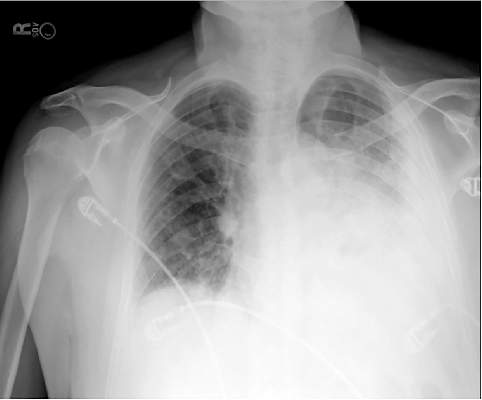
In recent years, the availability of portable ultrasound for bedside diagnostics and procedural guidance has revolutionized the practice of medicine, including the management of pleural effusions. When confronted with an obscured lower lobe on chest radiograph (Figure, left), clinicians were previously relegated to primitive bedside maneuvers, such as percussion or auscultation, to make critical decisions about the clinical management. Now, clinicians are able to look inside the body with point-of-care ultrasound and visually assess a pleural effusion before making any decisions. Point-of-care ultrasound has shifted the paradigm in the management of pleural effusions in several ways.
Ultrasound allows rapid detection and differentiation of pleural effusions from other pathologic findings.
Chest radiographs cannot accurately differentiate a pleural effusion from other common conditions, such as pneumonia, atelectasis, or an elevated hemidiaphragm. Ultrasound is the only bedside diagnostic modality that can rapidly differentiate these conditions within seconds and may reveal unsuspected findings, such as a mass or pericardial effusion.
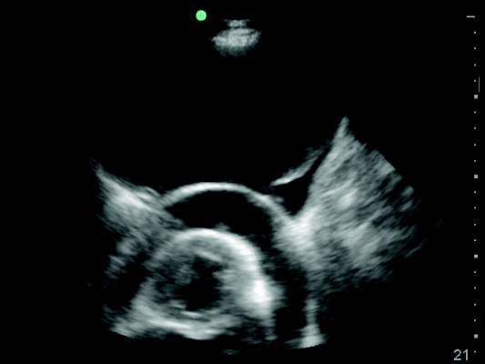
For example, the pleural ultrasound exam of a patient in the confirmed the presence of a large, left-sided pleural effusion (Figure, right) but also revealed an unsuspected large pericardial effusion (asterisk) that was causing hemodynamic compromise. The management of this patient shifted focus from the pleural effusion to the pericardial effusion, and urgent pericardiocentesis was performed. The sensitivity of ultrasound to detect a pleural effusion is proportional to the volume of fluid, reaching 100% with as little as 100 mL of fluid (Kalokairinou-Motogna et al. Med Ultra. 2010;12[1]:12). The diagnostic accuracy of ultrasound for detection of pleural effusions is comparable to CT scans of the chest and superior to portable chest radiographs (Lichtenstein et al. Anesthesiology. 2004;100[1]:9).
Ultrasound characterizes pleural effusions to determine the most appropriate management strategy.
Any clinician with basic ultrasonography skills can learn to evaluate pleural effusions and categorize them as simple or complex based on the sonographic appearance. Visualization of fibrinous stranding or loculations increases the probability of pleural fluid being exudative and often drives the decision to drain the fluid. The density and distribution of loculations can guide decisions about the most appropriate type of drainage procedure – thoracentesis versus tube thoracostomy versus surgical intervention. Use of color flow Doppler ultrasound allows clinicians to assess whether or not pleural fluid is free flowing and amenable to drainage, potentially saving the patient from an unnecessary attempt at drainage.
Ultrasound affords frontline clinicians the ability to streamline consultation with the most appropriate specialist based on the type of drainage procedure indicated and potentially prevent duplicate procedures on the same patient from different specialists.
Ultrasound reduces the risk of postprocedure complications from thoracentesis.
The risk of postthoracentesis pneumothorax was reported to be as high as 20%-39% before the routine use of point-of-care ultrasound (Grogan et al. Arch Int Med. 1990;150:873). Ultrasound guidance has been shown to increase procedural success rates and decrease the risk of postprocedure pneumothorax (2.7%), cost of hospitalization, and length of stay (Mercaldi et al. Chest. 2015;143[2]:532).
Regardless of the chest radiograph or CT scan findings, if the ultrasound exam reveals a scant volume of pleural fluid, or densely loculated pleural fluid, clinicians can avoid unnecessary attempts at bedside drainage, which likely partly accounts for the reduction in postprocedure pneumothorax. Use of ultrasound for needle site selection may prevent up to 10% of potential accidental organ punctures and increases accurate site selection by 26%, compared with chest radiograph and physical examination findings combined (Diacon et al. Chest. 2003;123:436).
Ultrasound facilitates patient-centered care.
Point-of-care ultrasound is the only new technology that has taken clinicians back to the bedside to spend more time with patients. Clinicians can simultaneously perform an ultrasound exam and converse with patients to gather a medical history. The ultrasound image serves as a tool to help patients understand their condition and facilitates shared decision making with clinicians at the bedside.
As more specialties have gained expertise in thoracic ultrasonography, the use of ultrasound guidance for thoracentesis has evolved to become the standard of care in many hospitals in the United States. Besides pulmonary specialists, several acute care specialists, including hospitalists, intensivists, and emergency medicine physicians, are routinely using point-of-care ultrasound to guide diagnostic decision making and procedures. Over the past 10 years, nearly a dozen procedure services led by internal medicine-trained hospitalists have been created at academic institutions that are routinely performing ultrasound-guided thoracenteses with low complication rates (Franco-Sadud et al. SGIM Forum. 2016;39[5]:13). More important, ultrasound is being used on the front lines to expeditiously evaluate pleural effusions and perform a diagnostic thoracentesis or consult with the appropriate subspecialist. Even though demonstration of competency in bedside procedures is no longer required for board certification in internal medicine, many internal medicine residency programs have incorporated diagnostic and procedural point-of-care ultrasound training into their education curriculum (Schnobrich et al. JGME. 2013;5[3]:498). Further, approximately 62% of medical schools report integrating ultrasound education in their medical student curriculum, and in coming years, most medical students will likely graduate with a basic skill set in point-of-care ultrasonography (Bahner et al. Academic Med. 2014;89[12]:1681). As point-of-care ultrasound education becomes integrated in training of physicians and other health-care providers, use of ultrasound to guide management of pleural effusions could become universally practiced and accepted as the new standard of care. Thus, it is plausible that a day will come in the near future when the sun will not set on an “un-ultrasound-ed” pleural effusion.
Dr. Franco-Sadud is with the section of hospital medicine/division of general internal medicine, Medical College of Wisconsin, Milwaukee, Wisconsin; Dr. Soni is with the section of hospital medicine and the section of pulmonary and critical care medicine, South Texas Veterans Health Care System and University of Texas Health Science Center, San Antonio.
The adage, “the sun should never set on an untapped pleural effusion,” was instilled in physicians for generations. However, anyone who practices medicine currently knows the sun often rises and sets several times before a pleural effusion is tapped. Why the change in mindset? Since the American Board of Internal Medicine removed the requirement for internal medicine residents to perform a minimum number of bedside procedures for certification, fewer graduating residents feel comfortable performing thoracentesis.
Additionally, the fear of litigation and institutional persecution from a postprocedure complication has caused many frontline clinicians to shy away from performing thoracentesis. Most important, we now appreciate that not all pleural effusions need to be tapped immediately, and the clinical decision making about the timing and technique to drain a pleural effusion is more complex than previously thought.
In recent years, the availability of portable ultrasound for bedside diagnostics and procedural guidance has revolutionized the practice of medicine, including the management of pleural effusions. When confronted with an obscured lower lobe on chest radiograph (Figure, left), clinicians were previously relegated to primitive bedside maneuvers, such as percussion or auscultation, to make critical decisions about the clinical management. Now, clinicians are able to look inside the body with point-of-care ultrasound and visually assess a pleural effusion before making any decisions. Point-of-care ultrasound has shifted the paradigm in the management of pleural effusions in several ways.
Ultrasound allows rapid detection and differentiation of pleural effusions from other pathologic findings.
Chest radiographs cannot accurately differentiate a pleural effusion from other common conditions, such as pneumonia, atelectasis, or an elevated hemidiaphragm. Ultrasound is the only bedside diagnostic modality that can rapidly differentiate these conditions within seconds and may reveal unsuspected findings, such as a mass or pericardial effusion.
For example, the pleural ultrasound exam of a patient in the confirmed the presence of a large, left-sided pleural effusion (Figure, right) but also revealed an unsuspected large pericardial effusion (asterisk) that was causing hemodynamic compromise. The management of this patient shifted focus from the pleural effusion to the pericardial effusion, and urgent pericardiocentesis was performed. The sensitivity of ultrasound to detect a pleural effusion is proportional to the volume of fluid, reaching 100% with as little as 100 mL of fluid (Kalokairinou-Motogna et al. Med Ultra. 2010;12[1]:12). The diagnostic accuracy of ultrasound for detection of pleural effusions is comparable to CT scans of the chest and superior to portable chest radiographs (Lichtenstein et al. Anesthesiology. 2004;100[1]:9).
Ultrasound characterizes pleural effusions to determine the most appropriate management strategy.
Any clinician with basic ultrasonography skills can learn to evaluate pleural effusions and categorize them as simple or complex based on the sonographic appearance. Visualization of fibrinous stranding or loculations increases the probability of pleural fluid being exudative and often drives the decision to drain the fluid. The density and distribution of loculations can guide decisions about the most appropriate type of drainage procedure – thoracentesis versus tube thoracostomy versus surgical intervention. Use of color flow Doppler ultrasound allows clinicians to assess whether or not pleural fluid is free flowing and amenable to drainage, potentially saving the patient from an unnecessary attempt at drainage.
Ultrasound affords frontline clinicians the ability to streamline consultation with the most appropriate specialist based on the type of drainage procedure indicated and potentially prevent duplicate procedures on the same patient from different specialists.
Ultrasound reduces the risk of postprocedure complications from thoracentesis.
The risk of postthoracentesis pneumothorax was reported to be as high as 20%-39% before the routine use of point-of-care ultrasound (Grogan et al. Arch Int Med. 1990;150:873). Ultrasound guidance has been shown to increase procedural success rates and decrease the risk of postprocedure pneumothorax (2.7%), cost of hospitalization, and length of stay (Mercaldi et al. Chest. 2015;143[2]:532).
Regardless of the chest radiograph or CT scan findings, if the ultrasound exam reveals a scant volume of pleural fluid, or densely loculated pleural fluid, clinicians can avoid unnecessary attempts at bedside drainage, which likely partly accounts for the reduction in postprocedure pneumothorax. Use of ultrasound for needle site selection may prevent up to 10% of potential accidental organ punctures and increases accurate site selection by 26%, compared with chest radiograph and physical examination findings combined (Diacon et al. Chest. 2003;123:436).
Ultrasound facilitates patient-centered care.
Point-of-care ultrasound is the only new technology that has taken clinicians back to the bedside to spend more time with patients. Clinicians can simultaneously perform an ultrasound exam and converse with patients to gather a medical history. The ultrasound image serves as a tool to help patients understand their condition and facilitates shared decision making with clinicians at the bedside.
As more specialties have gained expertise in thoracic ultrasonography, the use of ultrasound guidance for thoracentesis has evolved to become the standard of care in many hospitals in the United States. Besides pulmonary specialists, several acute care specialists, including hospitalists, intensivists, and emergency medicine physicians, are routinely using point-of-care ultrasound to guide diagnostic decision making and procedures. Over the past 10 years, nearly a dozen procedure services led by internal medicine-trained hospitalists have been created at academic institutions that are routinely performing ultrasound-guided thoracenteses with low complication rates (Franco-Sadud et al. SGIM Forum. 2016;39[5]:13). More important, ultrasound is being used on the front lines to expeditiously evaluate pleural effusions and perform a diagnostic thoracentesis or consult with the appropriate subspecialist. Even though demonstration of competency in bedside procedures is no longer required for board certification in internal medicine, many internal medicine residency programs have incorporated diagnostic and procedural point-of-care ultrasound training into their education curriculum (Schnobrich et al. JGME. 2013;5[3]:498). Further, approximately 62% of medical schools report integrating ultrasound education in their medical student curriculum, and in coming years, most medical students will likely graduate with a basic skill set in point-of-care ultrasonography (Bahner et al. Academic Med. 2014;89[12]:1681). As point-of-care ultrasound education becomes integrated in training of physicians and other health-care providers, use of ultrasound to guide management of pleural effusions could become universally practiced and accepted as the new standard of care. Thus, it is plausible that a day will come in the near future when the sun will not set on an “un-ultrasound-ed” pleural effusion.
Dr. Franco-Sadud is with the section of hospital medicine/division of general internal medicine, Medical College of Wisconsin, Milwaukee, Wisconsin; Dr. Soni is with the section of hospital medicine and the section of pulmonary and critical care medicine, South Texas Veterans Health Care System and University of Texas Health Science Center, San Antonio.

Patients with Epilepsy May Lack Essential Social Cognition Skills
Patients with epilepsy seem to have difficulty recognizing certain emotional states, according to a recent study that used video simulations to evaluate patients’ social cognition skills. When researchers administered the Awareness of Social Inference Test to 43 patients with focal epilepsy and 22 controls, using a video format, they found that neither group had trouble identifying positive emotional states like happiness; but patients with epilepsy had difficulty recognizing negative emotions such as anger, fear, and disgust. The study suggests that standard psychometric tools used to measure cognitive abilities in patients with epilepsy may need to be supplemented with a vehicle that evaluates social cognition.
Bujarski KA, Flashman L, Li Z, et al. Investigating social cognition in epilepsy using a naturalistic task. Epilepsia. 2016;57(9):1515-1520.
The Default Mode Network Plays Important Role in Pathology of Epilepsy
The default mode network (DMN), which connects brain regions such as precuneus/posterior cingulate cortex, medial prefrontal cortex, and medial, lateral, and inferior parietal cortex, plays an important role in temporal lobe epilepsy (TLE), according to a recent literature review. Among patients with TLE, the amplitude of the blood oxygenation-level dependent (BOLD) signal decreases during the interval between seizures. Investigators have also found that TLE patients have less anterograde connectivity from the anterior to the posterior DMN. Changes in the activity of the DMN in people with epilepsy, as well as several other neurological disorders, suggest that assessment of the network may help improve early detection and treatment, according to the researchers.
Mohan A, Roberto AJ, Mohan A, et al. The significance of the default mode network (DMN) in neurological and neuropsychiatric disorders: A review. Yale J Biol Med. 2016;89(1):49-57.
Using MRIs to Separate Rasmussen Encephalitis from Epilepsy
To help distinguish patients with Rasmussen encephalitis from patients with epilepsy not suffering from the syndrome, researchers performed quantitative volumetric MR imaging on 42 patients with Rasmussen syndrome and compared the readings to MRIs performed on 42 controls and 42 unaffected patients with epilepsy. Their analysis found that interhemispheric and frontal lobe ratios were the most effective way to differentiate Rasmussen encephalitis from the other 2 groups. They also found that the insula of Rasmussen encephalitis patients was significantly more atrophic, when compared with other cortical regions of the brain.
Wang Z, Krishnan B, Shattuck DW, et al. Automated MRI volumetric analysis in patients with Rasmussen syndrome [published online ahead of print September 8, 2016]. AJNR Am. J. Neuroradiol. 2016.
Long-term Intracranial Monitoring Reveals Circadian Pattern of Epileptic Discharges
Using the NeuroPace RNS system to monitor long-term epileptic-like activity, researchers have confirmed that there is a uniform circadian pattern to this brain activity. Studying 134 subjects, Spencer et al found the epileptiform activity peaked during normal sleeping hours. They also discovered a monophasic, nocturnally dominant rhythm in the neocortical areas of the brain and a more complex pattern, with a diurnal peak, in limbic sections of the brain. Some volunteers were also found to have a dual oscillator pattern to the brain activity, displaying a circadian and ultradian pattern.
Spencer D, Sun F, Brown S, Jobst, B, Wong V, Mirro E et al. Circadian and ultradian patterns of epileptiform discharges differ by seizure-onset location during long-term ambulatory intracranial monitoring. Epilepsia. 2016;57(9):1495-1502.