User login
Steroids could reduce death rate for TB patients with acute respiratory failure
Tuberculosis patients admitted to intensive care units with acute respiratory failure had significantly better survival at 90 days after treatment with corticosteroids and anti-TB drugs, compared with patients not treated with the steroids, according to a retrospective study.
An adjusted inverse probability of treatment weighted analysis using propensity scores revealed corticosteroid use to be independently associated with a significantly reduced 90-day mortality rate (OR = 0.47; 95% CI, 0.22-0.98). This statistical approach was used because it reduces selection bias and other potential confounding factors in a way that a multivariate analysis cannot, wrote Ji Young Yang, MD, of Busan (South Korea) Paik Hospital and Inje University College of Medicine in Busan.
Mortality rates were similar between the steroid-treated and non–steroid-treated groups (48.6% and 50%, respectively), and unadjusted 90-day mortality risk was not affected by steroid administration (odds ratio, 0.94; 95% CI, 0.46-1.92; P = .875), reported Dr. Yang and colleagues (Clin Infect Dis. 2016 Sep 8. doi: 10.1093/cid/ciw616).
The study involved the examination of records of 124 patients (mean age 62, 64% men) admitted to a single center over a 25-year period ending in 2014. Of these, 56.5% received corticosteroids, and 49.2% of the cohort died within 90 days.
The investigators acknowledged that their study was limited by various factors, including its small size, its use of data from a single center, and its lack of a standardized approach to steroid treatment.
“Further prospective randomized controlled trials will therefore be necessary to clarify the role of steroids in the management of these patients,” they wrote in their analysis. However, Dr. Yang and colleagues argued, in acute respiratory failure – a rare but dangerous complication in TB – “corticosteroids represent an attractive option because they can suppress cytokine expression and are effective in managing the inflammatory complications of extrapulmonary tuberculosis. Moreover, corticosteroids have been recently been shown to reduce mortality or treatment failure in patients with tuberculosis or severe pneumonia.”
Robert C. Hyzy, MD, director of the critical care medicine unit at the University of Michigan, Ann Arbor, said the findings “should be considered hypothesis generating.
“Clinicians should wait for prospective validation of this observation before considering the use of corticosteroids in hospitalized patients with tuberculosis,” he added.
Dr. Yang and colleagues disclosed no conflicts of interest or outside funding for their study.
Key clinical point: Corticosteroids used in combination with anti-TB treatment appeared to lower 90-day mortality in TB patients with ARF.
Major finding: Reduced 90-day mortality was associated with corticosteroid use (odds ratio, 0.47; 95% CI, 0.22-0.98; P = .049).
Data source: A retrospective cohort study of 124 patients admitted to intensive care units with TB and ARF in a single Korean center from 1989 to 2014.
Disclosures: The investigators reported no outside funding or conflicts of interest.
Midterm results of thoracic stenting for acute type B dissection promising
LAS VEGAS – Patients with acute, complicated type B aortic dissections are reported to have a greater than 50% likelihood of dying from their condition. Three-year results of the Valiant thoracic stent graft in the treatment of these dissections showed freedom from all-cause mortality of 79.4%, and a freedom from dissection-related mortality of 90%, according to Ali Azizzadeh, MD.
Dr. Azizzadeh presented the midterm results of the Medtronic Dissection US IDE trial of endovascular treatment with the Valiant Captivia thoracic stent graft (Medtronic) in acute, complicated type B aortic dissection patients at the 2016 Vascular Interventional Advances meeting.
One-year outcomes of the trial were reported last year in the Annals of Thoracic Surgery (2015 Sep;100:802-9).
Dr. Azizzadeh is a vascular surgeon at the Memorial Hermann Heart and Vascular Institute, Houston.
Between June 2010 and May 2012, 50 patients with acute, complicated type B aortic dissection were enrolled at 16 clinical sites in the United States in this multicenter, prospective, nonrandomized trial with a planned 5-year follow-up.
The primary safety endpoint was all-cause mortality within 30 days from the index procedure.
A total of 28 patients completed their 3-year follow-up. Through 3 years, there were no postindex ruptures or conversions to open surgical repair reported in the trial.
At 3 years, true lumen diameter over the stented region (or endograft segment) remained stable or increased in 92.3% of patients, according to Dr. Azizzadeh. False lumen diameter remained stable or decreased in 69.3% of patients, and the false lumen was partially or completely thrombosed in 75% of patients.
One death (from sepsis) occurred between years 2 and 3; and was adjudicated by the clinical events committee as unrelated to the device, the procedure, or the dissection.
Although these midterm results are encouraging, said Dr. Azizzadeh, longer-term outcomes are needed to assess the durability of the stent graft in this indication.
The trial was sponsored by Medtronic. Dr. Azizzadeh has consulted for and received research/trial funding from W.L. Gore & Associates and Medtronic.
AT VIVA16 LAS VEGAS
Key clinical point:
Major finding: Three-year results of the Valiant thoracic stent graft in the treatment of acute type B dissections showed freedom from all-cause mortality of 79.4%, and a freedom from dissection-related mortality of 90%.
Data source: Midterm results were presented from the multicenter, prospective, nonrandomized Medtronic Dissection US IDE trial.
Disclosures: The trial was sponsored by Medtronic. Dr. Azizzadeh has consulted for and received research/trial funding from W.L. Gore & Associates and Medtronic.
VIDEO: Is your patient clinically depressed, or is there something else?
Is your 42-year-old patient with well-controlled hypertension and type 2 diabetes dealing with a mood disorder that should be regarded as a psychiatric illness – or is she experiencing demoralization and grief?
In this installment of Mental Health Consult, the patient screens positive for depression but is ambivalent about taking antidepressants. In addition, the patient believes she has a number of coping resources that she can utilize. Finding out whether there is a need for an evidence-based psychotherapy, medication, or if other interventions are appropriate requires four key questions when taking a history.
Our expert panel from George Washington University, Washington, includes James L. Griffith, MD, chair of psychiatry and behavioral sciences; April Barbour, MD, MPH, director of the division of general internal medicine; and Lorenzo Norris, MD, medical director of psychiatric and behavioral services.
The video associated with this article is no longer available on this site. Please view all of our videos on the MDedge YouTube channel
wmcknight@frontlinemedcom.com
On Twitter @whitneymcknight
Intake of Vitamins and Minerals Is Inadequate for Most Americans: What Should We Advise Patients About Supplements?
Endovascular construction of arteriovenous fistulas shows promise
LAS VEGAS – Patients who had an arteriovenous fistula (AVF) created endovascularly using a new magnetic catheter system required fewer interventions and had fewer health care costs than patients whose AVF was created surgically, according to a late-breaking trial presented by Charmaine E. Lok, MD, at the 2016 Vascular Interventional Advances meeting.
The study compared AVF postcreation interventions between patients undergoing surgical (sAVF) creation and those whose fistula was created using a new endovascular AVF (endoAVF) system.
Medicare Standard Analytical Files were used to determine patient demographic and clinical characteristics and to identify and determine rates of sAVF postcreation interventions in patients with sAVF created from 2011 to 2013, according to Dr. Lok, a professor of medicine at the University of Toronto and senior scientist at the Toronto General Research Institute.
The rates of postcreation interventions per patient-year were determined based on patients’ outpatient and physician claims. Demographics and clinical information for patients with endoAVF were obtained from the single-arm Novel Endovascular Access Trial (NEAT) performed in Canada, Australia, and New Zealand.
The researchers determined the rates of postcreation interventions per patient-year from the trial based on patients’ outpatient and physician claims during specified follow-up.
Propensity score matching based on clinical and demographic factors was successful for comparing 60 Medicare patients who had surgical AVFs to NEAT patients. The matched surgical cohort had a significantly higher number of interventions than the endovascular cohort (3.4 vs. 0.6 per patient-year, respectively; P less than .0001). The associated average annual costs were $11,240 less for the endovascular patients, compared with the surgical patients.
In a breakdown of procedures, the endovascular cohort had lower event rates for angioplasty (0.04 vs. 0.93),respectively; thrombectomy (0.04 vs. 0.20); embolization/ligation of vein (0.13 vs. 0.1); revision (0.04 vs. 0.17); new AVF or transposition (0.11 vs. 0.30); catheter placement (0.11 vs. 0.43); vascular access–related infection (0.02 vs. 1.23), and arteriovenous graft placement (0.02 vs. 0.07), according to Dr. Lok.
The NEAT study assessed the FLEX system, which percutaneously creates a fistula in chronic kidney disease patients who require hemodialysis vascular access.
The FLEX system uses two catheters delivered percutaneously to an artery and a vein in proximity to each other in the arm. The catheters use magnets for alignment and a radio frequency system as an energy source. The catheters are magnetically aligned and an RF pulse creates an arteriovenous fistula endovascularly between the artery and vein and the catheters are then removed.
The technology used is not commercially available in the United States and is pending Food and Drug Administration review, according to Dr. Lok
The study was sponsored by TVA Medical. Dr. Lok has received honoraria from Maquet and W.L. Gore, and is a consultant for TVA Medical and W. L. Gore, and has received research funding from Maquet, Proteon, and TVA Medical.
Key clinical point: than patients whose AVF was created surgically
Major finding: A matched surgical cohort of patients with surgically constructed AVFs had a significantly higher number of interventions than an endovascular AVF cohort (3.4 vs. 0.6 per patient-year).
Data source: Researchers performed a propensity-matching analysis of Medicare patients to those patient who received an endovascularly created AVF in the NEAT trial.
Disclosures: The study was sponsored by TVA Medical. Dr. Lok has received honoraria from Maquet and W.L. Gore, and is a consultant for TVA Medical and W. L. Gore, and has received research funding from Maquet, Proteon, and TVA Medical.
Extended half-life clotting factors are safe, effective, and pricey
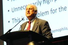
ORLANDO – Early experience with extended half-life clotting factor concentrates suggests these products are generally safe and effective, at least in the short term, according to hemophilia experts.
“In adolescent and adult patients, the extended half-life clotting factor concentrates demonstrate efficacy and safety in a variety of clinical settings, including prophylaxis, treatment of bleeds, and perisurgical hemostasis,” said Johnny Mahlangu, MD, director of the Haemophilia Comprehensive Care Centre in Johannesburg, South Africa.
Barriers to prophylaxis include “the need to inject the clotting factor concentrate at least two or three times a week, poor venous access, and, of course, poor adherence and compliance from our patients. Essentially, the extended half-life products were developed to mitigate these limitations,” he said.
Dr. Mahlangu and Guy Young, MD, a pediatric hematologist at Children’s Hospital Los Angeles, discussed results from published clinical trials of EHL products in children and adults.
The X factor
Extended half-life products currently available or in development include those created through fusion technology, including factor VIII products for treatment of hemophilia A, factor IX for treating hemophilia B, and factor VII products for treating factor VII deficiency (Alexander’s Disease).
More recent products created through pegylation technology include recombinant FVIII products.
Factor IX EHL CFCs
The three recombinant factor IX (rFIX) products that have completed phase III clinical trials are: nonacog beta pegol (N9-GP), rFIXFc (Alprolix), and rFIX-FP (Idelvion). The latter two are approved by the U.S. Food and Drug Administration; the former is waiting European and U.S. approval.
In clinical trials in adolescents and adults, these agents were associated with low median annualized bleeding rates, as follows:
• rFIXFc: 3.0 for a 50 IU/kg dose given every 7 days, and 1.4 for a 100 IU/kg dose given every 10 days
• rFIX-FP: 0.0 for a 40 IU/kg dose given every 7 days, and 1.08 for a 75 IU/kg dose given every 14 days
• Nonacog beta pegol: 2.93 for a 10 IU/kg dose and 1.0 for a 40 IU/kg dose, each given every 7 days.
All of these agents effectively treated bleeding episodes after one or two doses, Dr. Mahlangu said. The respective overall hemostatic efficacy rates were 97.2%, 96.7%, and 97.1%.
Safety analyses from published studies showed that no patients exposed to any of these agents developed inhibiting antibodies, although three patients treated with rFIXFc and nonacog beta pegol each developed noninhibitory antibodies.
There were no deaths from thromboembolic episodes and no drug-related serious adverse events.
Data are more limited for children treated with rFIX products, Dr. Young noted. In studies thus far, no children developed inhibitors, although this population had been heavily pretreated, he noted. Median annualized bleeding rates ranged from 1 to 3 and did not differ significantly between the products.
Factor VIII EHL CFCs
Four factor VIII EHL products are available or in development: rFVIIIFc (Eloctate); antihemophilic factor, pegylated (Adynovate); turoctocog alfa pegol (N8-GP); and BAY 94-9027. The former two agents are approved in the United States.
In adults and adolescents, these products have half-lives comparatively shorter than those seen with Factor IX products, the researchers noted.
Annualized bleeding rates for patients on prophylaxis with these agents were less than 4 bleeds per year, ranging from 1.3 to 3.6. In all, 96% of bleeds that did occur could be resolved with one or two injections of the extended half-life rFVIII products.
All patients had at least 50 exposures to these products, and none have developed inhibitors to date.
Only one study has been published to date of factor VIII products in children, comparing a standard half-life product with rFVIIIFc, Dr. Young said.
In this study, the patients were treated with a twice weekly, split-dose regimen. No patients developed inhibitors, and the mean annualized bleeding rate was a low 1.96.
Dr. Mahlangu disclosed research grants from Bayer, Biogen, CSL Behring, Novo Nordisk, and Roche, and speakers bureau participation for Amgen, Biotest, Biogen, CSL Behring, Novo Nordisk, and Sobi. Dr. Young disclosed honoraria and consulting fees from Baxalta, Bayer, Biogen, CSL Behring, and Novo Nordisk. Ellis J. Neufeld, MD, PhD, the invited discussant of the presentation, disclosed institutional grants from Baxalta, Novo Nordisk, and Octapharma, consulting/advising for those companies and for CSL Behring, Genentech, Hema Biologics, and Pfizer.
Improvements in half-life are incremental with the factor VIII products, but ground breaking with the factor IX products, allowing treatment intervals to be substantially prolonged. Pivotal trials of the several longer-acting products have been positive and generally convincing.
However, the pricing of these extended half-life products seems to reflect the so called “value proposition,” a concept from Big Pharma that puts a premium on convenience or novelty when determining the marketing price for a new drug. But the price may not always be commensurate with the clinical benefits patients derive from these newer agents.
How trials translate into treatment recommendations is complicated, and as Dr. Young and Dr. Mahlangu both made very clear, you need to consider that factor VIII and factor IX prolongation is entirely separate, even when the same technology is used, because the consequences are very different.
Ellis J. Neufeld, MD, PhD, is the associate chief of hematology/oncology at Boston Children’s Hospital, and was the invited discussant of the presentation.
ORLANDO – Early experience with extended half-life clotting factor concentrates suggests these products are generally safe and effective, at least in the short term, according to hemophilia experts.
“In adolescent and adult patients, the extended half-life clotting factor concentrates demonstrate efficacy and safety in a variety of clinical settings, including prophylaxis, treatment of bleeds, and perisurgical hemostasis,” said Johnny Mahlangu, MD, director of the Haemophilia Comprehensive Care Centre in Johannesburg, South Africa.
Barriers to prophylaxis include “the need to inject the clotting factor concentrate at least two or three times a week, poor venous access, and, of course, poor adherence and compliance from our patients. Essentially, the extended half-life products were developed to mitigate these limitations,” he said.
Dr. Mahlangu and Guy Young, MD, a pediatric hematologist at Children’s Hospital Los Angeles, discussed results from published clinical trials of EHL products in children and adults.
The X factor
Extended half-life products currently available or in development include those created through fusion technology, including factor VIII products for treatment of hemophilia A, factor IX for treating hemophilia B, and factor VII products for treating factor VII deficiency (Alexander’s Disease).
More recent products created through pegylation technology include recombinant FVIII products.
Factor IX EHL CFCs
The three recombinant factor IX (rFIX) products that have completed phase III clinical trials are: nonacog beta pegol (N9-GP), rFIXFc (Alprolix), and rFIX-FP (Idelvion). The latter two are approved by the U.S. Food and Drug Administration; the former is waiting European and U.S. approval.
In clinical trials in adolescents and adults, these agents were associated with low median annualized bleeding rates, as follows:
• rFIXFc: 3.0 for a 50 IU/kg dose given every 7 days, and 1.4 for a 100 IU/kg dose given every 10 days
• rFIX-FP: 0.0 for a 40 IU/kg dose given every 7 days, and 1.08 for a 75 IU/kg dose given every 14 days
• Nonacog beta pegol: 2.93 for a 10 IU/kg dose and 1.0 for a 40 IU/kg dose, each given every 7 days.
All of these agents effectively treated bleeding episodes after one or two doses, Dr. Mahlangu said. The respective overall hemostatic efficacy rates were 97.2%, 96.7%, and 97.1%.
Safety analyses from published studies showed that no patients exposed to any of these agents developed inhibiting antibodies, although three patients treated with rFIXFc and nonacog beta pegol each developed noninhibitory antibodies.
There were no deaths from thromboembolic episodes and no drug-related serious adverse events.
Data are more limited for children treated with rFIX products, Dr. Young noted. In studies thus far, no children developed inhibitors, although this population had been heavily pretreated, he noted. Median annualized bleeding rates ranged from 1 to 3 and did not differ significantly between the products.
Factor VIII EHL CFCs
Four factor VIII EHL products are available or in development: rFVIIIFc (Eloctate); antihemophilic factor, pegylated (Adynovate); turoctocog alfa pegol (N8-GP); and BAY 94-9027. The former two agents are approved in the United States.
In adults and adolescents, these products have half-lives comparatively shorter than those seen with Factor IX products, the researchers noted.
Annualized bleeding rates for patients on prophylaxis with these agents were less than 4 bleeds per year, ranging from 1.3 to 3.6. In all, 96% of bleeds that did occur could be resolved with one or two injections of the extended half-life rFVIII products.
All patients had at least 50 exposures to these products, and none have developed inhibitors to date.
Only one study has been published to date of factor VIII products in children, comparing a standard half-life product with rFVIIIFc, Dr. Young said.
In this study, the patients were treated with a twice weekly, split-dose regimen. No patients developed inhibitors, and the mean annualized bleeding rate was a low 1.96.
Dr. Mahlangu disclosed research grants from Bayer, Biogen, CSL Behring, Novo Nordisk, and Roche, and speakers bureau participation for Amgen, Biotest, Biogen, CSL Behring, Novo Nordisk, and Sobi. Dr. Young disclosed honoraria and consulting fees from Baxalta, Bayer, Biogen, CSL Behring, and Novo Nordisk. Ellis J. Neufeld, MD, PhD, the invited discussant of the presentation, disclosed institutional grants from Baxalta, Novo Nordisk, and Octapharma, consulting/advising for those companies and for CSL Behring, Genentech, Hema Biologics, and Pfizer.
AT WFH 2016 WORLD CONGRESS
Limit primary site radiation, but not craniospinal dose in medulloblastoma
BOSTON – For children with average-risk medulloblastoma, it is safe to limit the radiation boost to the posterior fossa, but reducing doses to the craniospinal axis results in worse outcomes among younger children, and is not advisable, according to results from the largest trial conducted in this population.
There were no significant differences in either 5-year event-free survival (EFS) or overall survival (OS) between children who received an involved field radiation therapy boost (boost to the tumor bed only) or a standard volume boost (to the whole posterior fossa) in a phase III randomized trial, reported lead author Jeff M. Michalski, MD, MBA, FASTRO, professor of radiation oncology at Washington University, St. Louis.
“We conclude that the survival rates and event-free survival rates following reduced boost volumes were comparable to standard radiation treatment volumes for the primary tumor site. This is the first trial to state definitively that there is no survival difference between these two approaches,” he said at a briefing following his presentation of the data in an oral abstract session at the annual meeting of the American Society for Radiation Oncology.
“However, the reduced craniospinal axis irradiation was associated with a higher event rate and worse survival. We believe that physicians can adopt smaller boost volumes to the posterior fossa in children with average-risk medulloblastoma, average risk being no evidence of spread at the time of diagnosis and near-complete or complete resection. But for all children, the standard of 23.4 Gy remains necessary to retain high-level tumor control,” he added.
Aggressive malignancy
Medulloblastoma, an aggressive tumor with the propensity to spread from the lower brain to the upper brain and spine, is the most common brain malignancy in children. The current standard of care for children with average-risk disease is surgical resection followed by systemic chemotherapy followed by irradiation to both the posterior fossa and to the craniospinal axis.
“Unfortunately, this strategy has significant negative consequences for the patients’ neurocognitive abilities, endocrine function, and hearing,” Dr. Michalski said.
The Children’s Oncology Group ACNS0331 trial was designed to determine whether reducing the volume of the boost from the whole posterior to the tumor bed only would compromise EFS and OS, and whether reducing the dose to the craniospinal axis from the current 23.4 Gy to 18 Gy in children aged 3-7 years would compromise survival measures.
The trial had two randomizations, both at the time of study enrollment. In the first, children from the ages of 3 to 7 years were randomly assigned to either low-dose craniospinal irradiation (18 Gy, 116 children) or to the standard dose (23.4 Gy, 110 children). All children aged 8 and older were assigned to receive the standard dose.
For the second randomization, all children were randomly allocated to either involved field RT boost or to standard volume boost to the whole posterior fossa.
Following radiation, all children were assigned to nine cycles of maintenance chemotherapy.
At a median follow-up of 6.6 years, 5-year EFS estimates for the primary site irradiation endpoint were 82.2% for 227 patients who received the involved-field radiation, compared with 80.8% for 237 patients who received whole posterior fossa irradiation.
Respective 5-year OS estimates were 84.1% and 85.2%. The upper limit of the hazard ratio confidence interval (CI) for the involved field therapy was lower than the prespecified limit, indicating that involved-field radiation was noninferior.
For the endpoint of low- vs. standard-dose craniospinal irradiation, the 5-year EFS estimates were 72.1% and 82.6%, respectively. The upper limit of the hazard ratio CI exceeded the prespecified limit, indicating that EFS was worse with the low-dose strategy.
Similarly, respective 5-year OS estimates were 78.1% and 85.9%, with the upper limit of the CI also higher than the prespecified upper limit.
When the investigators looked at the patterns of failure among the children in the two randomizations, they saw that there was no significant difference in the rate of isolated local failure between the involved-field or whole posterior fossa groups, or between the low-dose or high-dose craniospinal irradiation groups.
The finding that radiation volume to the primary site can be reduced is likely to be practice changing, said Geraldine Jacobson, MD, MPH, professor and chair of radiation oncology at West Virginia University, Morgantown. Dr. Jacobson moderated the briefing.
However, the worse survival with the low-dose craniospinal radiation “leaves the COG investigators with the challenge to explore other avenues for reducing toxicity of craniospinal irradiation,” she said.
Key clinical point: Primary site irradiation in children with medulloblastoma can be limited to the tumor bed rather than the whole posterior fossa.
Major finding: There were no differences in EFS or OS when radiation was limited to the tumor bed, but reduced dose to the craniospinal axis was associated with worse survival.
Data source: Randomized phase III trial of 690 children with average-risk medulloblastoma.
Disclosures: The National Cancer Institute funded the study. Dr. Michalski and Dr. Jacobson reported no relevant conflicts of interest.
Martina Mancini, PhD, and Graham Harker
The video associated with this article is no longer available on this site. Please view all of our videos on the MDedge YouTube channel
The video associated with this article is no longer available on this site. Please view all of our videos on the MDedge YouTube channel
The video associated with this article is no longer available on this site. Please view all of our videos on the MDedge YouTube channel
Boost dose pays off long term after surgery, WBRT for DCIS
BOSTON – Adding a boost dose after surgery and whole-breast radiation therapy (WBRT) for ductal carcinoma in situ offers a small but significant reduction in long-term risk of same-breast recurrence, according to results of a multicenter study.
An analysis of pooled data from 10 institutions showed that after 15 years of follow-up, 91.6% of women who had been treated with breast-conserving surgery, WBRT, and a boost dose remained free of ipsilateral breast tumor recurrence (IBTR), compared with 88% of women who received WBRT alone, reported Meena Savur Moran, MD, director of the breast cancer radiotherapy program at Smilow Cancer Hospital, Yale New Haven, Connecticut.
The absolute differences between the groups – 3% at 10 years, and 4% at 15 years – “don’t seem like a lot, but it is clinically important for patients, because what we’ve learned from the invasive cancer data is that this small, incremental decrease results in about a 40% less mastectomy rate for salvage mastectomies for recurrences,” she said.
It is common practice in many centers to give a radiation boost to the tumor bed of 8-16 Gy in 4-8 additional fractions following breast-conserving surgery and WBRT. This has been shown in invasive cancers to be associated with a statistically significant decrease in risk for IBTR in all age groups of about 4% at 20 years.
Yet because DCIS generally has an excellent prognosis with very few recurrences after whole-breast irradiation, it has been harder to determine whether radiation boosting would offer similar benefit to that seen in invasive cancers,
In an attempt to answer this question, investigators from 10 centers collaborated to create a DCIS database of patients treated with WBRT either with or without boost, and then analyzed the results.
The final cohort included 4,131 patients, far exceeding the minimum sample size the statisticians calculated would be needed to detect a benefit.
The median age of patients at the time of therapy was 56.1 years, median follow-up was 9 years, and the median boost dose was 14 Gy. In all, 4% of patients had positive tumor margins after surgery.
Rates of IBTR-free survival with boost vs. no boost were 97.1% vs. 96.3% at 5 years, 94.1% vs. 92.5% at 10 years, and, as noted before, 91.6% vs. 88% at 15 years (P = .0389).
In both univariate and in multivariate analysis controlling for tumor grade, comedo carcinoma, tamoxifen use, margin status, and age, IBTR was significantly associated with lower risk for recurrence.
The investigators also conducted a boost/no boost analysis stratified by margin status using both the National Surgical Adjuvant Breast and Bowel Project (NSABP) definition of negative margins as no ink on tumor, and the joint Society of Surgical Oncology, ASTRO, and American Society of Clinical Oncology guideline definition as less than 2 mm, and in each case found that among patients with negative margins, boosting offered a significant recurrence-free advantage.
Finally, they found that boosting offered a significant advantage both for patients 50 and older (P = .0073) and those 49 and younger (P = .0166).
“For DCIS, most of who treat breast cancer have used a boost, but we really extrapolated from the treatment from invasive breast cancer. Evidence for this practice until now in specific for DCIS has been lacking,” said Geraldine Jacobson, MD, MPH, professor and chair of radiation oncology at West Virginia University, Morgantown. Dr. Jacobson moderated the briefing.
She noted that the large sample size and long follow-up of the study was necessary for the treatment benefit to emerge.
AT THE ASTRO ANNUAL MEETING 2016
Key clinical point: This study shows a significant benefit for adding a boost radiation dose following breast-conserving surgery and whole-breast radiation for ductal carcinoma in situ (DCIS).
Major finding: After 15 years of follow-up, 91.6% of women who had received a boost dose were free of ipsilateral breast recurrence, compared with 88% of women who did not have a boost.
Data source: Analysis of pooled data on 4,131 patients with DCIS treated at 10 academic medical centers.
Disclosures: The study was supported by participating institutions. Dr. Moran and Dr. Jacobson reported no conflicts of interest.
Patient-specific model takes variability out of blood glucose average
Researchers have identified a patient-specific correction factor based on the age of a patient’s red blood cells that can improve the accuracy of average blood glucose estimations from hemoglobin A1c measures in patients with diabetes.
“The true average glucose concentration of a nondiabetic and a poorly controlled diabetic may differ by less than 15 mg/dL, but patients with identical HbA1c values may have true average glucose concentrations that differ by more than 60 mg/dL,” wrote Roy Malka, PhD, a mathematician and research fellow at Massachusetts General Hospital, Boston, and his associates.
In an article published in the Oct. 5 online edition of Science Translational Medicine, the researchers reported the development of a mathematical model for HbA1c formation inside the red blood cell, based on the chemical contributions of glycemic and nonglycemic factors, as well as the average age of a patient’s red blood cells.
Using existing continuous glucose monitoring (CGM) data from four different groups – totaling more than 200 diabetes patients – the researchers then personalized the parameters of the model to each patient to see the impact of patient-specific variation in the relationship between average concentration of glucose in the blood and HbA1c.
Finally, they applied the personalized model to data from each patient to determine its accuracy in estimating future blood glucose from future HbA1c measurements, and compared the blood glucose estimates with those made using the current standard method (Sci Transl Med. 2016, Oct 5. http://stm.sciencemag.org/lookup/doi/10.1126/scitranslmed.aaf9304).
This showed that their patient-specific model reduced the median absolute error in estimated average blood glucose from more than 15 mg/dL to less than 5 mg/dL, representing an error reduction of more than 66%.
“The model would require one pair of CGM-measured AG [average blood glucose] and an HbA1c measurement that would be used to determine the patient’s [mean red blood cell count (MRBC)],” the researchers wrote. “MRBC would then be used going forward to refine the future [average glucose (AG)] calculated on the basis of HbA1c.”
The researchers pointed out that work was still needed to calculate the duration of CGM that would allow sufficient calibration of MRBC. However, their analysis suggested that no more than 30 days would be needed, and significant improvements could be achieved within just 21 days.
In patients with stable monthly glucose averages, the prior month would be enough for calibration, and even a single week of stable weekly glucose averages might be sufficient.
“The improvement in AG calculation afforded by our model should improve medical care and provide for a personalized approach to determining AG from HbA1c.”
The ADAG study was supported by the American Diabetes Association, the European Association for the Study of Diabetes, Abbott Diabetes Care, Bayer Healthcare, GlaxoSmithKline, Sanofi-Aventis Netherlands, Merck, LifeScan, Medtronic MiniMed, and HemoCue. Two authors were supported by the National Institutes of Health, and Abbott Diagnostics. The authors are also listed as inventors on a patent application related to this work submitted by Partners HealthCare.
Key clinical point:
Major finding: A patient-specific model of the relationship between HbA1c and average blood sugar reduced the median absolute error in estimated average blood glucose from more than 15 mg/dL to less than 5 mg/dL, representing an error reduction of more than 66%.
Data source: Mathematical modeling validated via four sets of patient continuous glucose monitoring data.
Disclosures: The ADAG study was supported by the American Diabetes Association, the European Association for the Study of Diabetes, Abbott Diabetes Care, Bayer Healthcare, GlaxoSmithKline, Sanofi-Aventis Netherlands, Merck, LifeScan, Medtronic MiniMed, and HemoCue. Two authors were supported by the National Institutes of Health, and Abbott Diagnostics. The authors are also listed as inventors on a patent application related to this work submitted by Partners HealthCare.

