User login
Progressive back pain
The history and findings in this case are suggestive of advanced/metastatic breast cancer.
Breast cancer is the most frequently diagnosed life-threatening cancer and the second-leading cause of cancer-related deaths in women worldwide. In the United States, an estimated 287,850 new cases of invasive breast cancer were diagnosed in 2022 and 43,250 women died of the disease. Globally, approximately 2.3 million new diagnoses and 685,000 breast cancer–related deaths were reported in 2020.
Tumor size, nodal spread, and distant metastases (TNM) at the time of diagnosis are key prognostic factors. Immunohistochemistry tumor markers (ie, estrogen receptor [ER], progesterone receptor [PR], and HER2), as well as grade and Ki-67 expression, have also been shown to be independent predictors of breast cancer death and are used together with TNM to guide treatment decisions.
Despite advances in breast cancer diagnosis and treatment, metastatic recurrence remains a significant problem. Although the incidence of distance relapse is declining and survival times for patients with recurrent disease are improving, 20%-30% of patients with early breast cancer still die of metastatic disease. Metastatic breast cancer recurrence can arise months to decades after initial diagnosis and treatment.
According to the National Comprehensive Cancer Network (NCCN) guidelines, biopsy is a critical component of the workup for patients with recurrent or stage IV disease. This is because biopsy ensures accurate determination of metastatic/recurrent disease and tumor histology and enables biomarker determination and selection of appropriate treatment. Soft-tissue tumor biopsy is preferred over bone sites unless a portion of the biopsy can be protected from harsh decalcification solution to preserve more accurate evaluation of biomarkers. Determination of HR status (ER and PR) and HER2 status should be repeated in all cases when diagnostic tissue is obtained because ER and PR assays may be falsely negative or falsely positive, and there may be discordance between the primary and metastatic tumors. According to the NCCN panel, re-testing the receptor status of recurrent disease should be performed, particularly when it was previously unknown, originally negative, or not overexpressed.
Additionally, the staging evaluation of patients who present with recurrent or stage IV breast cancer should include history and physical exam; a complete blood cell count, liver function tests, chest diagnostic CT, bone scan, and radiography of any long or weight-bearing bones that are painful or appear abnormal on bone scan; diagnostic CT of the abdomen (with or without diagnostic CT of the pelvis) or MRI of the abdomen; and biopsy documentation of first recurrence whenever possible. The use of sodium fluoride PET or PET/CT for evaluating patients with recurrent disease is generally discouraged.
Presently, metastatic breast cancer remains incurable. However, in recent years, the treatment landscape for metastatic breast cancer has significantly advanced in all breast cancer subtypes, leading to improvements in progression-free survival and even overall survival in some cases. For example, newer, targeted approaches directly address mutation drivers and allow precise delivery of chemotherapeutic agents. Detailed guidance on the treatment of breast cancer can be found here and in the full NCCN guidelines.
Avan J. Armaghani, MD, Assistant Member, Department of Breast Oncology, Moffitt Cancer Center, University of South Florida, Tampa, FL.
Avan J. Armaghani, MD, has disclosed no relevant financial relationships.
Image Quizzes are fictional or fictionalized clinical scenarios intended to provide evidence-based educational takeaways.
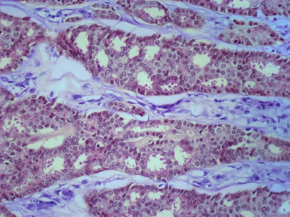
A 62-year-old nonsmoking woman presents with progressive moderate to severe back pain. The patient has a history of endometriosis and node-positive invasive ductal breast cancer, which was diagnosed 15 years ago. The tumor was hormone receptor (HR)–positive and human epidermal growth factor receptor 2 (HER2)–negative. After a lumpectomy, she received adjuvant chemotherapy, followed by radiation therapy and 5 years of adjuvant oral endocrine therapy. Physical examination reveals several large palpable nodes in the right axillary region; no abnormalities are noted in either breast or the left axillary region.
The patient is 5 ft 7 in and weighs 152 lb (BMI, 23.8). At her last visit, 3 years earlier, she weighed 176 lb. She states her weight loss has been unintentional and began about 6 months ago. The patient denies any respiratory or abdominal symptoms; she does report increasing fatigue, which she attributes to her back pain. Complete blood cell count values are within normal range, except for an elevated alkaline phosphatase level (215 IU/L).
A subsequent axillary lymph node ultrasound reveals several irregular hypoechoic masses in the right axilla of various sizes, the largest being 2.4 cm. PET, CT, and a bone scan were also performed and revealed multiple suspicious lesions in the spine and several pulmonary nodules.

Treat together: Tackle heart disease and obesity simultaneously
say the authors of a new state-of-the-art review.
“CVD and obesity are common conditions that frequently coexist. We cannot treat one of these conditions while ignoring the other,” Rosana G. Bianchettin, MD, of the division of cardiovascular diseases, Mayo Clinic, Rochester, Minn., and colleagues wrote in their review, recently published in the Journal of the American College of Cardiology.
The review outlines, for example, how obesity can impair common imaging tests used to diagnose heart disease, potentially reducing their accuracy.
And cardiac procedures such as percutaneous coronary intervention, open heart surgery, and revascularization all involve greater risk in the setting of obesity, while procedures such as valve replacement and heart transplantation carry a greater likelihood of failure.
Obesity can also alter drug pharmacokinetics and pharmacodynamics.
Weight reduction is an important part of the management of patients with cardiovascular disease and obesity, and “cardiac rehabilitation programs represent a potential opportunity for structured interventions,” the authors noted. However, “when other measures are insufficient, bariatric surgery can improve outcomes.”
They also advised against relying solely on body mass index (BMI) to assess adiposity: “It is prudent to investigate a range of complementary ... parameters alongside standard BMI calculations (accounting for age, race, and sex), including measures of central obesity, such as waist circumference, waist-to-hip ratio, and weight-to-height ratio.”
Excess fat acts as filter and can skew diagnostic results
“Obesity affects nearly all the diagnostic tests used in cardiology, such as ECG, CT scan, MRI, and echocardiogram,” senior author Francisco Lopez-Jimenez, MD, director of preventive cardiology at Mayo Clinic, explained in a statement.
The review includes a detailed table of these key obesity-related challenges. With electrocardiograms, for example, obesity can cause displacement of the heart, increased cardiac workload, and widening of the distance between the heart and the recording electrodes.
Obesity also lowers the sensitivity of exercise echocardiography, and use of CT coronary angiogram is completely precluded in people with a BMI above 40 kg/m2. In interventional radiology, there may be poor visualization of target areas.
“Excess fat acts as a kind of filter and can skew test readings to under- or overdiagnosis,” noted Dr. Lopez-Jimenez.
Therapeutic challenges: Drugs may work differently
A longer table in the review summarizes the therapeutic challenges involved in lifestyle modification, pharmacology, cardiac procedures, and other therapeutic measures for people with the two conditions.
Obesity can limit a person’s ability to exercise, for example, and smoking cessation may promote overeating and further weight gain.
Moreover, “tailoring pharmacotherapy is difficult because of unique pharmacokinetic and pharmacodynamic factors in people with obesity that alter distribution, metabolism, and elimination of drugs. Each drug also has special properties that must be considered when it is administrated,” the authors wrote.
Examples include the higher volume of distribution of lipophilic drugs in those with increased fat mass, alterations in liver metabolism, and difficulties with anticoagulant dosing.
Cardiac rehabilitation is an intervention opportunity
Although cardiac rehabilitation is “a cornerstone in secondary prevention” for people who have experienced a cardiac event, only 8% of such programs include formal in-house behavioral weight-loss programs.
But that could be remedied and expanded with the use of options such as home-based rehabilitation and telephone counseling, particularly in rural communities, Dr. Bianchettin and colleagues said.
“Motivated individuals will benefit from multicomponent approaches and should be encouraged to set specific, proximal, shared goals with their health care professional. A multitude of tools are available to support self-monitoring (e.g., smartphone applications, food diaries), and scheduled regular follow-up and feedback on progress can help to maintain motivation,” they wrote.
The bottom line, said Dr. Lopez-Jimenez: “Obesity is an important risk factor to address in patients with heart disease and it requires us to do something. ... The patient needs to know that their clinician can help them lose weight. Overall, weight-loss solutions come down to finding the right therapy for the patient.”
Dr. Bianchettin reported no relevant financial relationships. Dr. Lopez-Jimenez has reported conducting research related to 3D body assessment with Select Research, Mayo Clinic, and may benefit in the future if the technology is commercialized; he has not received any relevant monetary, financial, or other type of compensation to date, in relationship to this arrangement. He is a member of the scientific advisory board for Novo Nordisk.
A version of this article first appeared on Medscape.com.
FROM THE JOURNAL OF THE AMERICAN COLLEGE OF CARDIOLOGY
AI-assisted colonoscopy in IBD: Not all it’s cut out to be?
Within the rising tide of studies extolling the benefits of artificial intelligence for improving adenoma detection during colonoscopy comes new research suggesting the contrary, at least among people with inflammatory bowel disease (IBD).
Researchers retrospectively studied almost 1,000 colonoscopies before and after introduction of an AI system (GI Genius, Medtronic) at a tertiary medical center in Israel in which a large volume of endoscopies was performed. , and it was significantly higher for colonoscopies performed by gastroenterologists who had 5 or more years of experience, compared with the ADR for AI-assisted colonoscopies.
The lower ADR rate in AI-assisted procedures could be the result of an overreliance on the AI technology and shorter procedure times, which may have led to an underrecognition of adenomas, lead investigator Asaf Levartovsky, MD, said in an interview.
“AI is an aid to the endoscopist, not a replacement to the endoscopist,” added Dr. Levartovsky, a gastroenterologist at Sheba Medical Center, Tel Aviv.
The results were presented as a poster at the annual congress of the European Crohn’s and Colitis Organisation, held in Copenhagen and virtually.
Key findings
The use of AI has recently been shown to improve colorectal cancer screening overall, the authors note. ADR is a measure of the quality of screening colonoscopies. Detection rates were at least 20% among women and 30% among men, “indicative of adequate performance.”
The ADR for people with IBD can be lower than it is for average-risk patients, however, owing to a difference in age in the two populations and the presence of dysplasia-associated lesions, as opposed to sporadic adenomas, for patients with IBD, the researchers note. There is no consensus on an acceptable ADR target for patients with IBD, and the impact of AI-assisted colonoscopy in this patient population hasn’t been explored, they add.
To learn more, Dr. Levartovsky and colleagues compared 237 screening colonoscopies conducted in the 11 months before AI was introduced at the medical center in July 2021 to 759 colonoscopies performed in the 15 months after its introduction.
The pre-AI patient group and the AI patient group were similar (mean age, 44-45 years; about 55% men in each group). Crohn’s disease was more common than ulcerative colitis (63% in the pre-AI cohort and 57% in the AI-assisted cohort).
The ADR in the pre-AI group was 6.3%, compared with 4% in the AI-assisted group (P = .15). The distinction became significant, at 7.6% versus 3.8% (P = .035), when researchers evaluated colonoscopies performed by gastroenterologists who had 5 or more years of experience.
Total procedure time was longer for the patients in the pre-AI group, at 25 minutes, compared with 21 minutes in the AI-assisted group. This difference was statistically significant (P < .0001).
“I think this poster raises questions regarding the real-world utility of AI for adenoma detection [in patients with IBD],” Dr. Levartovsky said.
Dr. Levartovsky said he was not surprised by their findings, because they are similar to those reported in a recent article from his group, although this earlier study did not focus on patients with IBD.
The research had some limitations. The study was not case-control matched, and the pre-AI group was considerably smaller than the AI group.
Study design a factor
The study design could account for the difference in its findings, compared with research indicating that AI-assisted colonoscopies improve ADR, Cesare Hassan, MD, associate professor of gastroenterology at Humanitas University, Milan, said in an interview.
The study was retrospective, so researchers could not randomly assign people to the AI or the no-AI group. It therefore was not possible to ensure that the prevalence of disease was equivalent between the two groups, he said.
By comparison, the previous studies showing the benefits of AI-assisted colonoscopy with regard to ADR were randomized, controlled clinical trials, Dr. Hassan said.
The study was independently supported. Dr. Levartovsky and Dr. Hassan report no relevant financial relationships.
A version of this article originally appeared on Medscape.com.
FROM ECCO 2023
Two diets tied to lower Alzheimer’s pathology at autopsy
In a cohort of deceased older adults, those who had adhered to the Mediterranean-DASH Intervention for Neurodegenerative Delay (MIND) and Mediterranean diets for nearly a decade before death had less global Alzheimer’s disease–related pathology, primarily less beta-amyloid, at autopsy.
Those who most closely followed these diets had almost 40% lower odds of having an Alzheimer’s disease diagnosis at death. The findings offer one mechanism by which healthy diets protect cognition.
“While our research doesn’t prove that a healthy diet resulted in fewer brain deposits of amyloid plaques ... we know there is a relationship, and following the MIND and Mediterranean diets may be one way that people can improve their brain health and protect cognition as they age,” study investigator Puja Agarwal, PhD, of RUSH University Medical Center in Chicago, said in a statement.
The study was published online in Neurology.
Green leafy veggies key
The MIND diet was pioneered by the late Martha Clare Morris, ScD, a Rush nutritional epidemiologist, who died of cancer in 2020 at age 64.
Although similar, the Mediterranean diet recommends vegetables, fruit, and three or more servings of fish per week, whereas the MIND diet prioritizes green leafy vegetables, such as spinach, kale, and collard greens, along with other vegetables. The MIND diet also prioritizes berries over other fruit and recommends one or more servings of fish per week. Both diets recommend small amounts of wine.
The current study focused on 581 older adults who died while participating in the Rush Memory and Aging Project (MAP). All participants agreed to undergo annual clinical evaluations and brain autopsy after death.
Participants completed annual food frequency questionnaires beginning at a mean age of 84. The mean age at death was 91. Mean follow-up was 6.8 years.
Around the time of death, 224 participants (39%) had a diagnosis of clinical dementia, and 383 participants (66%) had a pathologic Alzheimer’s disease diagnosis at time of death.
The researchers used a series of regression analyses to investigate the MIND and Mediterranean diets and dietary components associated with Alzheimer’s disease pathology. They controlled for age at death, sex, education, APO-epsilon 4 status, and total calories.
Overall, both diets were significantly associated with lower global Alzheimer’s disease pathology (MIND: beta = –0.022, P = .034; and Mediterranean: beta = –0.007, P = .039) – specifically, with less beta-amyloid (MIND: beta = –0.068, P = .050; and Mediterranean: beta = –0.040, P = .004).
The findings persisted when the analysis was further adjusted for physical activity, smoking, and vascular disease burden and when participants with mild cognitive impairment or dementia at the baseline dietary assessment were excluded.
Individuals who most closely followed the Mediterranean diet had average beta-amyloid load similar to being 18 years younger than peers with the lowest adherence. And those who most closely followed the MIND diet had average beta-amyloid amounts similar to being 12 years younger than those with the lowest adherence.
A MIND diet score 1 point higher corresponded to typical plaque deposition of participants who are 4.25 years younger in age.
Regarding individual dietary components, those who ate seven or more servings of green leafy vegetables weekly had less global Alzheimer’s disease pathology than peers who ate one or fewer (beta = –0.115, P = .0038). Those who ate seven or more servings per week had plaque amounts in their brains corresponding to being almost 19 years younger in comparison with those who ate the fewest servings per week.
“Our finding that eating more green leafy vegetables is in itself associated with fewer signs of Alzheimer’s disease in the brain is intriguing enough for people to consider adding more of these vegetables to their diet,” Dr. Agarwal said in the news release.
Previous data from the MAP cohort showed that adherence to the MIND diet can improve memory and thinking skills of older adults, even in the presence of Alzheimer’s disease pathology.
Novel study, intriguing results
Heather Snyder, PhD, vice president of medical and scientific relations with the Alzheimer’s Association, noted that a number of studies have linked overall nutrition – especially a balanced diet low in saturated fats and sugar and high in vegetables – with brain health, including cognition, as one ages.
This new study “takes what we know about the link between nutrition and risk for cognitive decline a step further by looking at the specific brain changes that occur in Alzheimer’s disease. The study found an association of certain nutrition behaviors with less of these Alzheimer’s brain changes,” said Dr. Snyder, who was not involved in the study.
“This is intriguing, and more research is needed to test via an intervention if healthy dietary behaviors can modify the presence of Alzheimer’s brain changes and reduce risk of cognitive decline.”
The Alzheimer’s Association is leading a 2-year clinical trial known as US POINTER to study how targeting known dementia risk factors in combination may reduce risk of cognitive decline in older adults. The MIND diet is being used in US POINTER.
“But while we work to find an exact ‘recipe’ for risk reduction, it’s important to eat a heart-healthy diet that incorporates nutrients that our bodies and brains need to be at their best,” Dr. Snyder said.
The study was funded by the National Institutes of Health. Dr. Agarwal and Dr. Snyder have disclosed no relevant financial relationships.
A version of this article first appeared on Medscape.com.
FROM NEUROLOGY
High stress levels linked to cognitive decline
, a new study shows.
Individuals with elevated stress levels also had higher rates of diabetes, hypertension, and other cardiovascular disease (CVD) risk factors. But even after controlling for those risk factors, stress remained an independent predictor of cognitive decline.
The national cohort study showed that the association between stress and cognition was similar between Black and White individuals and that those with controlled stress were less likely to have cognitive impairment than those with uncontrolled or new stress.
“We have known for a while that excess levels of stress can be harmful for the human body and the heart, but we are now adding more evidence that excess levels of stress can be harmful for cognition,” said lead investigator Ambar Kulshreshtha, MD, PhD, associate professor of family and preventive medicine and epidemiology at Emory University, Atlanta. “We were able to see that regardless of race or gender, stress is bad.”
The findings were published online in JAMA Network Open.
Independent risk factor
For the study, investigators analyzed data from the Reasons for Geographic and Racial Differences in Stroke (REGARDS) study, a national population-based cohort of Black and White participants aged 45 years or older, sampled from the U.S. population.
Participants completed a questionnaire designed to evaluate stress levels when they were enrolled in the study between 2003 and 2007 and again about 11 years after enrollment.
Of the 24,448 participants (41.6% Black) in the study, 22.9% reported elevated stress levels.
Those with high stress were more likely to be younger, female, Black, smokers, and have a higher body mass index and less likely to have a college degree and to be physically active. They also had a lower family income and were more likely to have cardiovascular disease risk factors, such as hypertension, diabetes, and dyslipidemia.
Participants with elevated levels of perceived stress were 37% more likely to have poor cognition after adjustment for sociodemographic variables, cardiovascular risk factors, and depression (adjusted odds ratio, 1.37; 95% confidence interval, 1.22-1.53).
There was no significant difference between Black and White participants.
Damaging consequences
Researchers also found a dose-response relationship, with the greatest cognitive decline found in people who reported high stress at both time points and those who had new stress at follow up (aOR, 1.16; 95% CI, 0.92-1.45), compared with those with resolved stress (aOR, 1.03; 95% CI, 0.81-1.32) or no stress (aOR, 0.81; 95% CI, 0.68-0.97).
A change in perceived stress by 1 unit was associated with 4% increased risk of cognitive impairment after adjusting for sociodemographic variables, CVD risk factors, lifestyle factors, and depressive symptoms (aOR, 1.04; 95% CI, 1.02-1.06).
Although the study didn’t reveal the mechanisms that might link stress and cognition, it does point to stress as a potentially modifiable risk factor for cognitive decline, Dr. Kulshreshtha said.
“One in three of my patients have had to deal with extra levels of stress and anxiety over the past few years,” said Dr. Kulshreshtha. “We as clinicians are aware that stress can have damaging consequences to the heart and other organs, and when we see patients who have these complaints, especially elderly patients, we should spend some time asking people about their stress and how they are managing it.”
Additional screening
Gregory Day, MD, a neurologist at the Mayo Clinic, Jacksonville, Fla., said that the findings help fill a void in the research about stress and cognition.
“It’s a potentially important association that’s easy for us to miss in clinical practice,” said Dr. Day, who was not a part of the study. “It’s one of those things that we can all recognize impacts health, but we have very few large, well thought out studies that give us the data we need to inform its place in clinical decision-making.”
In addition to its large sample size, the overrepresentation of diverse populations is a strength of the study and a contribution to the field, Dr. Day said.
“One question they don’t directly ask is, is this association maybe due to some differences in stress? And the answer from the paper is probably not, because it looks like when we control for these things, we don’t see big differences incident risk factors between race,” he added.
The findings also point to the need for clinicians, especially primary care physicians, to screen patients for stress during routine examinations.
“Every visit is an opportunity to screen for risk factors that are going to set people up for future brain health,” Dr. Day said. “In addition to screening for all of these other risk factors for brain health, maybe it’s worth including some more global assessment of stress in a standard screener.”
The study was funded by the National Institute of Neurological Disorders and Stroke, the National Institutes of Health, and the National Institute on Aging. Dr. Kulshreshtha and Dr. Day report no relevant financial relationships.
A version of this article first appeared on Medscape.com.
From JAMA Network Open
Digital rectal exam fails as screening tool for prostate cancer
, say investigators reporting the PROBASE study.
The study compared risk-adapted screening measures in men who had prostate-specific antigen (PSA) measured at age 45 with those who had PSA measurements plus DRE at age 50.
The results show that as a solitary screening tool, 99% of DREs did not raise suspicion for prostate cancer, and among the 57 cases where DRE did raise suspicion, only three men were found to have cancer, all of which were low-grade, reported Agne Krilaviciute, PhD, from the German Cancer Research Center in Heidelberg, and colleagues.
“We also see that the cancer detection rate by PSA is four times higher compared to the DRE detection. Around 18% of the tumors are located in the part of the prostate where DRE cannot detect them,” she said in an oral presentation at the European Association of Urology Congress.
The investigators found that the majority of prostate cancers that occurred in this relatively young population were International Society of Urological Pathology grade 1 (Gleason score 3 + 3 = 6) or grade 2 (Gleason 3 + 4 = 7). DRE yields positive results in only about 12% of cases of ISUP grade 1 or 2, they noted.
“We conclude that DRE as a solitary screening test does not lead to a significant PCa [prostate cancer] detection rate in young men,” Dr. Krilaviciute said.
Falling by the wayside
The study adds to the growing body of evidence that DRE may not be especially helpful as either a screening tool or when used in active surveillance of men with prostate cancer.
An international consensus panel found that DRE could be safely skipped for active surveillance when MRI and other more accurate and objective measures, such as biomarkers, are available.
A prostate cancer expert who was not involved in the PROBASE study told this news organization that when he was in medical school, it would have been considered a serious lapse of practice not to perform a DRE, but that things have changed considerably over the past several years.
“We have PSA now, we have technology with MRI, and the yield of digital rectal examination is very low,” commented Julio Pow-Sang, MD, chief of the genitourinary oncology program at Moffitt Cancer Center in Tampa, Fla.
“Empirically, it’s very rare to find positive cancer through rectal exam in this day and age of PSA,” he said, adding that the examination itself is highly subjective, with varying results depending on the skills of the particular examiner.
“I think that in time, with good studies like this, digital rectal exam specifically for prostate cancer is going to slowly fade away,” Dr. Pow-Sang said.
PROBASE results
PROBASE was a randomized screening study enrolling men at age 45 to test a risk-adapted screening strategy using a baseline PSA value with the additional offer of DRE in a large subcohort of participants.
The study was conducted in Germany, and the authors note that the “German statutory early detection program recommends DRE as a stand-alone screening test starting annually at age 45.”
The PROBASE investigators enrolled 46,495 men from February 2014 through December 2019.
Among the first 23,194 men enrolled, 6,537 underwent DRE at enrollment without a study PSA test.
In this group, 6,480 DREs (99%) were not suspicious for cancer, and 57 (1%) were. Of those with suspected prostate cancer, 37 underwent biopsy and 20 did not. Of those biopsied, only two were found to have prostate cancer. This translated into a cancer detection rate of 0.03% for DRE.
After a median of 6.6 years of follow-up, only one additional case of ISUP grade 2 prostate cancer was detected among the 6,357 men who had DREs at enrollment, translating into a prostate cancer detection rate of .05%.
The investigators also looked at men who suspicious DRE findings at baseline. They assumed that a DRE-detectable tumor at age 45 would still be manifest 5 years later and should be detectable with PSA at age 50. Of the 57 men with initially suspicious findings, 11 returned for PSA screening but refused biopsy, and of this group only one had an elevated PSA level. He then underwent biopsy, but the findings were negative.
Of those who underwent biopsy on the basis of DRE, 16 had prostatitis, 14 had benign prostatic hyperplasia, 1 had high-grade prostatic intraepithelial neoplasia, 1 had atypical small acinar proliferation, and 3 had equivocal findings.
In total, the investigators found 24 tumors among men screened with DRE. Of these, 3 occurred in men with results deemed suspicious and 21 were in men with unsuspicious digital exams. All of the tumors were ISUP grade 1, 2, or 3 tumors.
Among 245 men who had biopsies for a PSA level equal to or higher than 3 ng/mL, primarily Prostate Imaging Reporting and Data System (PI-RADS) 3-5 tumors, DRE findings at the time of biopsy were unsuspicious in about 82% of cases, Dr. Krilaviciute said.
“We also used MRI data to determine what proportion of tumors would be potentially detectable by DRE. We estimated that around 18% of tumors are located in the upper part of the prostate, which is not detectable by DRE,” she said. “Even excluding those tumors, still the DRE detection rate is low in palpable tumors.”
Although DRE performed better in higher-grade tumors, 80% of the tumors in the PROBASE participants were ISUP grade 1 or 2 and were likely to be undetected by DRE, she added.
“In Germany, the recommendations for the screening still include 45-year-olds to go with annual DRE. The PROBASE trial allowed us to evaluate for the first time what was the diagnostic performance for DRE at such a young age, and we see that 99% of men undergoing DRE have no suspicious findings, and among the 1% of suspicious findings having cancers extremely unlikely,” she said.
The study was supported by Deutsche Krebshilfe (German Cancer Aid). Dr. Krilaviciute and Dr. Pow-Sang reported having no relevant conflicts of interest.
A version of this article first appeared on Medscape.com.
FROM EAU 2023
Gestational diabetes affects fetal lung development
Lung development in the fetus may be adversely affected by a mother’s gestational diabetes, based on data from in vivo, in vitro, and ex vivo studies.
Gestational diabetes mellitus (GDM) has recently been associated with fetal lung underdevelopment (FLUD) and delayed lung maturation that may lead to immediate respiratory distress in newborns and later chronic lung disease, Pengzheng Chen, PhD, of Shandong University, Jinan, China, and colleagues wrote.
Antenatal corticosteroids are considered an effective treatment for gestational fetal lung underdevelopment, but recent studies have shown adverse effects of these medications, and therefore more research is needed to identify the etiology and pathogenesis of FLUD induced by GDM, they said.
In a study published in the International Journal of Nanomedicine, the researchers collected umbilical cord blood samples from patients with GDM and matched controls at a single hospital in China.
“Using an ex vivo exosome exposure model of fetal lung explants, we observed the morphological alteration of lung explants and evaluated the expression of molecules involved in lung development,” the researchers wrote.
Fetal lung underdevelopment was more common after exposure to exosomes from the umbilical cord plasma of individuals with gestational diabetes mellitus, compared with exosomes from healthy controls.
The researchers also used mouse models to examine the effects of exosomes on fetal lung development in vivo. They found that exosomes associated with GDM impeded the growth, branching morphogenesis, and maturation of fetal lungs in mouse models. In addition, the expression of the apoptotic biomarkers known as BAX, BIM, and cleaved CASPASE-3 was up-regulated in GDMUB-exosomes and HG-exos groups, but the antiapoptotic protein BCL-2 was down-regulated; this further supported the negative impact of GDM exomes on fetal lung development, the researchers said.
The researchers then conducted miRNA sequencing, which showed that the miRNA in placenta-derived exosomes from GDM pregnancies were distinct from the miRNA in exosomes from healthy control pregnancies.
The study findings were limited by several factors including the impurity of the isolated placenta-derived exosomes from the umbilical cord blood plasma, which were not placenta specific, the researchers noted. Other limitations included the lack of data on different stages of lung development, and more research is needed to validate miRNAs and to explore the signally pathways involved in fetal lung development.
However, the study is the first known to demonstrate an adverse effect of GDM on fetal lung development via in vitro, ex vivo, and in vitro models, they said.
“These data highlight an emerging role of placenta-derived exosomes in the pathogenesis of fetal lung underdevelopment in GDM pregnancies, and provide a novel strategy for maternal-fetal communication,” they concluded.
The study received no outside funding. The researchers had no financial conflicts to disclose.
FROM THE INTERNATIONAL JOURNAL OF NANOMEDICINE
Urine test predicts future bladder cancer 12 years before symptoms
an international team of researchers claims.
The test, if validated in further studies, has the potential to serve as a cancer screening tool for individuals at elevated risk for bladder cancer due to genetics, smoking, or from environmental exposures to known carcinogens, and it could help to reduce the frequency of unnecessary cystoscopies, say urologists who were not involved in the research.
The test involved was performed using a next-generation sequencing assay (UroAmp, Convergent Genomics, based in San Francisco) that identifies mutations in 60 genes associated with bladder cancer. New research reported at the annual congress of the European Association of Urology described the screening model that focused on 10 key genes covered in the assay.
In training and validation cohorts, the urinary comprehensive genomic profiling test accurately predicted future bladder cancer in 66% of patient urine samples, including some that had been collected more than a decade prior to being tested, reported Florence Le Calvez-Kelm, PhD, MSc, from the International Agency for Research on Cancer, Lyon, France.
“Our results provide first evidence from a population-based cohort study of preclinical urothelial cancer detection with urinary comprehensive genomic profiling,” she told the meeting.
The results were consistent both in individuals with known risk factors for bladder cancer who were undergoing cystoscopy and in those with no evidence of disease, she said.
“Research of this nature is very encouraging, as it shows that our ability to identify molecular alterations in liquid biopsies such as urine that might indicate cancer is constantly improving,” commented Joost Boormans, MD, PhD, a urologist at the Erasmus University Medical Center, Rotterdam, Netherlands, and a member of the EAU Scientific Congress Office.
“While we do need to develop more accurate diagnostics, it’s unlikely that we’ll have a mass screening program for bladder cancer in the near future,” he continued. “Where a urine test for genetic mutations could show its value is in reducing cystoscopies and scans in bladder cancer patients who are being monitored for recurrence, as well as those referred for blood in their urine. A simple urine test would be far easier for patients to undergo than invasive procedures or scans, as well as being less costly for health services.”
Dr. Le Calvez-Kelm and colleagues had previously shown that promoter mutations in the gene encoding for the enzyme telomerase reverse transcriptase (TERT) identified in urine were “promising noninvasive biomarkers” for early detection of bladder cancer.
They found that TERT mutations in urine could predict which patients were likely to develop urothelial cancer with 48% sensitivity and 100% specificity.
In the study presented at EAU23, they hypothesized that uCGP of DNA in urine could offer enhanced sensitivity for early detection of urothelial cancer.
They first used the 60-gene assay to create a training set using urine samples from 46 patients with de novo urothelial cancer, 40 with recurrent cancer, and 140 healthy controls.
They then tested the model in two validation cohorts. The first validation cohort consisted of samples from 22 patients with de novo cancer, 48 with recurrent urothelial cancer, and 96 controls from a case-control study conducted at Massachusetts General Hospital, Boston, and Ohio State University, Columbus.
The second validation cohort included 29 patients from the prospective Golestan Cohort Study who subsequently developed urothelial cancer, with 98 controls.
In all, 10 genes were identified as optimal for inclusion in a screening model, which was trained to an overall sensitivity of 88% and a 97% sensitivity for high-grade tumors, with a specificity of 94%.
In the MGH/OSU validation cohort the sensitivity of the models was 71%, and the specificity was 94%. In the Golestan cohort, the sensitivity was 66%, with a specificity of 94%. This compared favorably with the performance of the TERT-only screening model, which, as noted before, had a sensitivity of 48%, albeit with 100% specificity.
“Interestingly, when we broke down the analysis according to the lag time between urine collection and diagnosis, sensitivity increased as the time to diagnosis decreased, so the closer we got to the diagnosis, the higher was the sensitivity,” Dr. Le Calvez-Kelm said.
When the analysis was limited to urothelial cancers diagnosed within 7 years of sample collection, the sensitivity for detecting preclinical cancer improved to 86%, compared with 57% for a test of TERT promoter mutations alone.
Among the patients in the Golestan cohort, uCGP-predicted positive results were associated with a more than eightfold higher risk for worse cancer-free survival, compared with uCGP-predicted negatives (hazard ratio 8.5, P < .0001).
“Of course, further studies are needed to validate this finding and to assess the clinical utility in other longitudinal cohorts,” Dr. Le Calvez-Kelm concluded.
A version of this article first appeared on Medscape.com.
FROM EAU23
CDC recommends screening all adults for hepatitis B
This is the first update to HBV screening guidelines since 2008, the agency said.
“Risk-based testing alone has not identified most persons living with chronic HBV infection and is considered inefficient for providers to implement,” the authors wrote in the new guidance, published in the CDC’s Morbidity and Mortality Weekly Report. “Universal screening of adults for HBV infection is cost-effective, compared with risk-based screening and averts liver disease and death. Although a curative treatment is not yet available, early diagnosis and treatment of chronic HBV infections reduces the risk for cirrhosis, liver cancer, and death.”
Howard Lee, MD, an assistant professor in the section of gastroenterology and hepatology at Baylor College of Medicine in Houston, agreed that risk-based screening has not been effective. A universal screening approach “is the way to go,” he said. With this new screening approach, patients can get tested without having to admit that they may be at risk for a chronic disease like HIV and HBV, which can be stigmatizing, said Dr. Lee, who was not involved with making these recommendations.
An estimated 580,000 to 2.4 million individuals are living with HBV infection in the United States, and two-thirds may be unaware they are infected, according to the CDC. The virus spreads through contact with blood, semen, and other body fluids of an infected person.
The guidance now recommends using the triple panel (HBsAg, anti-HBs, total anti-HBc) for initial screening.
“It can help identify persons who have an active HBV infection and could be linked to care; have resolved infection and might be susceptible to reactivation (for example, immunosuppressed persons); are susceptible and need vaccination; or are vaccinated,” the authors wrote.
Patients with previous HBV infection can have the infection reactivated with immunosuppressive treatments, Dr. Lee said, which is why detecting prior infection via the triple panel screening is important.
Women who are pregnant should be screened, ideally, in the first trimester of each pregnancy, regardless of vaccination status or testing history. If they have already received timely triple panel screening for hepatitis B and have no new HBV exposures, pregnant women only need HBsAg screening, the guidelines state.
The guidelines also specify that higher risk groups, specifically those incarcerated or formerly incarcerated, adults with current or past hepatitis C virus infection, and those with current or past sexually transmitted infections and multiple sex partners.
People who are susceptible for infection, refuse vaccination and are at higher risk for HBV should be screened periodically, but how often they should be screened should be based on shared decision-making between the provider and patient as well as individual risk and immune status.
Additional research into the optimal frequency of periodic testing is necessary, the authors say.
“Along with vaccination strategies, universal screening of adults and appropriate testing of persons at increased risk for HBV infection will improve health outcomes, reduce the prevalence of HBV infection in the United States, and advance viral hepatitis elimination goals,” the authors wrote.
The new recommendations now contrast with the 2020 screening guidelines issued by the U.S. Preventive Services Task Force (USPSTF) that recommend risk-based screening for hepatitis B.
“When that recommendation was published, the Task Force was aligned with several other organizations, including the CDC, in supporting screening for hepatitis B in high-risk populations — and importantly, we’re all still aligned in making sure that people get the care that they need,” said Michael Barry, MD, chair of the USPSTF, in an emailed statement. “The evidence on clinical preventive services is always changing, and the Task Force aims to keep all recommendations current, updating each recommendation approximately every 5 years.”
“In the meantime, we always encourage clinicians to use their judgment as they provide care for their patients — including those who may benefit from screening for hepatitis B — and to decide together with each patient which preventive services can best help them live a long and healthy life,” Dr. Barry said.
The American Association for the Study of Liver Diseases is currently updating their HBV screening recommendations, Dr. Lee said, and he expects other professional societies to follow the CDC recommendations.
“It’s not uncommon that we see the CDC or societies making recommendations and the USPSTF following along, so hopefully that’s the case for hepatitis B as well,” he said.
The authors reported no potential conflicts of interest.
A version of this article originally appeared on Medscape.com.
Are early childhood viral infections linked with asthma?
MARSEILLE, France – It is well known that viral infections, especially respiratory syncytial virus (RSV) and rhinovirus (RV), exacerbate symptoms of asthma. But could they also play a part in triggering the onset of asthma?
The link between RSV and RV infections in early childhood and the development of asthma symptoms is well established, said Camille Taillé, MD, PhD, of the department of respiratory medicine and the rare diseases center of excellence at Bichat Hospital, Paris. But getting asthma is probably not just a matter of having a viral infection at a young age or of having a severe form of it. Gene polymorphisms, immune system disorders, and preexisting atopy are also associated with the risk of asthma. This was the focus of the 27th French-language respiratory medicine conference, held in Marseille, France.
RV and RSV
Persons with asthma are vulnerable to certain viral respiratory infections, in particular the flu and RV, which can exacerbate asthma symptoms. Inhaled corticosteroids have an overall protective effect against viral-induced exacerbations. For worsening asthma symptoms during an epidemic or pandemic, there is no contraindication to inhaled or oral corticosteroids.
Young children from the time of birth to 4 years of age are particularly susceptible to viral respiratory infections. According to data from France’s clinical surveillance network, Sentinelles, from the period covering winter 2021-2022, the rate of incidence per 100,000 inhabitants was systematically greater for the 0 to 4-year age range than for older age ranges.
Of the most common viruses that infect young children, RV, the virus that causes the common cold, is a nonenveloped RNA virus from the enterovirus family. There are 160 types, which are classified into three strains (A, B, and C). Of those strains, A and C confer the most severe infections. The virus is highly variable, which makes developing a vaccine challenging. The virus circulates year round, usually peaking in the fall and at the end of spring. RSV is an RNA virus that is classed as a respiratory virus. It comprises two serotypes: type A and B. Almost all children will have been infected with RSV by the time they are 2 years old. Epidemics occur each year during winter or in early spring in temperate climates. Vaccines are currently being developed and will soon be marketed. A monoclonal antibody (palivizumab), which targets fusion proteins of the virus, is available as prophylactic treatment for at-risk children.
RSV infection
During an RSV infection, the severe inflammation of the bronchial and alveolar wall causes acute respiratory distress. “But not all infants will develop severe forms of bronchiolitis,” said Dr. Taillé. “The risk factors for the severe form of the illness are well known: being under 6 months of age, prematurity, comorbidities (neurovascular, cardiovascular, respiratory, etc.), history of a stay in a neonatal intensive care unit at birth, living in low socioeconomic status towns, and exposure to smoking.”
Asthma development
The issue of whether or not viral diseases cause asthma has been the subject of intense debate. The studies are starting to stack up, however. They seem to show that RSV or RV infections are associated with the risk of subsequent asthma development. “For example, in a study published in 2022,” said Dr. Taillé, “in children admitted with an RSV infection, 60% of those who had been admitted to neonatal intensive care presented with symptoms of asthma between 3 and 6 years of age, compared with 18% of those who had had a milder case of RSV (admitted to nonintensive care settings). A serious RSV infection is a risk factor for later development of asthma.”
However, the link between RSV and later onset of asthma is also seen in milder cases of the infection. The American COAST study was designed to examine the effect of childhood respiratory infections on the risk of developing asthma. Researchers followed 259 newborns prospectively for 1, 3, and 6 years. To qualify, at least one parent was required to have respiratory allergies (defined as one or more positive aeroallergen skin tests) or a history of physician-diagnosed asthma. Regular samples taken during infectious episodes identified a virus in 90% of cases.
“We now know that RSV is not the only pathogen responsible for bronchiolitis. RV is often found, now that it can routinely be detected by PCR tests,” said Dr. Taillé. In the COAST study, the onset of wheezing during an RSV or RV infection in children aged 0-3 years was associated with an increased risk of asthma at 6 years of age. Globally, 28% of children infected by either virus were deemed to have asthma at 6 years of age. “There is clearly a link between having had a respiratory virus like RV or RSV and getting asthma symptoms at 6 years of age,” said Dr. Taillé. “What’s more, the effect of RV is not changed in this study by allergic sensitization.”
Many articles have been published on this topic. The results of cohort studies, from Japan to Finland and the United States, Italy, and Australia, are consistent with each other. Persons who have contracted RV or RSV are more likely to suffer from recurrent wheezing or asthma, especially if the infection is contracted in infancy or if it is severe. “Some studies even suggest that viral-induced asthma is more severe,” said Dr. Taillé. “For example, a Scottish study ... showed that children with a previous history of RSV infection had more hospital admissions and required more medication than asthmatics with no history of an RSV infection, suggesting the link between a previous history of RSV infection and the development of a more severe form of asthma.”
Reaching adulthood
Few longitudinal cohorts explore this issue in adulthood. A relatively old study reported an increased rate of asthma among adults who had required hospital admission for bronchiolitis in early childhood, as well as the effect on respiratory function. A 2023 study of the effects of respiratory illnesses in childhood reported similar findings. The authors evaluated lung structure and function via CT scans of 39 patients aged 26 years and concluded that participants who had been infected with RSV in childhood presented with increased air trapping, which is suggestive of airway abnormalities, possibly linked to a direct effect of viruses on lung development.
Mechanisms of action
“The real question is understanding if it’s the virus itself that causes asthma, or if the virus is simply uncovering underlying asthma in predisposed children,” said Dr. Taillé. From 30% to 40% of children who have had RSV will go on to develop wheezing or asthma in childhood. This observation suggests that there are factors favoring the development of asthma after infection with RSV. It has been shown that there is a genetic predisposition for RV. The roles of cigarette smoke, air pollution, environmental exposures to allergens, rapid urbanization, low vitamin D levels, low maternal omega-3 long-chain polyunsaturated fatty acid levels, maternal stress, and depression have also been highlighted.
It would seem that RSV and RV are a bit different. RV is thought to be associated with the development of asthma and wheezing, especially in people with a preexisting atopy or a reduced interferon immune response, while RSV, which occurs at a younger age and among the most vulnerable populations, seems to act independently of a person’s predisposition to allergies. RV stands out from other viral factors, owing to its tendency to create a Th2-biased inflammatory environment and its association with specific risk genes in people predisposed to asthma development (CDHR3).
Dr. Taillé has disclosed no relevant financial relationships.
A version of this article first appeared on Medscape.com.