User login
Could guselkumab be a disease-modifying agent in plaque psoriasis?
SAN DIEGO – Could some of the monoclonal antibodies posting striking results in psoriasis trials be doing more than quelling symptoms?
At least some researchers think so, as evidenced by a brief discussion during AAD 2018 of the durable responses some guselkumab-treated patients achieved in the VOYAGE 2 trial.
“Isn’t this amazing?” asked Kristian Reich, MD, after listening to several late-breaking, solidly positive trials of monoclonal antibodies for plaque psoriasis. “I think it’s fantastic that we now have drugs that clear 50% or more of a patient’s psoriasis. We should not be taking this for granted.”
VOYAGE 2 was an active-comparator, placebo-controlled study that pitted guselkumab against adalimumab (Humira) and placebo in a crossover design. It enrolled about 900 patients with moderate to severe plaque psoriasis.
Patients were randomized to 28 weeks of treatment in three arms: guselkumab 100 mg (weeks 0 and 4, then every 8 weeks); placebo for 16 weeks, then guselkumab 100 mg at weeks 16 and 20; or adalimumab (80 mg at week 0, then 40 mg at week 1, and every 2 weeks through week 23).
At 28 weeks, a total of 375 Psoriasis Area and Severity Index (PASI) 90 treatment responders in the guselkumab arm were rerandomized to either stay on guselkumab (n = 193) or withdraw to placebo (n = 182) until they lost whatever response they had gained at that point.
Although PASI 90 responses were much better maintained in the guselkumab group that stayed on therapy, they did not fade quickly. And although 89% of the maintenance group maintained their PASI 90 response at 48 weeks, 37% of those in the withdrawal group had still maintained that 90% improvement over baseline by 48 weeks.
“Is this drug opening the door to disease modification? Is it doing something that allows disease control even if we stop the therapy? This is what we see happening when we stop the drug in PASI 90 responders. Yes, the disease is coming back, but the median time to recurrence is more than 3 months.”
The cytokine profiles of these patients appear to support this idea, Dr. Reich contended.
“In the first 28 weeks, when they were all receiving the drug, their IL-23, IL-17A, and IL-17F levels were all going down rapidly. But this is the interesting part. In some patients who maintained their PASI response after withdrawal, those cytokines continued to be suppressed. They rose in patients who lost response. We need to do more tests to understand what’s going on here, but I do think the door is opening to what I would call disease modification.”
“I admit, I did at one time believe this story about disease modification,” said Dr. Papp, founder and president of Probity Medical Research in Waterloo, Ont. “But now I think we are simply seeing a pharmacokinetic effect. How can you reconcile what is clearly a pharmacologic and mechanistic perspective with this suggestion that you’re modifying disease?”
Session moderator Hensin Tsao, MD, suggested that the answer might lie in some unknown in-between territory.
“We do see about 10%-20% of patients in whom drug-free remission is not explained by pharmacokinetics. In some patients, the drug is long gone, and they are still clear of disease – and we don’t know how to talk about those patients yet. But we do need to study them because, for those people, clearly it is not a [pharmacokinetic] issue.”
Dr. Reich disclosed financial relationships with numerous pharmaceutical companies, including Janssen, which manufactures guselkumab. Dr. Papp also disclosed multiple relationships with drug manufacturers.
SOURCE: Gordon K et al. AAD 2018 Abstract 6748.
REPORTING FROM AAD 2018
Key clinical point: Guselkumab shows some signs of having a disease-modifying effect in moderate to severe psoriasis after 28 weeks of treatment.
Major finding: A total of 37% of patients who withdrew from guselkumab at 28 weeks had still maintained PASI 90 improvement over baseline at 48 weeks.
Study details: An analysis of randomization to drug continuation vs. withdrawal in 375 patients with PASI 90 response to guselkumab in the VOYAGE 2 trial.
Disclosures: Dr. Reich disclosed financial relationships with numerous pharmaceutical companies, including Janssen, which manufactures guselkumab. Dr. Papp also disclosed multiple relationships with drug manufacturers.
Source: Gordon K et al. AAD 2018 Abstract 6748.

TNF inhibitors curb spinal x-ray progression in ankylosing spondylitis
MAUI, HAWAII – How long do patients with ankylosing spondylitis need to be on a tumor necrosis factor (TNF) blocker in order to experience clinically meaningful inhibition of spinal x-ray progression?
At least 2 years, Orrin M. Troum, MD, said at the 2018 Rheumatology Winter Clinical Symposium.
He cited a study by the rheumatologists of the Swiss Clinical Quality Management Program which he considers one of the recent highlights in rheumatologic imaging.
The mean increase in mSASSS was 0.9 units in 2 years. Prior use of a TNF inhibitor reduced the likelihood of progression by 50% in a multivariate analysis, observed Dr. Troum, a rheumatologist at the University of Southern California, Los Angeles, who is also in private practice in Santa Monica, Calif.
Responders to anti-TNF therapy as defined by an Ankylosing Spondylitis Disease Activity Score (ASDAS) of 2.1 or less at the beginning of a 2-year radiographic interval had a mean mSASSS progression of just 0.31 units in the next 2 years, compared with a 1.45-unit increase in nonresponders to anti-TNF therapy with an ASDAS score above 2.1.
Moreover, patients on anti-TNF therapy who achieved inactive disease status as defined by an ASDAS of 1.3 or less at the beginning of the next 2-year radiographic interval experienced essentially no radiographic progression during that interval, with a mean mSASSS increase of just 0.01 units as compared with a 0.52-unit increase in those with an ASDAS of 1.3-2.1. The inference, according to the investigators, is that the reduction in spinal x-ray progression associated with TNF inhibitor (TNFi) therapy was mediated by the biologic therapy’s inhibitory effect on disease activity.
“We present important clues concerning the period of time needed before the inhibitory effects can be objectified: around 2 years of continuous TNFi use, as there was no impact of TNFi treatment during a 2-year radiographic interval, while there was an effect if the treatment was started before this interval. ... Our study suggests that [an ASDAS of 1.3 or less] might be an adequate target, if the goal of treatment is inhibition of further spinal radiographic damage in addition to control of signs and symptoms, ” according to the investigators (Ann Rheum Dis. 2018 Jan;77[1]:63-9).
Dr. Troum reported serving as a consultant to and/or research grant recipient from more than half a dozen pharmaceutical companies.
EXPERT ANALYSIS FROM RWCS 2018
Consultation is key defense against sexual boundary violations in psychiatry
TAMPA, FLA. – Although there are very limited data on the risks, frequency, or consequences of sexual boundary violations in psychiatry, a personal experience evaluating, treating, or consulting in 300 such cases suggests that violators often consider their behavior to be justified, at least initially, according to an overview presented at the annual meeting of the American College of Psychiatrists.
“The capacity for individuals to rationalize their actions is just extraordinary,” said Glen O. Gabbard, MD, of the department of psychiatry and behavioral sciences at Baylor College of Medicine, Houston. He explained that many, if not all, violators are familiar with the reasons that sex between therapists and patients is unethical, but they typically consider their specific case exceptional.
These psychiatrists “will look you straight in the eye when telling you they are in love. They are convinced that there is nothing they can do. There is often no reasoning with these individuals,” Dr. Gabbard said.
Like many of these sexual boundary violations, those driven by the lovesickness syndrome typically reach a sexual relationship slowly. This is the reason that building a consultation into routine practice can be a critical defense against inappropriate treatment relationships, according to Dr. Gabbard. He suggested that it is important for psychiatrists to be open with the consultant therapist about all aspects of treatment, particularly ones that may be unflattering, and selecting a consultant who will not hesitate to challenge questionable behavior.
Self-monitoring questions represent another defense. Therapists will be able to recognize red flags when answering self-posed questions about whether there are aspects of any treatment or patient relationship they would not be willing to share with a colleague, whether there is any part of treatment that they would be unwilling to put in the patient’s chart, whether all treatment activities fit into a therapeutic plan, and whether everything they are doing meets community standards.
One obstacle to understanding the issue of sexual boundary violations is that it has remained “shrouded in secrecy despite a long history of transgressions,” according to Dr. Gabbard. There is very little reliable information about how often it occurs, the most common characteristics of therapists at risk for committing sexual boundary violations, or whether the incidence has been rising, falling, or has remained relatively constant.
In his anecdotal series of cases, Dr. Gabbard noted that 85% of the violators have been male therapists crossing sexual boundaries with female patients, but he cautioned that this might be a skewed sample.
“Males who have sex with female therapists often feel triumphant,” Dr. Gabbard said. This may explain why complaints by male patients against female therapists are relatively uncommon. However, citing several cases, Dr. Gabbard said that the general outrage is typically greater when a female therapist is the perpetrator.
Regardless of the circumstances, sexual relations with a patient are always a breach of fiduciary duty, Dr. Gabbard said. Nothing justifies this behavior. For example, a subsequent marriage between a therapist and his or her patient is not a mitigating proof of a justifiable romance. Rather, not least because of the unequal power dynamics between a therapist and his or her patient, Dr. Gabbard warned that such marriages have a strong potential for adverse long-term consequences for the well being of the patient.
Defenses are needed against sexual boundary violations, because sexual attraction involves complex dynamics with insidious effects on thought processes that are not always clear to the individuals involved. The line between flattery, charm, admiration, and friendliness can blur progressively into a sexualized relationship, particularly if physical contact, such as hugs, provides an opportunity to express sexual attraction.
Clinicians at risk of blurring these lines in the course of their efforts to help a patient should consider a key principle of lifeguarding, Dr. Gabbard said. He explained that lifeguards are taught to make sure they are safe before engaging in a rescue. This reason, according to Dr. Gabbard, is, “Drowning victims can take you with them.”
Dr. Gabbard reported no relevant conflicts of interest.
REPORTING FROM THE COLLEGE 2018
Women aged 35-39 years at greater risk for stillbirth
Women aged 35 years and older had a higher risk for giving birth before 34 weeks of gestation but only women aged 35-39 years had a higher risk for stillbirth, according to research published online in Obstetrics & Gynecology.
Line Elmerdahl Frederiksen of Aarhus (Denmark) University and her colleagues compared outcomes for women aged 35-39 years and women aged 40 years or older with outcomes for women aged 20-34 years.
The researchers analyzed 369,516 singleton pregnancies in women who had a first-trimester screening between 11 and 14 weeks of gestation at a public hospital in Denmark between January 2008 and December 2014.
“In this study, we were able to include detected chromosomal abnormalities and congenital malformations from an early gestational age, which in other studies would not have been available for assessment,” the authors wrote. The researchers also included miscarriage, birth before 34 weeks of gestation, and stillbirth outcomes in their analysis.
The researchers reported an increased risk for giving birth before 34 weeks of gestation for women aged 35-39 years (odds ratio, 1.25) and for women aged 40 years or older (OR, 1.66). The authors wrote that this finding was contradictory to previously reported data and noted that obstetric complications, which are known to increase with age, could explain their finding.
Women aged 35-39 years had a higher risk for stillbirth, compared with women aged 20-34 years (OR, 1.43), according to the study. However, the researchers did not observe a similar risk for women aged 40 years or older. “This counterintuitive finding could possibly occur because induction of labor for comorbidities is more likely in women older than 40 years,” wrote Ms. Frederiksen and her coauthors.
The researchers reported increased risk for both miscarriage and chromosomal abnormalities for women aged 35-39 years and women aged 40 years or older, adding to the previously published risk association for older women. Risk for congenital malformations did not appear to differ between maternal age groups, which was also consistent with previous data.
In a composite risk analysis, researchers found an increased risk for an adverse pregnancy outcome for women aged 35-39 years (OR, 1.29) and women aged 40 years or older (OR, 2.02).
The authors reported no potential conflicts of interest.
SOURCE: Frederiksen L et al. Obstet Gynecol. 2018 Feb 5. doi: 10.1097/AOG.0000000000002504.
FROM OBSTETRICS & GYNECOLOGY
Key clinical point: Women aged 35-39 years are at greater risk of stillbirth than women aged 40 years or older.
Major finding: Women aged 40 years and older were at greater risk of chromosomal abnormalities than women aged 20-34 years (3.83% vs. 0.56%; OR, 7.44).
Data source: A nationwide cohort study of 369,516 singleton pregnancies.
Disclosures: Ms. Frederiksen reported having no financial disclosures.
Source: Frederiksen L et al. Obstet Gynecol. 2018 Feb 5. doi: 10.1097/AOG.0000000000002504.
Ustekinumab quells aortic inflammation in patients with severe psoriasis
SAN DIEGO – A 12-week course of ustekinumab significantly reduced inflammation in the aorta – an effect on par with the benefit of statins – in patients with moderate to severe plaque psoriasis.
Whether or not the reduction in aortic inflammation will translate into a reduction in cardiovascular risk remains to be determined, but investigators are very encouraged by the results of the Vascular Inflammation in Psoriasis-Ustekinumab (VIP-U) study, Joel M. Gelfand, MD, said at the annual meeting of the American Academy of Dermatology.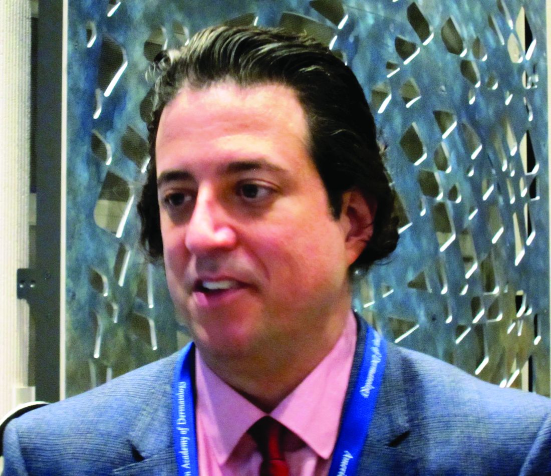
For the past decade, researchers have worked on the assumption that the chronic systemic inflammation of severe psoriasis overlaps with similar inflammatory pathways that drive atherosclerosis, plaques that rupture, and cardiovascular events.
Recently, they have employed fluorodeoxyglucose positron emission tomography (FDG-PET) to confirm some of this. Studies in 2011, 2015, and 2017 confirm that patients with severe plaque psoriasis can develop diffuse vascular inflammation with increased noncalcified coronary artery plaques. These more fragile plaques are the type that are prone to rupture and cause cardiovascular events, Dr. Gelfand said.
“In these studies, the more imaging signal we saw in the aorta, the higher the risk of a future cardiovascular event. In fact, we determined that patients with severe psoriasis can have increased aortic inflammation equivalent to a decade of aging. As your PASI [Psoriasis Area and Severity Index] score goes up, so does the amount of aortic inflammation. The anatomic consequence is that people develop a high risk of atherosclerotic plaques, and a higher rate of noncalcified coronary plaques that are more likely to lead to these events.”
The VIP-U study was conceived to examine whether calming psoriatic inflammation with ustekinumab (Stelara), an anti interleukin-12 and -23 antibody, could also improve vascular inflammation as measured by FDG-PET. The small study comprised 43 patients, half of whom received placebo and half of whom received two injections of ustekinumab: 45 or 90 mg depending on weight, at baseline and at week 4. There was an imaging assessment at week 12, after which the placebo patients crossed over to open-label ustekinumab every 2 weeks. Everyone was followed out to 64 weeks.
Dr. Gelfand reported the 12-week data; the 64-week data will be forthcoming later this year, he said.
The patients were typical for such a study, with a mean age of about 43 and a mean disease duration of 18 years. The mean body surface area was about 25%, and the mean PASI was 20. Most had been on prior therapy, including phototherapy, oral agents, and biologics.
Unsurprisingly, ustekinumab was significantly more effective than placebo in treating the psoriasis. At week 12, 10% of the placebo patients had achieved a PASI 75, compared with 77% of the ustekinumab patients. A Physicians Global Assessment (PGA) score of clear or almost clear occurred in 10% of those taking placebo and 64% of those taking the biologic.
However, the drug was also highly effective in reducing total aortic inflammation, Dr. Gelfand said. “In just 12 weeks, we saw a 6.6% reduction in total aortic inflammation among those taking ustekinumab, but a 12.1% increase in inflammation among those taking placebo. When you compare the delta, you see a highly statistically significant improvement in aortic inflammation in ustekinumab-treated patients, with the effect size similar to statin therapy.”
The benefit may be a class-associated one. Two recent similar studies of adalimumab, a tumor necrosis factor–alpha inhibitor, failed to find any improvement in vascular inflammation, compared with placebo.
“We need more information about the longer-term effects of ustekinumab treatment, as well as cardiometabolic biomarkers, and these are currently underway,” Dr. Gelfand said. “We also need additional trials to determine if this effect is due to the inhibition of IL-12, IL-23, or a combination. Cardiovascular studies will be necessary to fully determine the clinical implications of our findings.”
To refer a patient into the VIP studies, call 215-662-SKIN or email SkinVIP@upenn.edu.
The National Institutes of Health and University of Pennsylvania sponsored the study. Dr. Gelfand reported no financial disclosures.
SOURCE: Gelfand J et al. AAD 2018 Abstract 6645
REPORTING FROM AAD 2018
Key clinical point:
Major finding: Aortic inflammation decreased by 6.6% in those taking ustekinumab, but increased by 12.1% in those taking placebo.
Study details: The randomized trial comprised 43 patients.
Disclosures: The National Institutes of Health and University of Pennsylvania sponsored the study. Dr. Gelfand made no financial disclosures.
Source: Gelfand J et al. AAD 2018 Abstract 6645.
Badly behaved neutrophils are novel target in rheumatic diseases
MAUI, HAWAII – For most of her career as a pediatric rheumatologist and immunologist, Anne M. Stevens, MD, PhD, didn’t find neutrophils terribly interesting.
“I’m more of a T-cell person. T cells are very precise, they’re regulated, they’re very sophisticated cells. Neutrophils are like these bumbling idiots that just come in and hit everything in sight and cause all sorts of damage,” she observed at the 2018 Rheumatology Winter Clinical Symposium.
Recently, however, she’s changed her mind. Research in the past few years demonstrates that there’s a lot more to neutrophils than previously known. Neutrophils are not only the first-responder immune cells to acutely inflamed tissue, as has long been recognized, but they also play a huge role in initiating and perpetuating chronic inflammation. Indeed, neutrophils figure prominently in the pathogenesis of rheumatoid arthritis, systemic lupus erythematosus (SLE), antineutrophil cytoplasmic antibody (ANCA)–associated vasculitis, and perhaps in major nonautoimmune diseases as well, including atherosclerosis, type 1 diabetes, thrombosis, and some forms of kidney disease, explained Dr. Stevens, chief of pediatric rheumatology at the University of Washington, Seattle.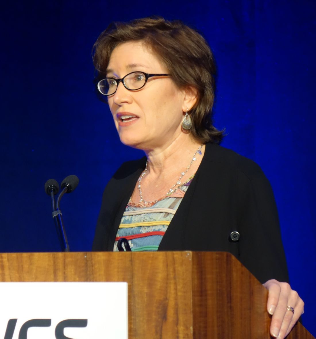
Indeed, it’s now clear that neutrophils can either die quietly or become activated in death by creating neutrophil extracellular traps, or NETs, in a process called NETosis. Even though the activated neutrophil is dead, its exteriorized NETs continue to function, grabbing and killing bacterial pathogens. But the NETs also attract immune cells. These NETs are long strands of sticky DNA containing chromatin, histones, elastase, myeloperoxidase, hypercitrullinated proteins, and other autoantigens. NETosis exposes these autoantigens to the immune system, with resultant generation of autoimmune responses in predisposed individuals.
Much detail has been learned about this process. For example, one type of neutrophil death results from lymphokine-activated killer cells releasing perforin, which pokes holes in the neutrophil cell membrane, allowing an influx of calcium. The inflow of calcium triggers activation of peptidyl arginine deiminases, and these enzymes in turn cause hypercitrullination of autoantigens, leading to formation of anti–citrullinated peptide autoantibodies (ACPAs). These ACPAs cause inflammation by inducing complement activation and binding to Fc gamma receptors on phagocytes.
Investigators at the Systemic Autoimmunity Branch of the National Institute of Arthritis and Musculoskeletal and Skin Diseases and several other research centers have shown that mixing RA synovial fibroblasts with NETs induces production of interleukin-6 and ACPAs. In a mouse model of RA, this leads to cartilage loss in joints, providing a plausible mechanistic explanation for joint damage in RA (Sci Immunol. 2017 Apr;2[10]:eaag3358).
Potential therapeutic strategies targeting NETosis
The various treatment approaches under study aim to either inhibit NET release, curb recruitment of neutrophils for activation, promote migration of neutrophils away from sites of inflammation, or foster efferocytosis.
Among the therapeutic possibilities are calcineurin inhibitors as a means of preventing the influx of calcium into neutrophils, peptidyl arginine deiminase inhibitors such as Cl-amidine to prevent hypercitrullination, complement component 5a-receptor antagonists to decrease NET formation, the myeloperoxidase inhibitor known for now as PF-1355, and N-acetyl cysteine to scavenge proinflammatory reactive oxygen species and thereby reduce NET release.
The key will be to develop highly selective agents that encourage well-behaved neutrophils; across-the-board blockade of neutrophil activity would be terribly immunosuppressive and likely dangerous.
“What if we could block neutrophil recruitment just to the organ we’re worried about? If I’m worried about nephritis, let’s just block neutrophil infiltration into the kidneys. There are drugs being developed that will block the specific types of integrins involved,” Dr. Stevens said.
She reported having no financial conflicts regarding her presentation.
EXPERT ANALYSIS FROM RWCS 2018
High objective response rate, OS seen with ATA129 in PTLD
SALT LAKE CITY, UTAH – An allogeneic off-the-shelf Epstein-Barr virus–targeted cytotoxic T lymphocyte–cell product known as ATA129 (tabelecleucel), is associated with a high response rate and a low rate of serious adverse events in patients with posttransplant lymphoproliferative disorder (PTLD), according to interim findings from an ongoing multicenter study.
The objective response rate at a median of 3.3 months among patients who were treated with ATA129 and who had sufficient follow-up to assess response was 80% in six patients treated following hematopoietic cell transplantation (HCT), and 83% in six who were treated after solid organ transplant (SOT), Susan E. Prockop, MD, reported at the combined annual meetings of the Center for International Blood & Marrow Transplant Research and the American Society for Blood and Marrow Transplantation.
Study participants included those with or without underlying immune deficiency with Epstein-Barr virus (EBV)–positive PTLD, EBV-positive lymphoma, EBV-positive hemophagocytic lymphohistiocytosis, or EBV viremia, and they had to have measurable disease. All had adequate organ function and performance status. The overall median age of the cohort was 41 years, and among the transplant recipients the median age was 24.5 years. They received a median of 5 weeks of therapy (2.1 months among post-HCT patients and 12.9 months among post-SOT patients), she said.
Patients in the trial underwent the adoptive T cell therapy with partially human leukocyte antigen (HLA)–matched ATA129 that shared at least 2 of 10 HLA alleles at high resolution, including at least 1 through which ATA129 exerted cytotoxicity, or “HLA restriction,” Dr. Prockop said, noting that the product was licensed and obtained breakthrough designation in February 2015.
The ATA129 dose was 1.6-2 million T cells/kg infused on days 1, 8, and 15 of every 35-day cycle. Those without toxicity were eligible to receive additional cycles, and patients with progressive disease after one cycle were allowed to switch to an ATA129 product with a different HLA restriction, she noted.
Treatment-emergent adverse events occurred in 21 patients, including 17 who experienced grade 3 or greater adverse events or serious adverse events. Six were treatment related; one of those was grade 3 or greater, and five were considered serious adverse events. One patient had a grade 5 treatment-emergent adverse event (disease progression); two in the post-HCT group experienced graft-versus-host disease (GVHD), including one with grade 3 skin GVHD after sun exposure, which resolved with topical therapy; and one had grade 4 GVHD of the gastrointestinal tract and liver. One patient had a tumor flare that resolved, Dr. Prockop said.
“The most common safety events were GI disorders in seven patients, infections and infestations in five patients, and general disorders and administration site conditions in four,” she said. “No events have been categorized as drug reactions.”
PTLD, an EBV-driven lymphoproliferative disorder, is a life-threatening condition typically involving aggressive, clonal, diffuse large B cell lymphomas. Survival without therapy is a median of 31 days, she explained. Patients at high risk have a mortality rate of 72%, and these included those over age 30 years, those with GVHD at the time of diagnosis, and those with extranodal disease, three or more sites of disease involved, or central nervous system disease.
Although some patients respond to single-agent rituximab (Rituxan) therapy, those with rituximab-refractory disease have a median overall survival of 16-56 days, she said.
SOT recipients who develop indolent PTLD may respond to reduction of immunosuppression. Two-year risk-based survival in these patients is 88% with zero or one risk factors, and 0% with three or more risk factors, which include older age, poor performance status at diagnosis, high lactate dehydrogenase, CNS involvement, and short time from transplant to development of PTLD.
Rituximab monotherapy response rates are 76% in those with early lesions, and 47% in those with high-grade lesions, she said.
“Two-year overall survival in this patient population is 33%, reflecting their eligibility for multiagent chemotherapy, although this approach comes with significant morbidity,” she added, noting that patients failing rituximab experience increased chemotherapy-induced treatment-related mortality, compared with other lymphoma patients.
The benefit-risk profile observed in this multicenter trial is favorable with maximum response rates being reached after two cycles of therapy, and the findings confirm those from prior single-center studies, she said, noting that based on those earlier findings in patients treated with both primary and third-party donor EBV-cytotoxic T lymphocytes, the therapy is now an established National Comprehensive Cancer Network guideline therapeutic alternative for PTLD.
“Further evaluation in rituximab-refractory PTLD is ongoing in phase 3 registration trials,” she said.
Atara Biotherapeutics sponsored the trial. Dr. Prockop reported having no disclosures.
sworcester@frontlinemedcom.com
SOURCE: Prockop S et al. BMT Tandem Meetings Abstract 21.
REPORTING FROM THE 2018 BMT TANDEM MEETINGS
Key clinical point:
Major finding: Overall 1-year survival was 90.3%.
Study details: Interim results in 23 patients from a multicenter study.
Disclosures: Atara Biotherapeutics sponsored the trial. Dr. Prockop reported having no disclosures.
Source: Prockop S et al. BMT Tandem Meetings Abstract 21.
Dual kinase inhibitor performs well in its first safety, efficacy study for atopic dermatitis
SAN DIEGO – A novel molecule that inhibits two major inflammatory pathways acquitted itself well in its first safety and efficacy clinical trial in patients with moderate to severe atopic dermatitis.
After 29 days, 100% of those taking the highest dose of the molecule, ASN002, achieved a 50% improvement in skin involvement.
“This is an interesting molecule,” Robert Bissonnette, MD, said at the annual meeting of the American Academy of Dermatology. “By targeting the entire JAK family, it inhibits cytokine signaling through IL-4 [interleukin-4], IL-13, IL-23, and thymic stromal lymphopoietin,” which plays a role in maturing T cells. The SYK inhibition targets IL-17, increases the terminal differentiation of keratinocytes, and inhibits B-cell signaling.
Dr. Bissonnette, president of Innovaderm Research presented the results in a late-breaking session at the meeting. Innovaderm designs, conducts, and analyzes dermatology clinical trials, and ran the study for Asana BioSciences.
In an Asana press release about the study results, CEO Sandeep Gupta, PhD, said that ASN002 “is the only oral compound in clinical development targeting JAK [including Tyk2] and SYK signaling, two clinically validated mechanisms.” He added that inhibition of JAK and SYK pathways “diminishes cytokine production and signaling including those mediated by Th2 and Th22 cytokines. Dysregulation of Th2 and Th22 cytokine pathways is implicated in the pathogenesis of atopic dermatitis.”
Safety was the primary endpoint in the phase 1b dose-ranging trial, but there were also several efficacy endpoints, Dr. Bissonnette said at the meeting. The study comprised 36 patients separated into three 12-patient groups. In each group, nine received the active drug every day, and three received a placebo. The first group received 20 mg ASN002 or placebo for 28 days, followed by the 14-day safety period. The next group received 40 mg ASN002 or placebo for 28 days, followed by the safety analysis. The third group followed the same treatment pattern, but received 80 mg of the drug.
All patients were aged 29-42 years. The Eczema Area and Severity Index (EASI) was imbalanced among the groups, ranging from 21 in the placebo group and 40-mg group, to 29 in the 20-mg and 80-mg groups. The Body Surface Area (BSA) index also differed between groups: 25% among the placebo patients, 45% in the 20-mg group, 32% in the 40-mg group, and 29% in the 80-mg group. The average Investigator Global Assessment (IGA) score was 3.
A pharmacokinetic analysis showed rapid uptake of the drug with a half-life of 10-14 hours depending on dose.
There were no concerning safety signals, Dr. Bissonnette said and no thromboembolic events, serious infections, or opportunistic infections. The most common adverse event was headache, which occurred at equal rates among the groups. The single serious adverse event was anxiety, which occurred in one patient taking 80 mg ASN002, 4 days after the medication was stopped, and was judged unrelated to ASN002.
There were no significant changes in any lab parameters, no changes in lipid profiles, and no hematologic abnormalities. One patient experienced an increase in creatinine phosphokinase, something that has been seen in other studies of JAK inhibitors, Dr. Bissonnette said.
The molecule performed well on secondary efficacy endpoints. By day 15, 63% of the 40-mg group and 75% of the 80-mg group had achieved EASI-50, and by day 29, these rates were 88% and 100%, respectively. These doses also performed well on the EASI-75 endpoint. By day 29, 63% of the 40-mg group and 50% of the 80-mg group had achieved a 75% improvement in EASI score.
The 40- and 80-mg groups also did well with regards to BSA improvement. By day 29, the 80-mg group had achieved a mean BSA reduction of 58%, and the 40-mg group a mean reduction of 64%. IGA tracked that improvement, with 25% of the 80-mg group and 38% of the 40-mg group achieving an IGA score of 1 or 0.
Itching responded well to ASN002, but here, the 20-mg dose threw investigators a bit of a curve ball. Pruritus decreased most in the 80-mg group (about 70% by the end of the study). But at 3 weeks, patients taking 20 mg and 40 mg were experiencing the same 30% decrease in itch. By 4 weeks, however, the scores had separated, with the 40-mg group landing at about a 40% reduction, and the 20-mg group rebounding to about a 20% reduction.
“I would say that this first trial of ASN002 showed clear efficacy in patients with moderate to severe atopic dermatitis, with rapid improvement itch and a large proportion of patients reaching EASI-50 as early as 2 weeks,” Dr. Bissonnette said.
Asana will soon initiate a phase 2b study of ASN002 in moderate to severe atopic dermatitis patients. Clinical studies in other dermatologic and autoimmune indications are under consideration, according to the company website.
Dr. Bissonnette is the president of Innovaderm Research, which was paid to run the ASN002 study.
SOURCE: Bissonnette R et al. AAD 2018, Abstract 6777
REPORTING FROM AAD 2018
Key clinical point: ASN002 calmed itch, improved skin involvement in moderate to severe atopic dermatitis.
Major finding: After 29 days, 100% of those taking 80-mg doses of ASN002 daily achieved a 50% improvement in EASI.
Study details: The randomized dose-escalation phase 1b study comprised 36 patients with moderate to severe atopic dermatitis.
Disclosures: Asana BioSciences sponsored the study. Dr. Bissonnette is CEO of Innovaderm Research, Montreal, which conducted the trial.
Source: Bissonnette R et al. AAD 2018, Abstract 6777.
Perfusion-only scan rules out PE in pregnancy
For pregnant women with suspected pulmonary embolism (PE), (CTPA), according to authors of a recent retrospective study.
Pulmonary embolism causes 9% of maternal deaths in the United States, according to the authors of the study, which was published online in the journal CHEST®. While it’s clear that perfusion scans yield lower radiation exposure than CTPA, to date, there has only been limited study of its diagnostic performance in women with suspected PE.
The low-dose perfusion scan offered comparable diagnostic efficacy while potentially limiting radiation exposure, according to the single-center cohort study.
The retrospective study included pregnant women (mean age, 27.3 years) who underwent imaging for pulmonary embolism at Montefiore Medical Center, New York, between 2008 and 2013. A total of 225 women underwent perfusion-only scans, while 97 underwent CTPA.
Chest pain and dyspnea were the most common symptoms for patients in both groups: 136 of the patients (60.4%) in the low-dose perfusion group reported chest pain versus 40 patients (41.2%) in the CTPA group. About half of the patients in both groups had dyspnea.
Tachycardia was found in 43 of patients (44.3%) who underwent CTPA, compared with 77 of patients (34.2% ) who underwent the diagnostic test involving less radiation exposure.
Imaging was negative for PE in 198 of the patients (88.0%) who were scanned with low-dose perfusion, while 84 of patients (86.6%) who had CTPAs were negative for PE. For both groups of patients, the percentage who had indeterminate imaging was 9.3%. Only one study participant had a deep vein thrombosis at the time she presented with PE symptoms.
The primary end point of the study, negative predictive value, was 100% for the perfusion-only group and 97.5% for CTPA, according to the report. It was determined by a diagnosis of venous thromboembolism within 90 days of evaluation.
Those “indistinguishable” negative predictive values suggest that low-dose perfusion scintigraphy performs comparably to CTPA, making it an appropriate first diagnostic modality for pregnant women who are suspected of having pulmonary embolism, Dr. Sheen and her colleagues wrote.
The negative predictive value was a particularly important endpoint to evaluate because pulmonary embolism is rare among pregnant women and most perfusion-only imaging is negative, the authors stated.
Of the women in the study, 252 (89%) of those who tested negative for PE – either by a low-dose perfusion scan or a CTPA – returned to the medical center for follow-up 90 days later. Thromboembolic events occurred in two of the women who previously had a negative CTPA, but none occurred in patients who had been tested for PE with low-dose perfusion scan. The two thromboembolic events were detected in women who were no longer pregnant.
Ten patients in the study (3.1%) were treated for pulmonary embolism, the authors reported. The PE diagnoses were based on four positive low-dose perfusion scans and six positive CTPAs “in conjunction with clinical suspicion.” These patients’ most common symptoms were chest pain and dyspnea, and one of these patients had recently been diagnosed with a deep vein thrombosis.
When perfusion defects are found, they should be interpreted cautiously, particularly in asthmatic patients, according to authors: “Segmental perfusion defects secondary to abnormal ventilation cannot be distinguished from PE without a ventilation scan,”they noted.
Three of the patients diagnosed with a PE had asthma. In a subanalysis of the 77 patients with asthma who participated in this study, the negative predictive values were 100% for both those who received a low-dose perfusion scan and those who received a CTPA. For patients in this subgroup, the negative rates of PE from low-dose perfusion scan and CTPA were 74.1% and 87.1%, respectively.
“Maternal-fetal radiation exposure should be of utmost importance when considering the choice of diagnostic test,” the authors wrote. “When available, [a low-dose perfusion scan] is a reasonable first choice modality for suspected pulmonary embolism in pregnant women with a negative chest radiograph.”
One study coauthor is on an advisory panel for Jubilant DraxImage, and another has a spouse who is a board member of Kyron Pharma Consulting. The remaining authors, including Dr. Sheen,reported no conflicts of interest.
SOURCE: Sheen JJ et al. Chest. 2018 Feb. doi: 10.1016/j.chest.2017.08.005.
Nirmal Sharma, MD, comments: During pregnancy, all radiation is bad radiation, but when it was really needed, we did use this low-radiation perfusion scan quite a bit at my past institution. This article definitely shines light on the utility/validity of this technique because most centers still use a computed tomographic pulmonary angiography study in pregnant females (with shielding methods) if suspicion of pulmonary embolism is high. The downside to low-dose perfusion scintigraphy is that it cannot be used in patients with grossly abnormal chest x-rays.

Nirmal Sharma, MD, comments: During pregnancy, all radiation is bad radiation, but when it was really needed, we did use this low-radiation perfusion scan quite a bit at my past institution. This article definitely shines light on the utility/validity of this technique because most centers still use a computed tomographic pulmonary angiography study in pregnant females (with shielding methods) if suspicion of pulmonary embolism is high. The downside to low-dose perfusion scintigraphy is that it cannot be used in patients with grossly abnormal chest x-rays.
Nirmal Sharma, MD, comments: During pregnancy, all radiation is bad radiation, but when it was really needed, we did use this low-radiation perfusion scan quite a bit at my past institution. This article definitely shines light on the utility/validity of this technique because most centers still use a computed tomographic pulmonary angiography study in pregnant females (with shielding methods) if suspicion of pulmonary embolism is high. The downside to low-dose perfusion scintigraphy is that it cannot be used in patients with grossly abnormal chest x-rays.
For pregnant women with suspected pulmonary embolism (PE), (CTPA), according to authors of a recent retrospective study.
Pulmonary embolism causes 9% of maternal deaths in the United States, according to the authors of the study, which was published online in the journal CHEST®. While it’s clear that perfusion scans yield lower radiation exposure than CTPA, to date, there has only been limited study of its diagnostic performance in women with suspected PE.
The low-dose perfusion scan offered comparable diagnostic efficacy while potentially limiting radiation exposure, according to the single-center cohort study.
The retrospective study included pregnant women (mean age, 27.3 years) who underwent imaging for pulmonary embolism at Montefiore Medical Center, New York, between 2008 and 2013. A total of 225 women underwent perfusion-only scans, while 97 underwent CTPA.
Chest pain and dyspnea were the most common symptoms for patients in both groups: 136 of the patients (60.4%) in the low-dose perfusion group reported chest pain versus 40 patients (41.2%) in the CTPA group. About half of the patients in both groups had dyspnea.
Tachycardia was found in 43 of patients (44.3%) who underwent CTPA, compared with 77 of patients (34.2% ) who underwent the diagnostic test involving less radiation exposure.
Imaging was negative for PE in 198 of the patients (88.0%) who were scanned with low-dose perfusion, while 84 of patients (86.6%) who had CTPAs were negative for PE. For both groups of patients, the percentage who had indeterminate imaging was 9.3%. Only one study participant had a deep vein thrombosis at the time she presented with PE symptoms.
The primary end point of the study, negative predictive value, was 100% for the perfusion-only group and 97.5% for CTPA, according to the report. It was determined by a diagnosis of venous thromboembolism within 90 days of evaluation.
Those “indistinguishable” negative predictive values suggest that low-dose perfusion scintigraphy performs comparably to CTPA, making it an appropriate first diagnostic modality for pregnant women who are suspected of having pulmonary embolism, Dr. Sheen and her colleagues wrote.
The negative predictive value was a particularly important endpoint to evaluate because pulmonary embolism is rare among pregnant women and most perfusion-only imaging is negative, the authors stated.
Of the women in the study, 252 (89%) of those who tested negative for PE – either by a low-dose perfusion scan or a CTPA – returned to the medical center for follow-up 90 days later. Thromboembolic events occurred in two of the women who previously had a negative CTPA, but none occurred in patients who had been tested for PE with low-dose perfusion scan. The two thromboembolic events were detected in women who were no longer pregnant.
Ten patients in the study (3.1%) were treated for pulmonary embolism, the authors reported. The PE diagnoses were based on four positive low-dose perfusion scans and six positive CTPAs “in conjunction with clinical suspicion.” These patients’ most common symptoms were chest pain and dyspnea, and one of these patients had recently been diagnosed with a deep vein thrombosis.
When perfusion defects are found, they should be interpreted cautiously, particularly in asthmatic patients, according to authors: “Segmental perfusion defects secondary to abnormal ventilation cannot be distinguished from PE without a ventilation scan,”they noted.
Three of the patients diagnosed with a PE had asthma. In a subanalysis of the 77 patients with asthma who participated in this study, the negative predictive values were 100% for both those who received a low-dose perfusion scan and those who received a CTPA. For patients in this subgroup, the negative rates of PE from low-dose perfusion scan and CTPA were 74.1% and 87.1%, respectively.
“Maternal-fetal radiation exposure should be of utmost importance when considering the choice of diagnostic test,” the authors wrote. “When available, [a low-dose perfusion scan] is a reasonable first choice modality for suspected pulmonary embolism in pregnant women with a negative chest radiograph.”
One study coauthor is on an advisory panel for Jubilant DraxImage, and another has a spouse who is a board member of Kyron Pharma Consulting. The remaining authors, including Dr. Sheen,reported no conflicts of interest.
SOURCE: Sheen JJ et al. Chest. 2018 Feb. doi: 10.1016/j.chest.2017.08.005.
FROM CHEST®
Key clinical point: In the evaluation of pregnant women with suspected pulmonary embolism, low-dose perfusion scintigraphy may offer diagnostic performance that’s comparable to CTPA.
Major finding: The negative predictive value of a pulmonary embolism was 100% for the low dose perfusion scan, compared with 97.5% for CTPA.
Study details: A retrospective, single-center cohort study including 322 pregnant women who underwent imaging studies for suspected pulmonary embolism.
Disclosures: One study coauthor is on an advisory panel for Jubilant DraxImage, and another has a spouse who is a board member of Kyron Pharma Consulting. The remaining authors, including Dr. Sheen,reported no conflicts of interest.
Source: Sheen JJ et al. Chest. 2018 Feb. doi: 10.1016/j.chest.2017.08.005.
Tryptophan depletion may explain high rate of eating disorders in women
TAMPA, FLA. – The far higher rate of eating disorders in women than men appears to be explained at least in part by a greater acute depletion of tryptophan, which is essential for the formation of serotonin, a key mediator of risk, according to a research review presented at the annual meeting of the American College of Psychiatrists.
“The specific vulnerability of women to eating disorders relates to the fact that women’s brains are much more sensitive to dietary intake of tryptophan than are men’s brains,” explained Allan S. Kaplan, MD, senior scientist at the Center for Addiction and Mental Health at the University of Toronto.
“Women are more likely than men to be dieting,” said Dr. Kaplan, walking through the evidence. “Low-calorie diets tend to be high in protein and low in cholesterol and fat. Such diets lead to tryptophan depletion and decreased serotonin synthesis in the brain. Because of lower levels of central serotonin, women are more vulnerable to mood and eating disorders than men.”
Not all women who diet may be vulnerable to this sequence of events. Genetics are likely to be a factor, according to Dr. Kaplan, who said, “Genes load the gun; the environment pulls the trigger.”
However, women do appear to be more susceptible for a number of reasons. For one, the mean rate of serotonin synthesis is 52% higher in normal males than normal females, giving them a greater buffer when dietary intake of tryptophan is low. For another, there is evidence that intake of nutrients most rich in tryptophan, particularly proteins, is typically lower in women than men.
The ratio of females to males for both anorexia nervosa and bulimia nervosa is about 10:1. Although the female-to-male ratio of binge eating is lower at 2:1, women dominate these psychiatric diagnoses. Several environmental factors associated with eating disorders are more closely associated with women than men, including a history of sexual or physical abuse and female preoccupation with body image, but acute tryptophan depletion may be an important factor participating in the translation of risk to an active disease, according to Dr. Kaplan.
Acute tryptophan deficiency may also explain why treatment of eating disorders with SSRIs has been disappointing. With low levels of tryptophan leading to serotonin depletion, “there is no substrate” for drugs administered to increase serotonin-mediated signaling, Dr. Kaplan explained.
Ensuring adequate dietary intake of tryptophan, which is “found mainly in high-protein animal foods,” may be important, even though Dr. Kaplan warned that achieving optimal levels of serotonin “can be challenging from food alone.” Nevertheless, behavioral therapies are commonly effective for eating disorders, presumably at least partially as a result of their ability to normalize diet.
Overall, the tryptophan hypothesis has provided a major shift in the understanding of eating disorders, according to Dr. Kaplan. Further studies are needed, but he said that the key message is that, “For women’s brains, you are what you eat.”
Dr. Kaplan reported no conflicts of interest relevant to this topic.
This story was updated on 2/25/2018.
EXPERT ANALYSIS FROM THE COLLEGE 2018