User login
A view from the bridge to transplant for PTCL
LA JOLLA, CALIF. – For patients with relapsed peripheral T-cell lymphoma, allogeneic stem cell transplants offer the best chance for achieving remission or even a cure, making the choice of therapies as bridges to transplant essential for getting there.
“The goal is to get to transplant with a curative intent. In our hands, that’s mostly allo[geneic] and mostly in the relapsed setting,” Steven M. Horwitz, MD, from the lymphoma service at Memorial Sloan Kettering Cancer Center, New York, said at the annual T-cell Lymphoma Forum. “The best bridge to transplant is the one that gets you across safely.”
“They’d finish ICE, get a 3- or 4-week break, get a transplant, leave the hospital 3 or 4 weeks later, and then usually by their first repeat scan, at least on average, those patients had already progressed, so we sort of cooled to the idea of auto transplant and started preferentially looking at allo if we were going to treat with curative intent in the relapsed setting,” he said.
In contrast to autologous SCT, the Memorial Sloan Kettering experience with allogeneic SCT for 65 patients with relapsed PTCL showed a 2-year overall survival rate of 59%, 2-year PFS rate of 48%, and a median PFS of 20.3 months. However, the rate of 1-year transplant-related mortality was still relatively high, at 17%, Dr. Horwitz acknowledged (ASH 2015. Abstract 4392).
An updated retrospective analysis of the center’s experience treating mature T-cell lymphoma patients with allogeneic SCT, also presented at the 2018 T-cell Lymphoma Forum, showed that disease status at transplant was one of the most important predictors of outcome. Median posttransplant PFS for patients in complete remission (CR) at the time of transplant was 61.3 months, compared with 11.4 months for patients in partial remission, 14 months for patients with stable disease, and 6.4 months for patients with disease progression (TCLF 2018. Abstract TM18_9).
“I think we can probably infer from [this] that CR not only gives you a better outcome with allo, but probably increases your chance that you get to an allo,” he said.
In the randomized phase 3 Lumiere study comparing the Aurora A kinase inhibitor alisertib with investigators’ choice of therapy in relapsed/refractory PTCL, alisertib was associated with a CR rate of 19%, whereas pralatrexate, gemcitabine, and romidepsin were associated with CR rates of 29%, 23%, and 33%, respectively, putting them on par with combination chemotherapy.
“I think many of us prefer some of the newer single agents because we’re really going for a durable maintenance of disease control rather than short-term bridge to transplant, but these drugs can provide adequate responses to transition over,” he said.
Better approaches by subtype?
The subtype of PTCL also appears to matter. Three approved agents for relapsed/refractory PTCL – belinostat (Beleodaq), romidepsin (Istodax), and pralatrexate (Folotyn) – are associated with CR rates of 11%, 15%, and 11%, respectively. But one PTCL subtype, anaplastic large cell lymphoma, appears particularly sensitive to treatment with brentuximab vedotin (Adcetris), with CR rates of 59%, Dr. Horwitz noted.
In a 2014 study, investigators reported that of the nine patients with anaplastic large cell lymphoma positive for anaplastic lymphoma kinase and treated with the anaplastic lymphoma kinase inhibitor crizotinib (Xalkori), all had a CR, with response durations stretching pasting 40 months in one patient, and past 30 months in two others (J Natl Cancer Inst. 2014 Feb;106[2]:djt378).
A different subtype, natural killer/T-cell lymphoma, was shown to be responsive to immunotherapy with pembrolizumab (Keytruda) in seven patients, with CRs in five and partial remissions in two. Responses to pembrolizumab in this PTCL subtype may be adequately long for getting patients to transplant, Dr. Horwitz said.
For some patients with angioimmunoblastic T-cell lymphoma, therapy with epigenetic modifying agents, such as decitabine or a combination of romidepsin and lenalidomide (Revlimid), with or without carfilzomib (Kyprolis), may also be effective bridges to transplant, based on the best available evidence.
Timing may also matter
Dr. Horwitz cautioned that for patients with cutaneous T-cell lymphoma, the investigational agent mogamulizumab, which was shown in the MAVORIC (Study of KW-0761 versus Vorinostat in Relapsed/Refractory CTCL) trial to offer significantly better PFS compared with vorinostat (Zolinza), also appears to increase the chance that patients will develop high-risk, potentially fatal graft vs. host disease posttransplant.
The risk appears to be slightly lower among patients who received the last dose of mogamulizumab more than 50 days before undergoing SCT, he noted.
Although there is no strong evidence to support it, Dr. Horwitz noted that the timing of most other therapies may also be important to the success of SCT. “I think we have seen that when patients have a big [long] break before transplant, the relapse rate is high, and I have a personal preference for using regimens that you can continue up close to transplant, because I think we lose [fewer] patients getting ready for that,” he said.
Dr. Horwitz had previously disclosed financial relationships with Celgene, Forty Seven, Huya Bioscience International, Infinity, Kyowa Hakko Kirin, Millennium, Seattle Genetics, and Takeda. The T-Cell Lymphoma Forum is held by Jonathan Wood & Associates, which is owned by the same company as this news organization.
EXPERT ANALYSIS FROM TCLF 2018
The T-cell repertoire in NSCLC: Therapeutic implications
SAN FRANCISCO – An analysis of the T-cell repertoire in nearly 400 patients with stage I-III non–small cell lung cancer (NSCLC) suggests that patients with a more tumor-focused repertoire have better outcomes.
The findings, which suggest that patients with fewer T cells and with lower clonality in tumor-adjacent normal lung tissue fare better, could have implications for the use of tumor-infiltrating lymphocyte (TIL) therapy and checkpoint blockade – and possibly other therapies – in these patients, according to Alexandre Reuben, PhD, of the University of Texas MD Anderson Cancer Center, Houston.
Studying the T-cell repertoire in the lung can be rather “messy,” because many T cells may be responding to the outside environment, but comparing findings in the normal lung with those in tumor tissue helps to clarify things, Dr. Reuben said at the ASCO-SITC Clinical Immuno-Oncology Symposium.
Why study the T-cell repertoire?
The successes seen with immune checkpoint blockade in recent years are largely a result of the ability of these therapies to enhance the antitumor T-cell response. Interestingly, checkpoint blockade works better in tumor types like lung cancer that have a high mutational load, Dr. Reuben said, explaining that this is largely attributable to the ability of the mutations to increase tumor immunogenicity through generation of tumor-specific antigens, which can then be targeted by the T-cell response.
This relationship between the mutational and neoantigen burden and patient outcomes has been described, but the role of the T-cell repertoire and how it relates to patient outcomes is less clear.
Hypothesizing that T-cell repertoire would be associated with survival in patients with NSCLC, he and his colleagues collected peripheral blood, normal lung, and tumor tissue, and performed T cell–receptor sequencing, among other analyses, in 398 patients.
“T cells recognize antigens through their T-cell receptor, as a result of which they undergo clonal expansion. Therefore, by sequencing the variable region of the T-cell receptor, one can gain insight into the T cells that are responding within the sample, as well as the overall T-cell repertoire,” he explained.
This provides information on T-cell density and richness, and thus on the diversity of the T-cell repertoire, he said, adding that it is possible to go beyond that and plot T cells based on their frequency in a sample to study clonality.
Since these T cells tend to expand clonally as a result of activation, uneven distribution would be associated with a reactive T-cell repertoire, as described by a high T-cell clonality.
An assessment to determine how the T-cell repertoire relates to clinical characteristics and response revealed a few interesting correlations. For example, adenocarcinomas tended to be more densely infiltrated than squamous cell carcinomas, smaller tumors were also more densely infiltrated by T cells than were their larger counterparts, and in smokers the T-cell repertoire appeared much more reactive than in nonsmokers.
“But ultimately, we didn’t see the [direct correlations between the T-cell repertoire and outcomes] we were hoping to see,” Dr. Reuben said, noting that this could be because of environmental influences.
“Obviously the lung is exposed to the outside environment, which could be masking some of the antitumor T-cell responses we were hoping to study,” he explained. “So we used a more holistic approach, integrating the peripheral blood and normal lung with tumor repertoire going forward.”
T cells in normal lung vs. tumor
Measuring the proportion of the T-cell repertoire that is shared in peripheral blood vs. normal lung and vs. tumor tissue showed that there is very little in common between them.
“However, when you compare the normal lung to the tumor, there’s much more homology in the T-cell repertoire,” he said, noting that, given the T-cell expansion resulting from antigenic stimulation, focusing on the most dominant cells in a sample highlights those most likely to be responding to antigens. “When we did that ... we saw even more of an enrichment in the homology between the normal lung and tumor T-cell repertoire, suggesting certain parallels in the ongoing immune responses across both these compartments.”
Further, T-cell density and diversity were actually higher in the tumor than in the normal lung in about two-thirds of patients, he said.
“However, surprisingly ... clonality appears to be much higher in the normal lung than in the tumor,” he added, noting that this was the case in about 75% of patients.
These findings raise three key questions:
Why is clonality higher in the normal lung?
T cells are not confined to a specific part of the host and are free to circulate, Dr. Reuben said.
“However, the closer you get to a site of inflammation, the higher the enrichment for T cells that are relevant to that specific site of inflammation, so you can use these statistical methods to enrich for T cells that are more relevant and try to subtract out T cells that are simply circulating through the organ,” he noted.
He and his colleagues used these methods and compared both normal lung and tumor to the peripheral blood, focusing only on clones that were statistically enriched in these two compartments “to really eliminate a lot of the background that may have been caused by the low-frequency T cells in these samples.”
When you look at the lung enriched T-cell repertoire between normal lung vs. tumor, the homology increases quite significantly, suggesting that by subtracting these T cells that are circulating through the host and not likely relevant to the antigenic response, you’re increasing the homology and further highlighting some of the aforementioned parallels in the ongoing immune responses between both sites, he said.
“Now if you look at clonality, there’s really no clear trend ... in the total T-cell repertoire or the enriched repertoire focusing on the normal lung, but if you look at the tumor, there’s a trend toward increased clonality in all patients – to the extent where you no longer see a difference in clonality between the normal lung and tumor, suggesting that this enrichment is allowing us to focus increasingly on T cells relevant to the antitumor response,” he added.
T-cell clonality is highly reliant on the ability of T cells to expand as a result of antigenic stimulation, and immune profiling showed that programmed cell death–1 (PD-1) and programmed death–ligand 1 (PD-L1) were higher within the tumor, suggesting that there is some dysfunction on both sides of this interaction, which could also explain the lower clonality originally seen within the tumor, he said.
Why is the T-cell repertoire so similar across normal lung and tumor (and what are these T cells really recognizing)?
“Well, we performed whole-exome sequencing and it’s really no surprise that mutational load is substantial in the tumor, but what was surprising was the amount of mutations we detected in the normal lung,” Dr. Reuben said.
The number was lower than in the tumor, though still considerable, and included a large proportion that were shared mutations between the normal lung and the tumor, he noted.
A closer look at the shared mutations showed that they correlated positively with the proportion of shared dominant T cells between the normal lung and the tumor, suggesting that some of the shared T cells may be targeting shared mutations between the normal lung and the tumor. The correlation was weak, but statistically significant, so while it doesn’t account for all of the overlap, it likely accounts for some of the homology, he said.
In a paper published last year, Mark M Davis, PhD, of Stanford (Calif.) University and his colleagues went beyond standard analysis of the T-cell repertoire and identified residues specific to certain antigens in order to classify T cells based on their likely reactivity. Dr. Reuben and his colleagues collaborated with that group to determine whether T cells were predominantly viral or nonviral.
“If you focus on the normal lung and tumor, you don’t see much of a trend. In some patients there are more viral motifs, and in others are more nonviral motifs, but what was striking was the enrichment for viral motifs that we saw when we focused on the T cells that were shared between the normal lung and tumor,” Dr Reuben said.
In fact, 88% of patients had more viral motifs within their shared T cells vs. only 33% in the normal lung and 30% in tumor.
“So T cells that are shared may be recognizing a combination of shared mutations and/or viruses,” he explained.
How does the T-cell repertoire relate to outcomes?
A focus on the normal lung showed that patients with fewer T cells and lower clonality had better outcomes.
“What does this mean? It suggests that potentially, in these patients, the immune response in the lung is less distracted by outside pathogens and agents unrelated to the tumor, potentially providing the opportunity for a more focused antitumor T-cell response,” Dr. Reuben said, concluding that “T-cell density is higher, but clonality is lower in tumor vs. normal lung, there’s a substantial overlap in the T-cell repertoire between the normal lung and the tumor (including many T cells which may be reactive to shared mutations and/or viruses), and it seems like a more tumor-focused T-cell repertoire in the lung may be associated with improved outcomes.”
In an interview, Dr. Reuben said the findings have certain therapeutic implications, because most current therapies target the T-cell response whether by design or consequence.
“Considering the large proportion of T cells found in lung tumors which are unrelated to tumor responses, expansion of the wrong T cells – whether these target viruses or shared mutations between the normal lung and tumor – could potentially offer no benefit to the patient, because it would likely not contribute to eradicating their tumor,” he explained. “Furthermore, targeting T cells (through checkpoint blockade or TIL therapy) that are reactive to shared mutations could increase the potential for toxicity within these patients. Therefore, a better understanding of the T-cell repertoire in the lung is necessary to increase the specificity and success rates of current immunotherapies.”
Invited discussant, Antoni Ribas, MD, of the University of California, Los Angeles, suggested that the finding of a substantial number of shared T-cells is likely a baseline phenomenon, and that on-therapy biopsies in patients who respond to treatment would better separate and expand the T cells that responded from those that did not.
In fact, Dr. Reuben and his colleagues have expanded their research in this manner.
“We are now studying this phenomenon longitudinally in patients receiving checkpoint blockade to see how these factors evolve over the course of therapy,” he said.
Dr. Reuben reported having no disclosures. Dr. Ribas owns stock in Advaxis, Arcus Ventures, Compugen, CytomX Therapeutics, Five Prime Therapeutics, FLX Bio, and Kite Pharma, and has served as a consultant or advisor for Amgen, Genentech/Roche, Merck, Novartis, and Pierre Fabre.
sworcester@frontlinemedcom.com
SOURCE: Reuben A et al. Clinical Immuno-Oncology Symposium Abstract 140.
REPORTING FROM THE CLINICAL IMMUNO-ONCOLOGY SYMPOSIUM
Key clinical point:
Major finding: Patients with fewer T cells and lower clonality in the normal lung had better outcomes.
Study details: An analysis of the T-cell repertoire in 398 patients with stage I-III NSCLC.
Disclosures: Dr. Reuben reported having no disclosures. Dr. Ribas owns stock in Advaxis, Arcus Ventures, Compugen, CytomX Therapeutics, Five Prime Therapeutics, FLX Bio, and Kite Pharma, and has served as a consultant or advisor for Amgen, Genentech/Roche, Merck, Novartis, and Pierre Fabre.
Source: Reuben A et al. Clinical Immuno-Oncology Symposium Abstract 140.
Consent and DNR orders
Question: Paramedics brought an unconscious 70-year-old man to a Florida hospital emergency department. The patient had the words “Do Not Resuscitate” tattooed onto his chest. No one accompanied him, and he had no identifications on his person. His blood alcohol level was elevated, and a few hours after his arrival, he lapsed into severe metabolic acidosis and hypotensive shock. The treating team decided to enter a DNR order, and the patient died shortly thereafter without benefit of cardiopulmonary resuscitation.
Which of the following is best?
A. An ethics consult may suggest honoring the patient’s DNR wishes, as it is reasonable to infer that the tattoo expressed an authentic preference.
B. It has been said, but remains debatable, that tattoos might represent “permanent reminders of regretted decisions made while the person was intoxicated.”
C. An earlier case report in the literature cautioned that the tattooed expression of a DNR request did not reflect that particular patient’s current wishes.
D. If this patient’s Florida Department of Health out-of-hospital DNR order confirms his DNR preference, then it is appropriate to withhold resuscitation.
E. All are correct.
ANSWER: E. The above hypothetical situation is modified from a recent case report in the correspondence section of the New England Journal of Medicine.1 It can be read as offering a sharp and dramatic focus on the issue of consent surrounding decisions to withhold CPR.
In 1983, the President’s Commission for the Study of Ethical Problems in Medicine supported DNR protocols (“no code”) based on three value considerations: self-determination, well-being, and equity.2
The physician is obligated to discuss with the patient or surrogate the procedure, risks, and benefits of CPR so that an informed choice can be made. DNR means that, in the event of a cardiac or respiratory arrest, no CPR efforts would be undertaken. DNR orders are not exclusive to the in-hospital setting, as some states, for example, Florida and Texas, have also enacted statutes that allow such orders to be valid outside the hospital.
Critics lament that problems – many surrounding the consent issue – continue to plague DNR orders.3 Discussions are often vague, and they may not meet the threshold of informed consent requirements, because they frequently omit risks and complications. A resident, rather than the attending physician, typically performs this important task. This is compounded by ill-timed discussions and wrong assumptions about patients’ preferences, which may in fact be ignored.4
Physicians sometimes extrapolate DNR orders to limit other treatments. Or, they perform CPR in contraindicated situations such as terminal illnesses, where death is expected, which amounts to “a positive violation of an individual’s right to die with dignity.” In some situations, physicians are known to override a patient’s DNR request.
Take the operating-room conundrum. There, the immediate availability of drugs, heightened skills, and in-place procedures significantly improve survival following a cardiopulmonary arrest. Studies show a 50% survival rate, versus 8%-14% elsewhere in the hospital. A Swedish study showed that 65% of the patients who had a cardiac arrest perioperatively were successfully resuscitated. When anesthesia caused the arrest, for example, esophageal intubation, disconnection from mechanical ventilation, or prolonged exposure to high concentrations of anesthetics, the recovery rate jumped to 92%.
Terminally ill patients typically disavow CPR when choosing a palliative course of action. However, surgery can be a part of palliation. In 1991, approximately 15% of patients with DNR orders had a surgical procedure, with most interventions targeting comfort and/or nursing care. When a terminally ill patient with a DNR order undergoes surgery, how should physicians deal with the patient’s no-code status, especially if an iatrogenic cardiac arrest should occur?
Because overriding a patient’s DNR wish violates the right of self-determination, a reasonable rule is to require the surgeon and/or anesthesiologist to discuss preoperatively the increased risk of a cardiac arrest during surgery, as well as the markedly improved chance of a successful resuscitation. The patient will then decide whether to retain his/her original DNR intent, or to suspend its execution in the perioperative period.5
What about iatrogenesis?
In 1999, David Casarett, MD, and Lainie F. Ross, MD, PhD, assessed whether physicians were more likely to override a DNR order if a hypothetical cardiac arrest was caused iatrogenically.6 Their survey revealed that 69% of physicians were very likely to do so. The authors suggested three explanations: 1) concern for malpractice litigation, 2) feelings of guilt or responsibility, and 3) the belief that patients do not consider the possibility of an iatrogenic cardiac arrest when they consent to a DNR order. Physicians may also believe a “properly negotiated DNR order does not apply to all foreseeable circumstances.”
However, some ethicists believe that an iatrogenic mishap does not make it permissible to override a patient’s prior refusal of treatment, because errors should not alter ethical obligations to respect a patient’s wishes to forgo treatment, including CPR.
Can a DNR order exist if it is against a patient’s wishes?7 In Gilgunn v. Massachusetts General Hospital, a 71-year-old diabetic woman with heart disease, breast cancer, and a hip fracture suffered two grand mal seizures and lapsed into a coma.8 Her daughter was the surrogate decision maker, and she made it clear that her mother always said she wanted everything done. After several weeks, the physicians decided that further treatment would be futile.
The chair of the ethics committee felt that the daughter’s opinion was not relevant because CPR was not a genuine therapeutic option and would be “medically contraindicated, inhumane, and unethical.” Accordingly, the attending physician entered a DNR order despite strong protest from the daughter. The patient died shortly thereafter without receiving CPR, and the daughter filed a negligence lawsuit against the hospital.
Still, there are state and federal statutes touching on DNR orders that warrant careful attention. For example, New York Public Health Law Section 2962, paragraph 1, states: “Every person admitted to a hospital shall be presumed to consent to the administration of cardiopulmonary resuscitation in the event of cardiac or respiratory arrest, unless there is consent to the issuance of an order not to resuscitate ...” This raises the question as to whether it is ever legally permissible in New York to enter a unilateral DNR order against the wishes of the patient.
And the federal “anti-dumping” law governing emergency treatment, widely known as EMTALA (Emergency Medical Treatment and Labor Act), requires all emergency departments to provide treatment necessary to prevent the material deterioration of the individual’s condition. This would always include the use of CPR unless specifically rejected by the patient or surrogate, as the law does not contain a “standard of care” or “futility” exception.9
Dr. Tan is emeritus professor of medicine and a former adjunct professor of law at the University of Hawaii. This article is meant to be educational and does not constitute medical, ethical, or legal advice. For additional information, readers may contact the author at siang@hawaii.edu.
References
1. N Engl J Med. 2017 Nov 30;377(22):2192-3.
2. President’s Commission for the Study of Ethical Problems in Medicine and Biomedical and Behavioral Research. Deciding to Forego Life-Sustaining Treatment. Washington, DC: Government Printing Office, 1983.
3. J Gen Intern Med. 2011 Jul;26(7):791-7.
4. JAMA. 1995 Nov 22-29;274(20):1591-8.
5. Hawaii Med J. 2001 Mar;60(3):64-7.
6. N Engl J Med. 1997 Jun 26;336(26):1908-10.
7. Tan SY. Futility and DNR Orders. Internal Medicine News, March 21, 2014.
8. Gilgunn v. Mass. General Hosp. No. 92-4820 (Mass. Super Ct. Apr. 21, 1995).
9. In re Baby K, 16 F.3d 590 (4th Cir. 1994).
Can case management cut hypertension’s consequences?
MONTREAL – who received phone calls, care coordination, and coaching from a nurse case manager, according to a retrospective population-based cohort study of almost 85,000 patients with hypertension.
The reduction yielded a hazard ratio for all-cause mortality of 0.504, with a number needed to treat (NNT) of 25 (P less than .05).
Service utilization also decreased for those participating in the intervention, compared with those receiving usual care: Hospitalizations fell by 7 per 100 patient-years, with greater reductions seen in emergency department, specialist, and primary care utilization (P less than .05 for all).
At the time the study was conceived, the Hong Kong Hospital Authority cared for about 200,000 hypertensive patients, of whom more than 40% hadn’t achieved the target blood pressure of less than 140/90 mm Hg, said Dr. Yu.
Dr. Yu of the department of family medicine and primary care at the University of Hong Kong said there are challenges in bringing more hypertension patients into good blood pressure control in Hong Kong. These include the “idiosyncratic practice” of some frontline physicians, who also often have limited time for patient consultations and only limited access to the services of allied health professionals to help them in their work. Patient adherence, she said, is also an issue.
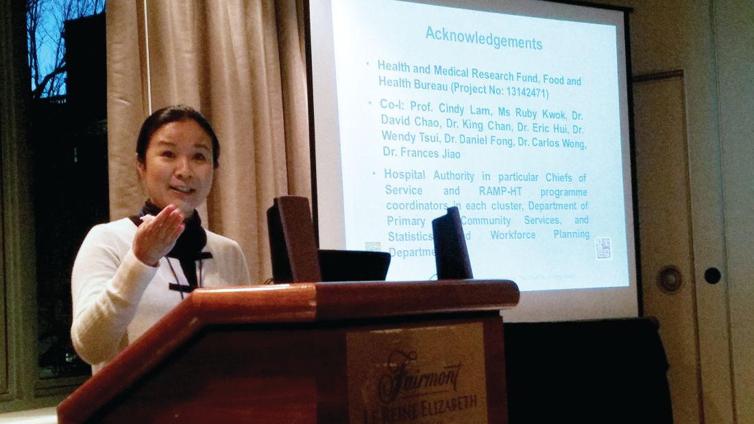
To tackle these persistent high rates of patients whose blood pressures remained too high, Dr. Yu and her colleagues at the Hospital Authority launched the Risk Assessment and Management Program – Hypertension (RAMP-HT) in 2011. The program, she said, is an “evidence-based, structured, protocol-driven, multidisciplinary program” that includes risk assessment and screening for complications, and uses a risk-guided management approach.
Patients in RAMP-HT received interventions according to a matrix for risk management of patients with hypertension. Patients with a blood pressure between 140/90 and 160/100 mm Hg who were assessed as being low and medium risk according to the Joint British Societies guidelines for cardiovascular risk continued to receive management from their primary care physician. High-risk patients with blood pressure in this range also received a statin if their low-density lipoprotein cholesterol level was suboptimal.
Patients whose blood pressure was at least 160/100 mm Hg were followed by a RAMP-HT nurse. For those with this degree of blood pressure elevation who were already on at least three antihypertensive medications, specialty appointments were also arranged.
Other targeted interventions were also available to participants, including the services of dietitians and physical therapists for those with a body mass index (BMI) of at least 27.5 kg/m2; smoking cessation and mental health services were also available, as appropriate.
After 3 years, those participating in the RAMP-HT program (n = 79,116) were compared with those in the usual care group (n = 43,901). In both arms, adult patients with complete data and without preexisting cardiovascular disease, diabetes, or end-stage renal disease were included. In each group, about 58% of participants were female, and the mean age was about 65 years.
Primary outcome measures included the incidence of cardiovascular disease, an outcome that included coronary heart disease, heart failure, and stroke; end-stage renal disease; and all-cause mortality. The significant reductions in these measures for the RAMP-HT group remained after multivariable analysis accounted for sex, age, smoking status, renal function, lipid values, BMI, comorbidities, and antihypertensive and lipid-lowering medication use.
The reduced care utilization seen among RAMP-HT participants also persisted after multivariable analysis for these potential confounders.
Dr. Yu said the systematic, protocol-driven program was a primary strength of RAMP-HT. The key to the program was use of nurses to provide patient education and physicians and allied health resources only as needed, she said; the program reinforced the importance of self-management and adherence because patients heard a unified message from many different health care professionals.
However, lifestyle factors such as diet and exercise weren’t tracked, and the retrospective study design introduced the potential for some bias, she said. In ongoing work, the long-term efficacy and cost-effectiveness of the RAMP-HT program are being tracked.
Dr. Yu reported that the study was funded by the Hong Kong Health and Medical Research Fund. She reported no relevant conflicts of interest.
SOURCE: Yu, Esther et al. NAPCRG 2017, Abstract HY33.
MONTREAL – who received phone calls, care coordination, and coaching from a nurse case manager, according to a retrospective population-based cohort study of almost 85,000 patients with hypertension.
The reduction yielded a hazard ratio for all-cause mortality of 0.504, with a number needed to treat (NNT) of 25 (P less than .05).
Service utilization also decreased for those participating in the intervention, compared with those receiving usual care: Hospitalizations fell by 7 per 100 patient-years, with greater reductions seen in emergency department, specialist, and primary care utilization (P less than .05 for all).
At the time the study was conceived, the Hong Kong Hospital Authority cared for about 200,000 hypertensive patients, of whom more than 40% hadn’t achieved the target blood pressure of less than 140/90 mm Hg, said Dr. Yu.
Dr. Yu of the department of family medicine and primary care at the University of Hong Kong said there are challenges in bringing more hypertension patients into good blood pressure control in Hong Kong. These include the “idiosyncratic practice” of some frontline physicians, who also often have limited time for patient consultations and only limited access to the services of allied health professionals to help them in their work. Patient adherence, she said, is also an issue.
To tackle these persistent high rates of patients whose blood pressures remained too high, Dr. Yu and her colleagues at the Hospital Authority launched the Risk Assessment and Management Program – Hypertension (RAMP-HT) in 2011. The program, she said, is an “evidence-based, structured, protocol-driven, multidisciplinary program” that includes risk assessment and screening for complications, and uses a risk-guided management approach.
Patients in RAMP-HT received interventions according to a matrix for risk management of patients with hypertension. Patients with a blood pressure between 140/90 and 160/100 mm Hg who were assessed as being low and medium risk according to the Joint British Societies guidelines for cardiovascular risk continued to receive management from their primary care physician. High-risk patients with blood pressure in this range also received a statin if their low-density lipoprotein cholesterol level was suboptimal.
Patients whose blood pressure was at least 160/100 mm Hg were followed by a RAMP-HT nurse. For those with this degree of blood pressure elevation who were already on at least three antihypertensive medications, specialty appointments were also arranged.
Other targeted interventions were also available to participants, including the services of dietitians and physical therapists for those with a body mass index (BMI) of at least 27.5 kg/m2; smoking cessation and mental health services were also available, as appropriate.
After 3 years, those participating in the RAMP-HT program (n = 79,116) were compared with those in the usual care group (n = 43,901). In both arms, adult patients with complete data and without preexisting cardiovascular disease, diabetes, or end-stage renal disease were included. In each group, about 58% of participants were female, and the mean age was about 65 years.
Primary outcome measures included the incidence of cardiovascular disease, an outcome that included coronary heart disease, heart failure, and stroke; end-stage renal disease; and all-cause mortality. The significant reductions in these measures for the RAMP-HT group remained after multivariable analysis accounted for sex, age, smoking status, renal function, lipid values, BMI, comorbidities, and antihypertensive and lipid-lowering medication use.
The reduced care utilization seen among RAMP-HT participants also persisted after multivariable analysis for these potential confounders.
Dr. Yu said the systematic, protocol-driven program was a primary strength of RAMP-HT. The key to the program was use of nurses to provide patient education and physicians and allied health resources only as needed, she said; the program reinforced the importance of self-management and adherence because patients heard a unified message from many different health care professionals.
However, lifestyle factors such as diet and exercise weren’t tracked, and the retrospective study design introduced the potential for some bias, she said. In ongoing work, the long-term efficacy and cost-effectiveness of the RAMP-HT program are being tracked.
Dr. Yu reported that the study was funded by the Hong Kong Health and Medical Research Fund. She reported no relevant conflicts of interest.
SOURCE: Yu, Esther et al. NAPCRG 2017, Abstract HY33.
REPORTING FROM NAPCRG 2017
Key clinical point:
Major finding: The hazard ratio for all-cause mortality was 0.504 for patients in the intervention arm.
Study details: A retrospective population-based cohort study of almost 85,000 Hong Kong patients with hypertension.
Disclosures: The study was funded by the Hong Kong Health and Medical Research Fund. Dr. Yu reported no relevant financial disclosures.
Source: Yu E et al. NAPCRG 2017, Abstract HY33.
Radiation exposure in MICU may exceed recommended limit
according to results of a recent observational study.
These “substantial” radiation doses in some patients suggest that efforts are warranted to “justify, restrict and optimize” the use of radiological resources when possible, said Sudhir Krishnan, MD, of the Cleveland Clinic, and his coauthors.
The retrospective, observational study included 4,155 adult admissions to a medical intensive care unit (MICU) at an academic medical center in 2013. Investigators calculated the cumulative effective dose (CED) of radiation based on reported ionizing radiological studies for each patient.
With a median length of stay of just 6.4 days, a total of 131 admissions (3%) accrued a CED of radiation of at least 50 millisieverts (mSv), the annual limit recommended by the National Commission on Radiation Protection, and 47 of those patients (1%) accrued a CED of radiation of at least 100 mSv, the 5-year cumulative exposure limit, the authors reported.
These findings suggest that “MICU patients could be subjected to radiation doses in a matter of days that are equivalent to or more than [the] CED observed in patients with chronic diseases and patients with trauma,” wrote Dr. Krishnan and his coauthors.
As hypothesized, patients with higher severity of illness scores (APACHE III scores) received a higher CED of radiation, according to the report. Using a multivariable linear regression model, investigators found that higher CED was predicted by higher APACHE III scores, sepsis, longer MICU stay, and gastrointestinal disorders and bleeding.
CT scans were the most common source of radiation exposure in patients who exceeded a 50 mSv of radiation, accounting for 49% of the total accrued dose, with interventional radiology accounting for 38%, authors reported.
Despite concerns about “the statistical risk of latent radiogenic cancer,” radiologic studies performed in the critically ill have the potential to reduce morbidity and mortality, the authors acknowledged in a discussion of the results.
“This understandably shifts the risk-benefit ratio towards radiation exposure,” the researchers wrote. “However, complacency in this regard cannot be entirely justified.”
Of the patients in the study who were exposed to a CED of at least 50 mSv, 81% survived the hospital admission and could be subjected to even more radiation as a part of ongoing medical care, they noted.
“Robust tools for monitoring CED prospectively per episode of clinical care, counseling patients exposed to high doses of radiation, and prospective studies exploring radiogenic risk associated with medical radiation are urgently required,” the authors said.
Dr. Krishnan and his coauthors reported no significant conflicts of interest.
SOURCE: Krishnan S et al. Chest. 2018 Feb 4. doi: 10.1016/j.chest.2018.01.019.
FROM CHEST
Key clinical point: Patients admitted to MICUs may be exposed to doses of radiation that are substantial and may exceed federal occupational health limits.
Major finding: In a short span of time (median 6.4 days length of stay), 3% of MICU patients received a cumulative dose of radiation that exceeded the U.S. recommended limit, and 1% accrued enough exposure to exceed the 5-year cumulative limit.
Data source: A retrospective, observational study including 4,155 adult admissions to the MICU at an academic medical center in 2013.
Disclosures: The study authors reported no significant conflicts of interest.
Source: Krishnan S et al. Chest. 2018 Feb 4. doi: 10.1016/j.chest.2018.01.019.
Colorectal cancer deaths projected for 2018
Approximately 50,630 deaths from colorectal cancer are predicted for the year in the United States by the American Cancer Society (ACS) in its Cancer Facts & Figures 2018, based on analysis of 2001-2015 data from the National Center for Health Statistics.
Nationally, the colorectal cancer death rate has been declining for decades, but hidden inside that long-term trend are a couple of competing ones: From 2006 to 2015, mortality dropped 2.9% a year for those aged 55 years and older but increased by 1% annually for adults aged 55 and under, the ACS said.
Incidence rates for colon cancer and rectal cancer showed a similar trend: From 2005 to 2015 they were down by 3.8% (colon) and 3.5% (rectal) a year for adults aged 55 and older but rose 1.4% and 2.4%, respectively, for adults younger than 55. Accurate statistics on colon and rectal cancer deaths are not available separately “because many deaths from rectal cancer are misclassified as colon cancer on death certificates,” the ACS said.
Therapeutic horseback riding may lower veterans’ PTSD symptoms
Veterans with posttraumatic stress disorder might benefit from therapeutic horseback riding, a small study suggests.
“Our findings provide empirical evidence that [therapeutic horseback riding] is effective at improving coping skills and in lessening one’s difficulty with emotional regulation, especially with longer riding interventions,” wrote Rebecca A. Johnson, PhD, of the University of Missouri, Columbia, and her associates.
Overall, 57 veterans were recruited and 28 enrolled in the randomized trial at baseline. Those individuals were randomized into two groups: a wait-list control group and a treatment group. Eventually, all of the veterans participated in the therapeutic riding program. Meanwhile, the riding center staff were not aware of which veterans had been assigned to either group. The Professional Association of Therapeutic Horsemanship, a nonprofit group that promotes equine-related activities for people with special needs, selected the horses that were used in the study. During the data collection periods, PTSD symptoms were measured via the PTSD Checklist–Military Version, or PCL-M. This self-report measure asks patients about problems in response to “stressful military experiences,” the researchers wrote. The Coping Self-Efficacy Scale and the Difficulties in Emotion Regulation Scale were among the other instruments used.
While riding, the results showed, participants had a statistically significant decrease in PTSD symptoms over the course of the 6-week program. “,” Dr. Johnson and her associates wrote. “Further detailed examination showed that participants had a 66.7% likelihood of having lower PTSD scores at 3 weeks, and an 87.5% likelihood at 6 weeks.”
Anecdotally, some of the veterans wanted to continue therapeutic riding after the end of the program, and they were able to do so.
“We conclude that [therapeutic horseback riding] shows promise as a beneficial intervention for veterans with PTSD, but did not measure functional ability,” they wrote.
FROM MILITARY MEDICAL RESEARCH
Debunking Acne Myths: Does Wearing Makeup Cause Acne?
Myth: Wearing makeup causes acne breakouts
Acne breakouts caused by makeup and other skin care products, known as acne cosmetica, typically resolve when patients stop using pore-clogging products; however, the overall impact of cosmetics on the development of acne lesions is considered to be negligible. Many cosmetics are not inherently comedogenic and can be used safely by patients in combination with proper skin care techniques.
Although dermatologists may be inclined to discourage makeup use during acne treatment or breakouts due to its potential to aggravate the patient’s condition, research has shown that treatment results and quality of life (QoL) scores associated with makeup use in acne patients may improve when patients receive instruction on how to use skin care products and cosmetics effectively. In one study of 50 female acne patients, 25 participants were instructed on how to use skin care products and cosmetics, and the other 25 participants received no specific instructions from dermatologists. After 4 weeks of treatment with conventional topical and/or oral acne medications, the investigators concluded that use of skin care products did not negatively impact acne treatment, and the group that received application instructions showed more notable improvements in QoL scores versus those who did not. In another study, the overall number of acne eruptions decreased over a 2- to 4-week period in female acne patients who were trained by a makeup artist to apply cosmetics while undergoing acne treatment. These results suggest that acne patients who wear makeup may benefit from a conversation with their dermatologist about what products and skin care techniques they can use to minimize exacerbation of or even improve their condition.
When choosing makeup that will not cause or exacerbate acne breakouts, patients should look for packaging that indicates the product will not clog pores and is oil-free, noncomedogenic, and/or nonacnegenic. Some makeup products are specifically formulated to help camouflage redness and pimples, which can help improve quality of life and self-esteem in acne patients who otherwise may be self-conscious about their appearance. Mineral-based cosmetics containing powdered formulas of silica, titanium dioxide, and zinc oxide can be used to absorb oil, camouflage redness, and prevent irritation. Anti-inflammatory ingredients and antioxidants also are used in some makeup products to reduce skin irritation and promote barrier repair. Additional cosmetic ingredients that can affect the mechanisms of acne pathogenesis and may contribute to a decrease in acne lesions include nicotinamide, lactic acid, triethyl acetate/ethyllineolate, and prebiotic plant extracts.
Makeup should be applied gently to avoid irritating the skin. It also is important to remind patients not to share their makeup brushes and applicators and to clean them weekly to ensure that bacteria, dead skin cells, and oil are not spread to the skin, which can lead to new breakouts. Although patients may be compelled to scrub the skin to remove makeup, a mild cleanser should be gently applied using the fingertips and rinsed off with lukewarm water to minimize skin irritation. Any makeup remaining on the skin after washing should be gently removed with an oil-free makeup remover.
Hayashi N, Imori M, Yanagisawa M, et al. Make-up improves the quality of life of acne patients without aggravating acne eruptions during treatments. Eur J Dermatol. 2005;15:284-287.
I have acne! is it okay to wear makeup? American Academy of Dermatology website. https://www.aad.org/public/diseases/acne-and-rosacea/makeup-with-acne. Accessed February 13, 2018.
Korting HC, Borelli C, Schöllmann C. Acne vulgaris. role of cosmetics [in German]. 2010;61:126-131.
Matsuoka Y, Yoneda K, Sadahira C, et al. Effects of skin care and makeup under instructions from dermatologists on the quality of life of female patients with acne vulgaris. J Dermatol. 2006;33:745-752.
Proper skin care lays the foundation for successful acne and rosacea treatment. American Academy of Dermatology website. https://www.aad.org/media/news-releases/proper-skin-care-lays-the-foundation-for-successful-acne-and-rosacea-treatment Published July 31, 2013. Accessed February 13, 2018.
Myth: Wearing makeup causes acne breakouts
Acne breakouts caused by makeup and other skin care products, known as acne cosmetica, typically resolve when patients stop using pore-clogging products; however, the overall impact of cosmetics on the development of acne lesions is considered to be negligible. Many cosmetics are not inherently comedogenic and can be used safely by patients in combination with proper skin care techniques.
Although dermatologists may be inclined to discourage makeup use during acne treatment or breakouts due to its potential to aggravate the patient’s condition, research has shown that treatment results and quality of life (QoL) scores associated with makeup use in acne patients may improve when patients receive instruction on how to use skin care products and cosmetics effectively. In one study of 50 female acne patients, 25 participants were instructed on how to use skin care products and cosmetics, and the other 25 participants received no specific instructions from dermatologists. After 4 weeks of treatment with conventional topical and/or oral acne medications, the investigators concluded that use of skin care products did not negatively impact acne treatment, and the group that received application instructions showed more notable improvements in QoL scores versus those who did not. In another study, the overall number of acne eruptions decreased over a 2- to 4-week period in female acne patients who were trained by a makeup artist to apply cosmetics while undergoing acne treatment. These results suggest that acne patients who wear makeup may benefit from a conversation with their dermatologist about what products and skin care techniques they can use to minimize exacerbation of or even improve their condition.
When choosing makeup that will not cause or exacerbate acne breakouts, patients should look for packaging that indicates the product will not clog pores and is oil-free, noncomedogenic, and/or nonacnegenic. Some makeup products are specifically formulated to help camouflage redness and pimples, which can help improve quality of life and self-esteem in acne patients who otherwise may be self-conscious about their appearance. Mineral-based cosmetics containing powdered formulas of silica, titanium dioxide, and zinc oxide can be used to absorb oil, camouflage redness, and prevent irritation. Anti-inflammatory ingredients and antioxidants also are used in some makeup products to reduce skin irritation and promote barrier repair. Additional cosmetic ingredients that can affect the mechanisms of acne pathogenesis and may contribute to a decrease in acne lesions include nicotinamide, lactic acid, triethyl acetate/ethyllineolate, and prebiotic plant extracts.
Makeup should be applied gently to avoid irritating the skin. It also is important to remind patients not to share their makeup brushes and applicators and to clean them weekly to ensure that bacteria, dead skin cells, and oil are not spread to the skin, which can lead to new breakouts. Although patients may be compelled to scrub the skin to remove makeup, a mild cleanser should be gently applied using the fingertips and rinsed off with lukewarm water to minimize skin irritation. Any makeup remaining on the skin after washing should be gently removed with an oil-free makeup remover.
Hayashi N, Imori M, Yanagisawa M, et al. Make-up improves the quality of life of acne patients without aggravating acne eruptions during treatments. Eur J Dermatol. 2005;15:284-287.
I have acne! is it okay to wear makeup? American Academy of Dermatology website. https://www.aad.org/public/diseases/acne-and-rosacea/makeup-with-acne. Accessed February 13, 2018.
Korting HC, Borelli C, Schöllmann C. Acne vulgaris. role of cosmetics [in German]. 2010;61:126-131.
Matsuoka Y, Yoneda K, Sadahira C, et al. Effects of skin care and makeup under instructions from dermatologists on the quality of life of female patients with acne vulgaris. J Dermatol. 2006;33:745-752.
Proper skin care lays the foundation for successful acne and rosacea treatment. American Academy of Dermatology website. https://www.aad.org/media/news-releases/proper-skin-care-lays-the-foundation-for-successful-acne-and-rosacea-treatment Published July 31, 2013. Accessed February 13, 2018.

Acne is linked to higher chances of major depression
according to a retrospective cohort analysis published as a research letter.
“The onset of acne, when patients present for treatment because of active disease, is associated with a greater risk of developing depression” wrote Isabelle A. Vallerand, an MD/PhD student at the University of Calgary (Alta.), and her associates. “Although the severity of acne was not assessed directly in the current study, this finding suggests a potential dose/response relationship such that more active disease may lead to a greater risk of depression.”
In total, 134,437 acne patients and 1,731,608 patients without acne were identified from THIN. Over a 15-year follow-up, the probability of developing MDD was 18.5% among patients with acne, and 12% for those without acne. This risk was much higher within the first year after diagnosis (adjusted hazard ratio, 1.63; 95% confidence interval, 1.33-2), which subsequently decreased.
The researchers found that patients with acne tended to be younger (67.6% vs. 22.8%), female (58.6% vs. 48.6%), and of a higher socioeconomic status (24.4% vs. 22.1%) compared with patients without acne. Those with acne tended to smoke (58.4% vs. 48.6%) and to have comorbidities (17.2% vs. 13.8%). Conversely, acne patients were less likely to use alcohol (17% vs. 39%) and less likely to be obese (2.3% vs. 6.6%) (all P less than .001).
Although these results are promising, there are several limitations that could have influenced the study findings. The misclassification of patients with acne and MDD could have occurred if patients did not present themselves to a physician for treatment. Another limitation was that isotretinoin was the only acne treatment that was considered by the researchers. Considering that treatment has been shown to reduce depressive symptoms, the researchers believe that their estimates are conservative.
“Given the tremendous burden of MDD and its temporal association with active acne, it is critical that physicians monitor mood symptoms in patients with acne and initiate prompt MDD management or seek consultation from a psychiatrist when needed” wrote Ms. Vallerand and her colleagues.
Ms. Vallerand received funding for this study from the Alberta Innovates Health Solutions MD-PhD Studentship and from the Mach-Gaensslen Foundation of Canada. None of the other authors had disclosures to report.
SOURCE: Vallerand I et al. Br J Dermatol. 2018 Feb 7. doi: 10.1111/bjd.16099.
FROM BRITISH JOURNAL OF DERMATOLOGY
Key clinical point: Patients with acne have a much higher chance of developing major depressive disorder (MDD).
Major finding: The chance of developing MDD was 18.5% in patients with acne.
Study details: Analysis of retrospective cohort data obtained from the Health Improvement Network (THIN) between 1986 and 2012.
Disclosures: Ms. Vallerand received funding for this study from the Alberta Innovates Health Solutions MD-PhD Studentship and from the Mach-Gaensslen Foundation of Canada. None of the other authors had disclosures to report.
Source: Vallerand I et al. Br J Dermatol. 2018 Feb 7. doi: 10.1111/bjd.16099.
Are you as frustrated with medical care as we are?
How did the experience of office visits get to be so frustrating for both patients and doctors? Let’s put it under the microscope and examine it.
To medicine’s credit, it realized the value of looking for diseases before symptoms occurred, such as using mammograms to detect breast cancer and controlling blood pressure and blood sugar to avert comorbidities.
Today, a doctor looks at the computer screen and checks off when a mammogram was done and whether blood pressure and blood sugar are controlled. “Authorities” believe that good health is achieved by performing positive checkoffs to questions like this. This definition of quality care is, in reality, “quantity care” and can be tied to physician compensation. Physicians who did not adequately meet Physician Quality Reporting System requirements have received letters informing them that their Medicare Part B payments for 2018 will be reduced by 3%.
Many seasoned clinicians recognize that practicing good medicine involves more than following a computer printout of tests and treatments based on the patient’s symptoms, more than plugging into the diagnostic and prescription mills that are part of today’s managed care system. Making the correct diagnosis requires a carefully taken history, listening to the patients’ stories of their journeys into and through illness, and using a bio-psychosocial-spiritual approach.
Getting to know the patient as a person requires that the doctor and patient take a journey together. In that journey, when the doctor empathizes with the patient and understands what makes the patient tick, the doctor can empower the patient – giving the patient a fuller understanding of their medical conditions, greater participation in the diagnostic work-up and in treatments, and hope for success – all leading to better outcomes.
Doctors are frustrated with the current medical assembly-line system. A study has shown that physicians spend 2 hours on electronic health records and clerical work for every hour they provide direct patient care (Ann Intern Med. 2016;165[11]:753-60). Nearly half of physicians now report that they are “burned out” by the demand to achieve the quantitative requirements on the one hand and their inability to minister to the needs of their patients on the other hand. Patients are also frustrated by the system as they cope with health insurance and costs, with the short time allocated for office visits, and with a fragmented and impersonal medical system. Patients feel that they are little more than a source of information for boxes to be checked off by the physician whose eyes are forced to be on the computer and the clock.
How can we begin to integrate these measures of quality into “quantity medicine” and make the experience of medical visits less frustrating for doctors and patients? How can we reward the skills that recognize that the course of an illness is influenced by patients’ emotions and thoughts related to their problems, their supportive or stressful relationships with others, and the context within which they conceptualize their lives – particularly their religious and spiritual beliefs about life’s purpose and challenges and attitudes toward death?
Caring for patients requires a more sophisticated approach than seeing patients as computer checkoffs. Office visits need to focus on the patient who has the symptoms, not just the symptoms the patient has.
Isn’t it time to make patient-centered care a reality and not just a slogan? If this speaks to you, then what should you do? Even though solutions may not be simple, we should not be deterred from finding patient-centered systems since patients and doctors are unhappy with today’s system. Why not have patients grade their office visits?
While this approach has its shortcomings, and isn’t the only solution, it does place the patient at the center of the process, answering questions about whether the doctor listened to them, heard their concerns, and presented a reasonable plan to help them get better.
In addition, all those involved with medical care should be involved in the process to replace today’s deficient system. The nation’s main organizations representing physicians should propose solutions to support patient-centered care. Individual physicians should become involved, speaking up and sending articles and letters to medical journals and the lay press.
Patients should be empowered to open up a public discussion – in print and broadcast media – on how they want to improve their own medical experiences and the quality of their health care.
It’s worth it. It’s our health.
Dr. Banner is a practicing internist in Philadelphia and chair emeritus of the Albert Einstein Medical Center Medical Ethics Committee. Dr. Benor is a psychiatric psychotherapist in the United States and a wholistic psychotherapist in Canada. Dr. Reiser is adjunct professor, University of Texas School of Public Health, Austin, and the UT Austin Plan II Honors Program, and teaches medical history, medical ethics, and public policy. The authors are indebted to Benjamin Sharfman, PhD, and Jane Brown, PhD, for their important roles in creating this article.