User login
MedPAC: Medicare hospital readmissions program is working
WASHINGTON – The Medicare Hospital Readmissions Reduction Program is working, according to an original analysis of Medicare claims data presented at a meeting of the Medicare Payment Advisory Commission.
“First, readmissions declined,” MedPAC staff member Jeff Stensland, PhD, said during a congressionally mandated staff report to the commissioners. “Second, while observation stays increased, they did not fully offset the decrease in readmissions. Third, while [emergency department] visits also increased, those increases appear to largely be due to factors other than the readmission program. And fourth, in addition, all the evidence we examined suggests that the readmissions program did not result in increased mortality.”
including a reduction in readmissions and patients spending less time in the hospital with “at least equal outcomes,” Dr. Stensland said at the meeting.
Taxpayers benefited from a $2 billion reduction in spending on readmissions, which will “help extend the viability of the Medicare Trust Fund.” He noted that improvements to the program will be discussed at future MedPAC meetings.
Not all MedPAC commissioners agreed with the staff analysis.
David Nerenz, PhD, of the Henry Ford Health System, Detroit, also was not convinced the program was having an impact, noting that hospital readmissions began to decline even before the program started.
In looking at a graph presented that showed this trend, “I was impressed by the fact that the trend line started coming down all the way to the left side of the graph, and what my eye was impressed with was more just the continuation rather than a change, so I guess I feel cautious saying the program had certain effects because they certainly don’t jump off the graph visually,” Dr. Nerenz said. “I’m not disputing the numbers, but to say just as a clear unqualified conclusion the program reduced readmissions, I’m not so sure.”
It is likely premature to make any firm conclusions about how effectively this program decreases unnecessary utilization of hospitals. However, it is heartening to know that it did not increase mortality. The one variable that would best control readmissions is patient education. What constitutes an emergency requiring hospital evaluation and potential admission is often not explained to the patient by you and me. 
It is likely premature to make any firm conclusions about how effectively this program decreases unnecessary utilization of hospitals. However, it is heartening to know that it did not increase mortality. The one variable that would best control readmissions is patient education. What constitutes an emergency requiring hospital evaluation and potential admission is often not explained to the patient by you and me.
It is likely premature to make any firm conclusions about how effectively this program decreases unnecessary utilization of hospitals. However, it is heartening to know that it did not increase mortality. The one variable that would best control readmissions is patient education. What constitutes an emergency requiring hospital evaluation and potential admission is often not explained to the patient by you and me.
WASHINGTON – The Medicare Hospital Readmissions Reduction Program is working, according to an original analysis of Medicare claims data presented at a meeting of the Medicare Payment Advisory Commission.
“First, readmissions declined,” MedPAC staff member Jeff Stensland, PhD, said during a congressionally mandated staff report to the commissioners. “Second, while observation stays increased, they did not fully offset the decrease in readmissions. Third, while [emergency department] visits also increased, those increases appear to largely be due to factors other than the readmission program. And fourth, in addition, all the evidence we examined suggests that the readmissions program did not result in increased mortality.”
including a reduction in readmissions and patients spending less time in the hospital with “at least equal outcomes,” Dr. Stensland said at the meeting.
Taxpayers benefited from a $2 billion reduction in spending on readmissions, which will “help extend the viability of the Medicare Trust Fund.” He noted that improvements to the program will be discussed at future MedPAC meetings.
Not all MedPAC commissioners agreed with the staff analysis.
David Nerenz, PhD, of the Henry Ford Health System, Detroit, also was not convinced the program was having an impact, noting that hospital readmissions began to decline even before the program started.
In looking at a graph presented that showed this trend, “I was impressed by the fact that the trend line started coming down all the way to the left side of the graph, and what my eye was impressed with was more just the continuation rather than a change, so I guess I feel cautious saying the program had certain effects because they certainly don’t jump off the graph visually,” Dr. Nerenz said. “I’m not disputing the numbers, but to say just as a clear unqualified conclusion the program reduced readmissions, I’m not so sure.”
REPORTING FROM MEDPAC
CV risk factors go undiagnosed, untreated in many psoriatic patients
A significant proportion of patients with psoriasis and psoriatic arthritis (PsA) are underdiagnosed and undertreated for cardiovascular risk factors (CVRF), according to Lihi Eder, MD, of the University of Toronto, and her associates.
In a cross-sectional analysis published in the Journal of Rheumatology, researchers examined 2,254 patients (58.9% with PsA, 41.1% with psoriasis only) from eight centers in Canada, the United States, and Israel. They found that 1,017 of the patients had hypertension (PsA: 48.5%, psoriasis: 40.2%), including 233 who were not previously diagnosed with hypertension and were not taking any blood pressure–lowering medications (PsA: 19.9%, psoriasis: 39.1%). Many patients had low adherence to hypertension treatment recommendations: A total of 602 (PsA: 55.9%, psoriasis: 64.8%) were untreated or undertreated. Undertreatment of hypertension occurred in 60.9% patients with cardiovascular disease or diabetes mellitus.
“In this large international study, we found significant gaps in screening and treating CVRF in patients with psoriasis and PsA,” the researchers concluded. “Although questions exist regarding the optimal treatment targets for CVRF in psoriatic patients, adherence by physicians to, at a minimum, the general treatment recommendations for primary CV prevention is warranted.”
SOURCE: Eder L et al. J Rheumatol. 2018 Feb 1. doi: 10.3899/jrheum.170379.
FROM JOURNAL OF RHEUMATOLOGY
Fetal alcohol spectrum disorders incidence exceeds previous estimates
, according to a cross-sectional study.
Philip A. May, PhD, of the University of North Carolina, Gillings School of Global Public Health, and his coauthors assessed 6,639 first-grade children from four communities in the Rocky Mountain, Midwestern, Southeastern, and Pacific Southwestern regions of the United States.
In their report, published Feb. 6 in JAMA, they identified 222 cases of fetal alcohol spectrum disorders, representing conservative prevalence estimates ranging from 11.3 to 50 cases per 1,000 children (JAMA 2018;319[5]:474-82. doi: 10.1001/jama.2017.21896).
Of these children, 27 met the criteria for fetal alcohol syndrome, 104 met the criteria for partial fetal alcohol syndrome, and 91 met the criteria for alcohol-related neurodevelopmental disorder.
“These prevalence estimates are consistent with mounting evidence that harmful fetal alcohol exposure is common in the United States today and highlight the public health burden due to fetal alcohol spectrum disorders,” the authors wrote.
The finding was much higher than previous estimates of the prevalence of fetal alcohol spectrum disorders in the United States. The authors pointed out that routine surveillance methods may have previously underestimated the prevalence because so many children are either misdiagnosed or not diagnosed at all. But even two previous single-site, active-case ascertainment studies conducted in the United States found prevalence rates of 10 and 24 per 1,000 children.
“This consortium study, to our knowledge, was the first to apply active case ascertainment, common methodology, a single classification system, and expert in-person evaluation for a continuum of fetal alcohol spectrum disorders including alcohol-related neurodevelopmental disorder to a large number of children from communities across the United States,” the authors wrote. “These data have highlighted the need for a larger study with broader representation of U.S. communities with general population samples.”
Only 2 of the 222 children had actually been previously diagnosed with a fetal alcohol spectrum disorder, “although many parents and guardians were aware of the learning and behavioral challenges facing their children,” the researchers noted.
“These data confirm that missed diagnoses and misdiagnoses of children are common,” the authors wrote, pointing to a previous study in U.S. schoolchildren that found only one in seven children identified as having fetal alcohol syndrome had been previously diagnosed.
The assessment of fetal alcohol spectrum disorder was based on four domains: growth, dysmorphology, neurodevelopment, and prenatal alcohol exposure – the latter being assessed by interviewing the mother in person or by telephone.
The lowest prevalence – 11.3 per 1,000 children – was seen in the Midwestern community, while the highest – 50 per 1,000 – was in the Rocky Mountain community.
The main weakness of the study was that no individual sample included the entire eligible population because of differing policies on access to children for recruitment and variability in willingness to consent.
“Consent rates for screening ranged from 36.9% to 92.5% in individual samples and overall consent rates for screening averaged only 59.9% of eligible children,” the authors wrote. “If nonconsented children differed from consented, this could have biased prevalence estimates in either direction.”
The researchers acknowledged that the absence of a definitive biomarker for fetal alcohol spectrum disorder meant it was impossible to know for certain that the observed deficits were actually the result of fetal alcohol exposure, and that the prevalence estimates may therefore be overestimated.
The study was funded by grants from the National Institute on Alcohol Abuse and Alcoholism. One author declared grant support from the National Institute on Alcohol Abuse and Alcoholism and personal fees and honorarium from the Alcohol Center. No conflicts of interest were declared.
SOURCE: May, P et al. JAMA 2018;319[5]:474-82. doi: 10.1001/jama.2017.21896.
The finding of higher-than-expected prevalence estimates for fetal alcohol spectrum disorder has significant implications for clinicians and for public health. Many cases are misdiagnosed or not diagnosed, which may be the result of unknown or unconfirmed prenatal alcohol exposure, overlapping diagnostic criteria with other neurodevelopmental disorders, high rates of comorbidity, and the presence of a number of different clinical diagnostic guidelines. There is, therefore, a need for a universal diagnostic approach and the identification of novel and reliable biomarkers for detecting fetal alcohol effects.
In addition, the high prevalence of these disorders points to a need for better education of girls and women of childbearing age about the detrimental effects of alcohol consumption during pregnancy. Primary care clinicians should routinely include appropriate screening for alcohol use in all women of childbearing age in preconceptual health promotion and in contraceptive counseling and refer anyone identified as having an alcohol use disorder to substance abuse programs.
Shannon Lange, MPH, Jurgen Rehm, PhD, and Svetlana Popova, PhD, are with the Institute for Mental Health Policy Research at the Centre for Addiction and Mental Health in Toronto and the University of Toronto. These comments are taken from an accompanying editorial (JAMA 2018, 319[5]:448-9. doi: 10.1001/jama.2017.21895). No conflicts of interest were declared.
, according to a cross-sectional study.
Philip A. May, PhD, of the University of North Carolina, Gillings School of Global Public Health, and his coauthors assessed 6,639 first-grade children from four communities in the Rocky Mountain, Midwestern, Southeastern, and Pacific Southwestern regions of the United States.
In their report, published Feb. 6 in JAMA, they identified 222 cases of fetal alcohol spectrum disorders, representing conservative prevalence estimates ranging from 11.3 to 50 cases per 1,000 children (JAMA 2018;319[5]:474-82. doi: 10.1001/jama.2017.21896).
Of these children, 27 met the criteria for fetal alcohol syndrome, 104 met the criteria for partial fetal alcohol syndrome, and 91 met the criteria for alcohol-related neurodevelopmental disorder.
“These prevalence estimates are consistent with mounting evidence that harmful fetal alcohol exposure is common in the United States today and highlight the public health burden due to fetal alcohol spectrum disorders,” the authors wrote.
The finding was much higher than previous estimates of the prevalence of fetal alcohol spectrum disorders in the United States. The authors pointed out that routine surveillance methods may have previously underestimated the prevalence because so many children are either misdiagnosed or not diagnosed at all. But even two previous single-site, active-case ascertainment studies conducted in the United States found prevalence rates of 10 and 24 per 1,000 children.
“This consortium study, to our knowledge, was the first to apply active case ascertainment, common methodology, a single classification system, and expert in-person evaluation for a continuum of fetal alcohol spectrum disorders including alcohol-related neurodevelopmental disorder to a large number of children from communities across the United States,” the authors wrote. “These data have highlighted the need for a larger study with broader representation of U.S. communities with general population samples.”
Only 2 of the 222 children had actually been previously diagnosed with a fetal alcohol spectrum disorder, “although many parents and guardians were aware of the learning and behavioral challenges facing their children,” the researchers noted.
“These data confirm that missed diagnoses and misdiagnoses of children are common,” the authors wrote, pointing to a previous study in U.S. schoolchildren that found only one in seven children identified as having fetal alcohol syndrome had been previously diagnosed.
The assessment of fetal alcohol spectrum disorder was based on four domains: growth, dysmorphology, neurodevelopment, and prenatal alcohol exposure – the latter being assessed by interviewing the mother in person or by telephone.
The lowest prevalence – 11.3 per 1,000 children – was seen in the Midwestern community, while the highest – 50 per 1,000 – was in the Rocky Mountain community.
The main weakness of the study was that no individual sample included the entire eligible population because of differing policies on access to children for recruitment and variability in willingness to consent.
“Consent rates for screening ranged from 36.9% to 92.5% in individual samples and overall consent rates for screening averaged only 59.9% of eligible children,” the authors wrote. “If nonconsented children differed from consented, this could have biased prevalence estimates in either direction.”
The researchers acknowledged that the absence of a definitive biomarker for fetal alcohol spectrum disorder meant it was impossible to know for certain that the observed deficits were actually the result of fetal alcohol exposure, and that the prevalence estimates may therefore be overestimated.
The study was funded by grants from the National Institute on Alcohol Abuse and Alcoholism. One author declared grant support from the National Institute on Alcohol Abuse and Alcoholism and personal fees and honorarium from the Alcohol Center. No conflicts of interest were declared.
SOURCE: May, P et al. JAMA 2018;319[5]:474-82. doi: 10.1001/jama.2017.21896.
FROM JAMA
Key clinical point: The prevalence of fetal alcohol spectrum disorder in first-grade children in the United States may be as high as 1 in 20.
Major finding: The prevalence of fetal alcohol spectrum disorder ranged from 11.3 to 50 children per 1,000.
Data source: Cross-sectional study of 6,639 first-grade children.
Disclosures: The study was funded by grants from the National Institute on Alcohol Abuse and Alcoholism. One author declared grant support from the National Institute on Alcohol Abuse and Alcoholism and personal fees and honorarium from the Alcohol Center. No conflicts of interest were declared.
Source: May, P et al. JAMA 2018;319[5]:474-82. doi: 10.1001/jama.2017.21896.
How Will You Champion Lung Health in 2018?
Our CHEST Foundation grantees are doing amazing research and community service projects that are paving the way for change and improvements in chest medicine. How will you help champion lung health?
“This award carries great importance to me as a young clinician who is in the early phase of my career. I’m driven by my passion for researching this disease (PAH). This award helps establish me as a strong clinical researcher – where I see my career heading. It also helps us identify those clues that can lead to changing how this disease state is treated. Everything starts with an idea.”
Sandeep Sahay, MD, FCCP
Houston Methodist Hospital – Houston, Texas
CHEST Foundation Research Grant in Pulmonary Arterial Hypertension
Title: Alterations of Estrogen Metabolism in the Development of Portopulmonary Hypertension
“This grant has allowed me to do this project, period. Having support from the CHEST Foundation automatically gives me credibility at my new institution. As I would meet with people to discuss my project, they would see that a big organization is supporting me, and that is the outside validation to show that this must be a useful project. The grant really helps me hit the ground running, and plants the seed to help us do a larger project in the future.”
Drew Harris, MD
Yale University – New Haven, Connecticut
CHEST Foundation Research Grant in Asthma
Title: Utilizing Medical-Legal Partnership to Promote Asthma Health Equity
“I recently completed my MD, and because of this grant, I am able to do a completely independent research study. I’ve also been recently short-listed for a clinical lecturer post at my university...which positions me to be the lead for quantitative imaging should I receive the post. This grant added gravitas to my project, and, without it, I don’t think I would have had as big of a boost.”
Diana Crossley, MBChB
University Hospital Birmingham – Birmingham, England
CHEST Foundation and the Alpha-1 Foundation Research Grant in Alpha-1 Antitrypsin Deficiency
Title: Functional Magnetic Resonance Lung Imaging Using Inhaled Hyperpolarised 129Xenon: A Pilot Study of the Clinical Utility in Alpha One Antitrypsin Deficiency (AATD).
“Because of this grant, we are able to be effective teachers to Haitian pediatricians, so they can more effectively intervene and save childrens’ lives. We are able to translate these critical care materials into French and provide the best opportunity for learning to our colleagues there.”
Adam Silverman, MD

Connecticut Children’s Medical Center – Hartford, Connecticut
CHEST Foundation Community Service Grant Honoring D. Robert McCaffree, MD, Master FCCP
Title: Haitian Pediatric Critical Care Collaborative Training Course
The CHEST Foundation is accepting grant applications now until March 31 in the following areas:
• CHEST Foundation Research Grant in Lung Cancer – $50,000 - $100,000 2-year grant*
• CHEST Foundation Research Grant in Asthma - $15,000 - $30,000 1-year grant*
• CHEST Foundation Research Grant in Pulmonary Arterial Hypertension - $25,000 - $50,000 1-year grant*
• CHEST Foundation and the Alpha-1 Foundation Research Grant in Alpha-1 Antitrypsin Deficiency - $25,000 1-year grant
• CHEST Foundation Research Grant in Pulmonary Fibrosis – $50,000 1-year grant
• CHEST Foundation Research Grant in Chronic Obstructive Pulmonary Disease – $50,000 1-year grant
• CHEST Foundation Research Grant in Venous Thromboembolism - $15,000 – $30,000 1-year grant*
• CHEST Foundation Research Grant in Nontuberculous Mycobacteria Disease - $30,000 1-year grant
• CHEST Foundation Research Grant in Women’s Lung Health - $10,000 1-year grant
• CHEST Foundation Research Grant in Cystic Fibrosis – $15,000 – $30,000 1-year grant
• CHEST Foundation Community Service Grant Honoring D. Robert McCaffree, MD, Master FCCP - $2,500- $15,000 1-year grant
*Amount contingent on funding.
Go to chestfoundation.org/grants to apply now.
Our CHEST Foundation grantees are doing amazing research and community service projects that are paving the way for change and improvements in chest medicine. How will you help champion lung health?
“This award carries great importance to me as a young clinician who is in the early phase of my career. I’m driven by my passion for researching this disease (PAH). This award helps establish me as a strong clinical researcher – where I see my career heading. It also helps us identify those clues that can lead to changing how this disease state is treated. Everything starts with an idea.”
Sandeep Sahay, MD, FCCP
Houston Methodist Hospital – Houston, Texas
CHEST Foundation Research Grant in Pulmonary Arterial Hypertension
Title: Alterations of Estrogen Metabolism in the Development of Portopulmonary Hypertension
“This grant has allowed me to do this project, period. Having support from the CHEST Foundation automatically gives me credibility at my new institution. As I would meet with people to discuss my project, they would see that a big organization is supporting me, and that is the outside validation to show that this must be a useful project. The grant really helps me hit the ground running, and plants the seed to help us do a larger project in the future.”
Drew Harris, MD
Yale University – New Haven, Connecticut
CHEST Foundation Research Grant in Asthma
Title: Utilizing Medical-Legal Partnership to Promote Asthma Health Equity
“I recently completed my MD, and because of this grant, I am able to do a completely independent research study. I’ve also been recently short-listed for a clinical lecturer post at my university...which positions me to be the lead for quantitative imaging should I receive the post. This grant added gravitas to my project, and, without it, I don’t think I would have had as big of a boost.”
Diana Crossley, MBChB
University Hospital Birmingham – Birmingham, England
CHEST Foundation and the Alpha-1 Foundation Research Grant in Alpha-1 Antitrypsin Deficiency
Title: Functional Magnetic Resonance Lung Imaging Using Inhaled Hyperpolarised 129Xenon: A Pilot Study of the Clinical Utility in Alpha One Antitrypsin Deficiency (AATD).
“Because of this grant, we are able to be effective teachers to Haitian pediatricians, so they can more effectively intervene and save childrens’ lives. We are able to translate these critical care materials into French and provide the best opportunity for learning to our colleagues there.”
Adam Silverman, MD
Connecticut Children’s Medical Center – Hartford, Connecticut
CHEST Foundation Community Service Grant Honoring D. Robert McCaffree, MD, Master FCCP
Title: Haitian Pediatric Critical Care Collaborative Training Course
The CHEST Foundation is accepting grant applications now until March 31 in the following areas:
• CHEST Foundation Research Grant in Lung Cancer – $50,000 - $100,000 2-year grant*
• CHEST Foundation Research Grant in Asthma - $15,000 - $30,000 1-year grant*
• CHEST Foundation Research Grant in Pulmonary Arterial Hypertension - $25,000 - $50,000 1-year grant*
• CHEST Foundation and the Alpha-1 Foundation Research Grant in Alpha-1 Antitrypsin Deficiency - $25,000 1-year grant
• CHEST Foundation Research Grant in Pulmonary Fibrosis – $50,000 1-year grant
• CHEST Foundation Research Grant in Chronic Obstructive Pulmonary Disease – $50,000 1-year grant
• CHEST Foundation Research Grant in Venous Thromboembolism - $15,000 – $30,000 1-year grant*
• CHEST Foundation Research Grant in Nontuberculous Mycobacteria Disease - $30,000 1-year grant
• CHEST Foundation Research Grant in Women’s Lung Health - $10,000 1-year grant
• CHEST Foundation Research Grant in Cystic Fibrosis – $15,000 – $30,000 1-year grant
• CHEST Foundation Community Service Grant Honoring D. Robert McCaffree, MD, Master FCCP - $2,500- $15,000 1-year grant
*Amount contingent on funding.
Go to chestfoundation.org/grants to apply now.
Pembrolizumab nets durable responses in sorafenib-treated advanced HCC
SAN FRANCISCO – The immune checkpoint inhibitor pembrolizumab is active and well tolerated when used as second-line therapy for hepatocellular carcinoma, according to results of the KEYNOTE-224 trial.
The 104 patients treated on the single-arm, open-label phase 2 trial had advanced hepatocellular carcinoma (HCC) previously treated with sorafenib (Nexavar), the standard of care for first-line therapy. All received monotherapy with pembrolizumab (Keytruda), an anti–programmed death 1 antibody.
A quarter of patients experienced grade 3 or worse treatment–related adverse events, but only about 7% stopped treatment because of such events.
“Pembrolizumab treatment demonstrated efficacy in advanced HCC previously treated with sorafenib as evidenced by durable responses and promising progression-free and overall survival,” said Andrew X. Zhu, MD, PhD, director of liver cancer research at Massachusetts General Hospital, Boston, and a professor of medicine at Harvard Medical School, Boston. “The safety profile was generally comparable to what has been established for pembrolizumab monotherapy in other indications, and no viral flares were seen in this cohort.”
The investigators are analyzing data for biomarkers such as programmed death ligand 1, alpha-fetoprotein, and microsatellite instability, to identify predictors of benefit, he added. In addition, a phase 3 randomized trial evaluating pembrolizumab versus placebo as second-line therapy in this population (KEYNOTE-240) is under way.
Session cochair Shishir K. Maithel, MD, of Emory University, Atlanta wondered whether the findings are robust enough to justify use of immune checkpoint inhibitors earlier in the course of HCC today.
“Do we have to wait for the trials that look at immunotherapy in the first-line setting before we start using immunotherapy in the first-line setting?” he asked. “I’ve spoken with many people, and it seems like, based on these data in the second line, people have actually moved immunotherapy to the first line, given the relatively modest responses to sorafenib.”
“It’s fair to say the ongoing first-line trial is needed to definitively demonstrate whether there is an overall survival benefit,” Dr. Zhu replied. “As of now, nivolumab [Opdivo] is approved in the second-line setting. If you are really evidence based, we still have to follow the guideline. But having said that, in selected settings, it’s up to the treating physician’s consideration and mutual agreement and discussion with the patient. I think decisions will be made regardless of what you say or I say.”
Patients treated in the KEYNOTE-224 trial, sponsored by Merck, had stopped sorafenib because of progression (80%) or intolerance (20%). Some 21.2% were hepatitis B virus–positive and 26% were hepatitis C virus–positive.
Additional findings showed that median time to response with pembrolizumab was 2.1 months, corresponding to the timing of the first planned response assessment, Dr. Zhu said at the symposium, which was sponsored by the American Gastroenterological Association, the American Society for Clinical Oncology, the American Society for Radiation Oncology, and the Society of Surgical Oncology.
The disease control rate was 61.5%. Waterfall plots showed a similar pattern of responses regardless of whether patients had hepatitis B virus infection, hepatitis C virus infection, or no hepatitis virus infection.
The rate of grade 3 or worse adverse events was 25%; the rate of events leading to discontinuation was just 6.7%, and the rate of events leading to death was 1.0%, reported Dr. Zhu. The most common events of any grade were pruritus (21.2%) and fatigue (12.5%).
In terms of hepatic-related events, 2.9% of patients developed immune-mediated hepatitis on pembrolizumab, but none experienced a viral flare.
Dr. Zhu disclosed that he has a consulting or advisory role with Merck. The trial was sponsored by Merck.
SOURCE: Zhu AX et al. GI Cancers Symposium Abstract 209
REPORTING FROM THE 2018 GI CANCERS SYMPOSIUM
Key clinical point:
Major finding: The overall response rate with pembrolizumab was 16.3%, and median duration of response was 8.2 months.
Data source: A single-arm phase 2 trial among 104 patients with advanced HCC previously treated with sorafenib (KEYNOTE-224).
Disclosures: Dr. Zhu disclosed that he has a consulting or advisory role with Merck. The trial was sponsored by Merck.
Source: Zhu AX et al. GI Cancers Symposium, Abstract 209
MDedge Daily News: Are STEMI success rates slowing?
The video associated with this article is no longer available on this site. Please view all of our videos on the MDedge YouTube channel
Are STEMI success rates slowing? Will TAVR replace SAVR in low-risk aortic valve replacement? The key role alcohol and opioids play in suicides, and why your psoriasis patients need to meet a real PEST.
Listen to the MDedge Daily News podcast for all the details on today’s top news.

Meningococcal Arthritis Masking as Possible Myeloma
For a group of clinicians in Australia, the diagnosis of meningococcal arthritis was “straightforward” except for abnormal serum total protein, anemia, and immunoglobulin results, which suggested their patient might have a hematological disorder such as myeloma.
The patient came to the hospital after 4 days of worsening knee and arm pain so severe he could not stand. His knees and both wrists showed swelling but no palpable lymphadenopathy or hepatosplenomegaly. The patient’s medical history showed he was taking no regular medications.
Joint aspiration grew Neisseria meningitidis. The patient’s blood tests showed hemoglobin 126 g/dL, white blood cell count 15.3 x 10⁹/L, an unusually high total protein level (100 g/L), and an IgM kappa paraprotein band of 45 g/L on protein electrophoresis. A computed tomography scan showed widespread lymphadenopathy, hepatosplenomegaly, and multilevel thoracic vertebral collapse. A bone marrow biopsy showed evidence of a lymphocytic infiltrate, with lymphoplasmacytoid differentiation.
The histology best fitted a diagnosis of nodal marginal zone lymphoma with plasmacytic differentiation, the clinicians say. Having ruled out other possibilities, they settled on non-Hodgkin lymphoma.
Initially, the suspicion was that the patient had septic arthritis due to Staphylococcus aureus (the most common organism isolated in septic arthritis), and he was given piperacillin/tazobactam. That was changed to flucloxacillin and then to ceftriaxone after the result of N meningitidis. The patient also was treated with rituximab and bendamustine for the lymphoma with a complete remission.
Meningococcal infection presenting as septic arthritis in the case of invasive meningococcemia is rare, the clinicians say, but primary meningococcal arthritis is even rarer. The case highlights the important aspect that “diagnosis of one condition can lead to diagnosis of another”—in this case, the lymphoma-weakened immune system led to the symptoms of polyarthropathy and the diagnosis of primary meningococcal arthritis. The clinicians also cited a case of a patient who presented with meningococcal meningitis and arthritis who was found to have an underlying Waldenström disease, and a patient whose HIV was diagnosed again after the patient presented with meningococcal arthritis symptoms.
The clinicans say such cases underscore the importance of screening for an underlying impaired immune response in patients presenting with rare conditions such as meningococcal arthritis.
Why Fruit Flies (and People) Sleep Differently
Why do some people need more—or less—sleep than others do? Scientists from the National Heart, Lung, and Blood Institute say they have identified a group of genes that might help answer that question.
Using 13 generations of fruit flies bred to be long sleepers (18 hours a day) or short sleepers (3 hours a day), the researchers compared genetic data and identified 126 differences among 80 genes that seem to be associated with sleep duration. According to the researchers, the genetic differences were tied to several important developmental and cell signaling pathways, and some had known functions in brain development, learning, and memory.
The lifespans of the 2 groups of long and short sleepers did not differ from those of the flies with normal sleeping patterns. That suggests, the researchers say, that there are few physiologic consequences of being an extremely long or short sleeper.
The study is “an important step toward solving one of the biggest mysteries in biology: the need to sleep,” says study leader Susan Harbison, PhD. “The involvement of highly diverse biologic processes in sleep duration may help explain why the purpose of sleep has been so elusive.”
Favorable results with chidamide in rel/ref NKTCL
LA JOLLA, CA—Results of a phase 2 study suggest chidamide can produce durable responses in patients with relapsed/refractory natural killer/T-cell lymphoma (NKTCL).
The overall response rate was 57.2% in these patients, and the complete response (CR) rate was 28.6%.
Seven of 14 evaluable patients were still receiving chidamide and still in response at last follow-up. For one patient, this was 50 weeks from initiating treatment with chidamide.
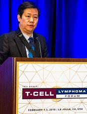
“The response is quite promising and encouraging,” said study investigator Huiqiang Huang, MD, PhD, of Sun Yat-sen University Cancer Center in Guangzhou, China.
“In terms of safety, the toxicity is mild to moderate.”
Dr Huang presented these results at the 10th Annual T-cell Lymphoma Forum.
This investigator-sponsored trial enrolled patients with relapsed/refractory non-Hodgkin lymphoma, but Dr Huang presented results in NKTCL patients only.
There were 15 NKTCL patients, most of whom were male (n=12). Their median age was 41 (range, 17-65). All 15 had an ECOG status of 0 or 1, 9 had stage I/II disease, and 6 had B symptoms.
Nine patients had Epstein-Barr virus (EBV) DNA levels of at least 1000 copy/mL at baseline, and 5 patients had lactate dehydrogenase levels of at least 245 U/L.
The patients had a median of 2 prior systemic therapies (range, 1-3), and 2 patients had undergone a transplant.
Efficacy
Patients received chidamide at 2 doses—10 mg daily or 30 mg twice a week. Dr Huang said both doses were effective against the lymphoma types studied, but the 30 mg twice-weekly dose appeared to be more effective for patients with NKTCL.
Fourteen NKTCL patients were evaluable for efficacy, and the median follow-up was 17.6 weeks (range, 2.6-50).
The overall response rate was 68.2% (6/14), and the CR rate was 28.6% (4/14). The disease control rate was 71.4%, meaning 10 of 14 patients had a CR, partial response (PR), or stable disease (SD).
Dr Huang noted that response was associated with elevated H3 acetylation level.
The median time to response was 5.25 weeks (range, 1.1-6.6).
As for duration of response, the 4 complete responders were still on treatment and in CR at last follow-up, which was 22.7 weeks, 38.1 weeks, 41.3 weeks, and 50 weeks, respectively, from treatment initiation.
Three of the 4 partial responders were still on treatment and in PR at 14.1 weeks, 26.9 weeks, and 32 weeks, respectively. Two patients with SD were still on treatment and in SD at 15.3 weeks and 15.9 weeks, respectively.
Three patients progressed while on treatment and died. A fourth patient died 2.6 weeks after treatment initiation.
Safety
Dr Huang noted that adverse events (AEs) were similar with the 2 dose groups. However, patients who received 30 mg biweekly had a higher incidence of gastrointestinal AEs.
Overall, the most common AEs were hematologic—anemia, thrombocytopenia, etc.—but dose reductions allowed for quick resolution of these AEs, according to Dr Huang.
AEs included:
- Lymphopenia—10 grade 1/2 and 1 grade 3/4
- Anemia—9 grade 1/2 and 3 grade 3/4
- Thrombocytopenia—7 grade 1/2 and grade 3/4
- Leukopenia—7 grade 1/2 and 6 grade 3/4
- Increased alanine aminotransferase—7 grade 1/2
- Neutropenia—6 grade 1/2 and 7 grade 3/4
- Increased aspartate aminotransferase—5 grade 1/2
- Hypoalbuminemia—4 grade 1/2
- Nausea—4 grade 1/2 and 1 grade 3/4
- Vomiting—3 grade 1/2
- Mucositis—2 grade 1/2
- Fatigue—2 grade 1/2
- Epistaxis—2 grade 1/2
- Abdominal distension—1 grade 1/2
- Loss of appetite—1 grade 1/2
- Diarrhea—1 grade 1/2
- Hyperbilirubinemia—1 grade 1/2
- Fever—1 grade 1/2 and grade 3/4
- Pain—1 grade 1/2
- Cough—1 grade 1/2
- Constipation—1 grade 1/2.
Dr Huang said EBV reactivation was not confirmed in this study. An elevated EBV DNA load was only observed in 2 patients with progressive disease. ![]()
LA JOLLA, CA—Results of a phase 2 study suggest chidamide can produce durable responses in patients with relapsed/refractory natural killer/T-cell lymphoma (NKTCL).
The overall response rate was 57.2% in these patients, and the complete response (CR) rate was 28.6%.
Seven of 14 evaluable patients were still receiving chidamide and still in response at last follow-up. For one patient, this was 50 weeks from initiating treatment with chidamide.
“The response is quite promising and encouraging,” said study investigator Huiqiang Huang, MD, PhD, of Sun Yat-sen University Cancer Center in Guangzhou, China.
“In terms of safety, the toxicity is mild to moderate.”
Dr Huang presented these results at the 10th Annual T-cell Lymphoma Forum.
This investigator-sponsored trial enrolled patients with relapsed/refractory non-Hodgkin lymphoma, but Dr Huang presented results in NKTCL patients only.
There were 15 NKTCL patients, most of whom were male (n=12). Their median age was 41 (range, 17-65). All 15 had an ECOG status of 0 or 1, 9 had stage I/II disease, and 6 had B symptoms.
Nine patients had Epstein-Barr virus (EBV) DNA levels of at least 1000 copy/mL at baseline, and 5 patients had lactate dehydrogenase levels of at least 245 U/L.
The patients had a median of 2 prior systemic therapies (range, 1-3), and 2 patients had undergone a transplant.
Efficacy
Patients received chidamide at 2 doses—10 mg daily or 30 mg twice a week. Dr Huang said both doses were effective against the lymphoma types studied, but the 30 mg twice-weekly dose appeared to be more effective for patients with NKTCL.
Fourteen NKTCL patients were evaluable for efficacy, and the median follow-up was 17.6 weeks (range, 2.6-50).
The overall response rate was 68.2% (6/14), and the CR rate was 28.6% (4/14). The disease control rate was 71.4%, meaning 10 of 14 patients had a CR, partial response (PR), or stable disease (SD).
Dr Huang noted that response was associated with elevated H3 acetylation level.
The median time to response was 5.25 weeks (range, 1.1-6.6).
As for duration of response, the 4 complete responders were still on treatment and in CR at last follow-up, which was 22.7 weeks, 38.1 weeks, 41.3 weeks, and 50 weeks, respectively, from treatment initiation.
Three of the 4 partial responders were still on treatment and in PR at 14.1 weeks, 26.9 weeks, and 32 weeks, respectively. Two patients with SD were still on treatment and in SD at 15.3 weeks and 15.9 weeks, respectively.
Three patients progressed while on treatment and died. A fourth patient died 2.6 weeks after treatment initiation.
Safety
Dr Huang noted that adverse events (AEs) were similar with the 2 dose groups. However, patients who received 30 mg biweekly had a higher incidence of gastrointestinal AEs.
Overall, the most common AEs were hematologic—anemia, thrombocytopenia, etc.—but dose reductions allowed for quick resolution of these AEs, according to Dr Huang.
AEs included:
- Lymphopenia—10 grade 1/2 and 1 grade 3/4
- Anemia—9 grade 1/2 and 3 grade 3/4
- Thrombocytopenia—7 grade 1/2 and grade 3/4
- Leukopenia—7 grade 1/2 and 6 grade 3/4
- Increased alanine aminotransferase—7 grade 1/2
- Neutropenia—6 grade 1/2 and 7 grade 3/4
- Increased aspartate aminotransferase—5 grade 1/2
- Hypoalbuminemia—4 grade 1/2
- Nausea—4 grade 1/2 and 1 grade 3/4
- Vomiting—3 grade 1/2
- Mucositis—2 grade 1/2
- Fatigue—2 grade 1/2
- Epistaxis—2 grade 1/2
- Abdominal distension—1 grade 1/2
- Loss of appetite—1 grade 1/2
- Diarrhea—1 grade 1/2
- Hyperbilirubinemia—1 grade 1/2
- Fever—1 grade 1/2 and grade 3/4
- Pain—1 grade 1/2
- Cough—1 grade 1/2
- Constipation—1 grade 1/2.
Dr Huang said EBV reactivation was not confirmed in this study. An elevated EBV DNA load was only observed in 2 patients with progressive disease. ![]()
Disease burden impacts survival, toxicity after CAR T-cell therapy

Adults with relapsed or refractory acute lymphoblastic leukemia (ALL) have better outcomes if they have a low disease burden when receiving chimeric antigen receptor (CAR) T-cell therapy, according to research published in NEJM.
The final analysis of a phase 1 trial showed that patients with a low disease burden at baseline had superior event-free survival (EFS) and overall survival (OS) after therapy, compared to patients with a high disease burden.
Patients with a low disease burden also had a lower rate of cytokine release syndrome (CRS) and neurotoxic events.
“This is the longest follow-up study of people with ALL treated with CAR therapy,” said study author Jae Park, MD, of Memorial Sloan Kettering Cancer Center in New York, New York.
“With the long follow-up, we were able to demonstrate, for the first time, that patients with a lower disease burden benefited the most from CAR therapy, with significantly improved survival and reduced toxicity.”
The study included 53 adults with ALL who had a median age of 44 (range, 23-74).
They were heavily pretreated, with 68% receiving CAR T-cell therapy as a third or later salvage treatment. Thirty-six percent of patients had received an allogeneic transplant, and 23% had primary refractory disease.
In this trial, patients received a single infusion of 19-28z CAR T cells after conditioning chemotherapy. The maximum follow-up time was 5.5 years, with a median follow-up of 29 months.
In all, 83% of patients achieved a complete response. The median EFS was 6.1 months, and the median OS was 12.9 months.
The median EFS was significantly longer for patients with a low disease burden (<5% bone marrow blasts) compared to a high disease burden (≥5% bone marrow blasts or extramedullary disease)—10.6 months and 5.6 months, respectively (P=0.01).
The same was true for the median OS, which was 20.1 months in patients with a low disease burden and 12.4 months in those with a high disease burden (P=0.02).
For the entire study population, the rate of CRS was 85%, and 26% of patients had severe CRS. One patient died of severe CRS and multi-organ failure before the researchers began modifying the dose of CAR T cells according to the pretreatment disease burden.
Severe CRS occurred in 41% of patients with a high disease burden and 5% of those with a low disease burden.
In the entire study population, 2% of patients had grade 2 neurotoxic effects, 36% had grade 3, 6% had grade 4, and none had grade 5. The rate of severe neurotoxicity was 42%.
Neurotoxic effects occurred in 59% of patients with a high disease burden and 14% of those with a low disease burden.
“Among all of the clinical and disease factors we examined, pretreatment disease burden was the strongest predictor of long-term outcome after CAR therapy,” Dr Park said. “Our data supports the incorporation of CAR therapy in an earlier treatment setting in ALL, when the disease volume is small, so as to achieve the greatest long-term efficacy and lowest toxicity.”
This work was supported by Juno Therapeutics, the National Institutes of Health, the Carson Family Charitable Trust, the Emerald Foundation, the Mr. and Mrs. Goodwyn Commonwealth Fund, the Terry Fox Run for Cancer Research organized by the Canadian Association of New York, Kate’s Team, William Laurence and Blanche Hughes Foundation, the Center for Experimental Therapeutics at Memorial Sloan Kettering, and the Lake Road Foundation. ![]()