User login
Is patient suicide in psychiatry a medical error?
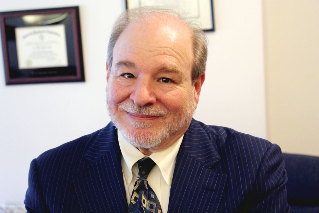
When Rodney Vivian, MD, a psychiatrist in Cincinnati, was sued for medical malpractice after a psychiatric inpatient died by suicide, he recalls being naive about the process and how difficult it would be. “I was thinking that truth and common sense would prevail. How stupid I was,” he said.

Although Dr. Vivian, who was at the time the medical director of a hospital psychiatric unit in Ohio, was found not liable in two appeals, the legal process dragged on for 6 years, creating an emotional roller coaster of sadness, fear, vulnerability, and anxiety.
“The lawsuit took a big chunk out of me, and there was a sense of unfairness. It was incredibly humiliating and destructive; and it did not make me a better person or psychiatrist,” Dr. Vivian said.
Dr. Vivian is just one of the many psychiatrists who have had their world turned upside down after a patient suicide. When such events occur, grief-stricken families often point the finger at the treating psychiatrist. Although lawsuits are rare in psychiatry, patient suicide can lead to a myriad of emotional, legal, and career consequences.
Tyler Black, MD, child and adolescent psychiatrist and assistant clinical professor at the University of British Columbia, Vancouver, likens patient suicide to “a nuclear bomb” but emphasizes the importance of not classifying such events as a medical error or assigning blame.
“Starting with the assumption that suicide is always avoidable is not evidence based,” Dr. Black said.
Although patient suicide can occur across medicine, the odds are alarmingly high in psychiatry.
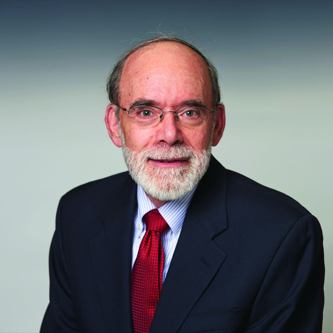
“There’s at least a 50-50 chance that a psychiatrist is going to face the suicide of a patient,” said Eric Plakun, MD, medical director/CEO at the Austen Riggs Center, Stockbridge, Mass. a hospital-based facility that offers a continuum of psychiatric treatment. Quoting forensic psychiatrist Robert Simon, Dr. Plakun said: “There are two kinds of psychiatrists – those who have had a patient die by suicide, and those who will.”
Research from 2015 shows that, among specialists, psychiatrists are among the least likely to be sued. A 2007/2008 Physician Survey from the American Medical Association showed that 22.2% of psychiatrists had been sued for malpractice; the probability that they would face a claim each year was only 2.6%. However, failure to prevent suicide is one of the top reasons for lawsuits.
One report from 2008 suggests that 20%-68% of psychiatrists will lose a patient to suicide. A report cowritten by Dr. Plakun in 2005 notes that about one in six psychiatric interns and one in three psychiatric residents will experience a patient suicide some time during their training. The authors added that 50% of all psychiatrists will have a patient die by suicide during their career. That risk stays at about 50% for future patients even after a clinician experiences the death of a previous patient.
Although mental health professionals prevail in up to 80% of suicide-related malpractice cases, such events are still emotionally devastating for everyone involved.
When a patient dies by suicide, it is a huge event, he noted. “It fuels a lot of fear and a lot of guilt, worry, and sadness.”
Paul S. Appelbaum, MD, past president of the American Psychiatric Association and Dollard Professor of Psychiatry, Medicine, and Law at Columbia University, New York, noted that patient suicide will happen.
The problem with many administrators who talk about a target of “zero suicide” is that when suicide does occur, it can lead to the erroneous conclusion that someone is to blame, said Dr. Appelbaum, who is also director of the Center for Law, Ethics, and Psychiatry at Columbia University. “That’s not necessarily true and contributes to finger-pointing.”
Stopping the blame game
Dr. Black’s first experience as a psychiatry resident was arriving at the hospital and finding the body of a patient who had died by suicide by hanging. Although he did not know the patient, Dr. Black said he had a strong emotional response that was coupled with an intense and sometimes confusing reaction by the hospital administration, including what he called “nonsensical banning” of pencils on the ward.

Dr. Black is now the medical director of emergency psychiatry at BC Children’s Hospital in Vancouver and specializes in suicidology and emergency/crisis youth mental health care. He said during a recent live chat on Twitter that he does not predict suicide but instead “assesses risk,” meaning he examines potential risk factors in his patients.
“If systems and administrators (and consulting doctors) could recognize this, the ‘blame game’ would severely decrease. From the advocacy end, we have to stop seeing suicide as a ‘medical error,’ ” Dr. Black tweeted.
“There’s a strong administrative push, especially in the face of suicide, to dive into the [occurrence] as if it must be that an error was made,” he said in an interview.
To help counteract any potential finger-pointing, Dr. Black created a free-to-download patient risk assessment document called the Assessment of Suicide and Risk Inventory (ASARI) for use at every patient visit.
“ASARI was designed to walk an assessor through their thinking process such that they can put all of their thoughts down on one piece of paper. It makes it a better communication document, and it’s definitely better medicolegal documentation,” he said.
Dr. Appelbaum noted that, although having documentation is beneficial, “I don’t think that you necessarily need to separate actions that are ‘protective’ from actions that are intended to help a patient.”
However, he pointed out that, if a psychiatrist conforms to or exceeds the standard of care, including conducting appropriate suicide risk assessments, developing an appropriate treatment plan, and keeping comprehensive documentation, these measures “should provide an effective defense to claims of malpractice or negligence.”
‘Horrendous event’
Dr. Vivian said that, during his 40-year career in psychiatry, there have been about 12 “office patients” who died by suicide. However, nothing prepared him for the fallout from a lawsuit.
In 2010, a patient who had overdosed was transferred to the psychiatric unit of Mercy Health–Clermont (Ohio) Hospital, where Dr. Vivian was the admitting physician. Although the hospital staff was ordered to check on her every 15 minutes, her husband found her unconscious from a hanging attempt when he came to visit the next evening. After she was transferred to the ICU, she was taken off life support and died a few days later.
“It was a horrendous event,” Dr. Vivian said.
The family sued the hospital, and the matter was settled out of court without Dr. Vivian’s knowledge. The family also filed a separate lawsuit against Dr. Vivian, which went to trial 3 years later.
“My insurance company’s claims person was very supportive and wanted me to not settle. She agreed that I didn’t do anything wrong and that I needed to face this,” he added.
In the first trial, a jury found Dr. Vivian not liable. Six months later, the plaintiff’s attorney filed an appeal. A year after the first trial, the court of appeals also came back with a new ruling in his favor and, in a subsequent appeal, the Ohio Supreme Court also ruled in his favor.
Dr. Vivian noted that there really are no winners in these situations. “Even though the jury ruled in my favor, there was never a sense of ‘success.’ I could never feel good about what happened.” He was told the insurance company spent more than $300,000 on his defense.
Although he no longer performs psychiatric inpatient admissions, Dr. Vivian continues to work in private practice and provides psychiatric consultation to patients at a local medical center.
“I consider my work as a blessing in my life, and I continue to learn from my patients,” he said.
‘Will I be sued?’
Dr. Appelbaum noted there is a difference between a malpractice claim that may be filed and a “payout” to plaintiffs because of a negotiated resolution of a case or an award that is made at trial.
Malpractice insurers may raise the rates of a physician who has been found at fault in one or more legal actions in which financial settlements have been paid out, he said.
The issue in any malpractice case is whether the psychiatrist met the standard of care, which is traditionally defined as “skill and learning that is ordinarily possessed and exercised by members of that profession in good standing.”
“No physician is expected to be the guarantor of a good outcome of a case. Sometimes things go wrong. Merely because there’s a bad outcome, merely because a suicide has occurred, doesn’t mean that the psychiatrist was negligent,” Dr. Appelbaum said.
He believes all large centers should have a “clear-cut plan” in place to assist clinicians in the event of a patient suicide. Such plans should help in dealing with stress from losing a patient and should provide guidance about how to handle any potential lawsuit.
For those worried that a patient’s suicide will shadow them through their career, Dr. Appelbaum said that it can happen, especially in cases involving a financial settlement against the clinician.
Such cases must be reported to the national practitioner data bank, where they can be accessed by any licensing body in any state when physicians apply for a medical license.
In addition, Dr. Appelbaum pointed out that licensure, medical staff, and malpractice applications typically require disclosure of a history of successful or unsuccessful claims filed against a physician. Although that may be limited to the past 10 years, the requirement can go on indefinitely.
Beware how you share
Dr. Plakun noted that there is a sense of isolation for a clinician in cases of patient suicide and that physicians often turn inward. He added that, although it is important to talk with others, in institutions, this is best done in a “peer-review, protected space” – and perhaps with a lawyer present.
However, Dr. Appelbaum warned that sharing information, even in this type of setting, may not offer legal protection. Talking to others in order to get some emotional support is permitted once the statute of limitations for filing a claim has lapsed or if a claim has been closed.
Discussing a case of patient suicide with peers prior to that can have serious legal implications, he added. Colleagues can be called to testify in any resulting legal case and disclose what was said during such conversations.
“The typical advice that a risk manager, a claims manager, or an attorney would give to a clinician is, don’t talk to other people about it other than the lawyer or claims manager who’s dealing with the case,” he noted.
That said, there are three general exceptions to this rule. These include attorney-client privilege, any matters discussed with the physician’s own therapist, and, “depending on the state, there are varying protections for what’s considered ‘peer review.’ ”
For instance, when hospitals implement a formal review process after an event, what is said during discovery may be protected. However, not all states have such protection. That’s why it is important to understand what the law is in your particular state, said Dr. Appelbaum.
Support for psychiatrists
Kaz J. Nelson, MD, psychiatrist and associate professor at the University of Minnesota, Minneapolis, also works with high-risk populations, including those with acute suicidality and self-injury.
During a recent chat on patient suicide, Dr. Nelson tweeted: “Sadly in our field, suicide is not an IF but a WHEN. Don’t keep the inevitable shame and sadness to yourself.”
Dr. Nelson agreed with Dr. Black that it’s important to look into these occurrences as a quality improvement measure, but not as a way to assign blame. Preparing for potential patient loss “and having very solid, very supportive, very inclusive ‘postvention’ procedures” is critical, she noted. “When you don’t have these policies and procedures in place and have them very transparent, it creates a culture of silence around the issue.”
Dr. Plakun reiterated the importance of not staying silent. “We can’t simply surrender to the idea of not talking about patient suicide. We have to find a way to speak.”
A version of this story originally appeared on Medscape.com.
When Rodney Vivian, MD, a psychiatrist in Cincinnati, was sued for medical malpractice after a psychiatric inpatient died by suicide, he recalls being naive about the process and how difficult it would be. “I was thinking that truth and common sense would prevail. How stupid I was,” he said.
Although Dr. Vivian, who was at the time the medical director of a hospital psychiatric unit in Ohio, was found not liable in two appeals, the legal process dragged on for 6 years, creating an emotional roller coaster of sadness, fear, vulnerability, and anxiety.
“The lawsuit took a big chunk out of me, and there was a sense of unfairness. It was incredibly humiliating and destructive; and it did not make me a better person or psychiatrist,” Dr. Vivian said.
Dr. Vivian is just one of the many psychiatrists who have had their world turned upside down after a patient suicide. When such events occur, grief-stricken families often point the finger at the treating psychiatrist. Although lawsuits are rare in psychiatry, patient suicide can lead to a myriad of emotional, legal, and career consequences.
Tyler Black, MD, child and adolescent psychiatrist and assistant clinical professor at the University of British Columbia, Vancouver, likens patient suicide to “a nuclear bomb” but emphasizes the importance of not classifying such events as a medical error or assigning blame.
“Starting with the assumption that suicide is always avoidable is not evidence based,” Dr. Black said.
Although patient suicide can occur across medicine, the odds are alarmingly high in psychiatry.
“There’s at least a 50-50 chance that a psychiatrist is going to face the suicide of a patient,” said Eric Plakun, MD, medical director/CEO at the Austen Riggs Center, Stockbridge, Mass. a hospital-based facility that offers a continuum of psychiatric treatment. Quoting forensic psychiatrist Robert Simon, Dr. Plakun said: “There are two kinds of psychiatrists – those who have had a patient die by suicide, and those who will.”
Research from 2015 shows that, among specialists, psychiatrists are among the least likely to be sued. A 2007/2008 Physician Survey from the American Medical Association showed that 22.2% of psychiatrists had been sued for malpractice; the probability that they would face a claim each year was only 2.6%. However, failure to prevent suicide is one of the top reasons for lawsuits.
One report from 2008 suggests that 20%-68% of psychiatrists will lose a patient to suicide. A report cowritten by Dr. Plakun in 2005 notes that about one in six psychiatric interns and one in three psychiatric residents will experience a patient suicide some time during their training. The authors added that 50% of all psychiatrists will have a patient die by suicide during their career. That risk stays at about 50% for future patients even after a clinician experiences the death of a previous patient.
Although mental health professionals prevail in up to 80% of suicide-related malpractice cases, such events are still emotionally devastating for everyone involved.
When a patient dies by suicide, it is a huge event, he noted. “It fuels a lot of fear and a lot of guilt, worry, and sadness.”
Paul S. Appelbaum, MD, past president of the American Psychiatric Association and Dollard Professor of Psychiatry, Medicine, and Law at Columbia University, New York, noted that patient suicide will happen.
The problem with many administrators who talk about a target of “zero suicide” is that when suicide does occur, it can lead to the erroneous conclusion that someone is to blame, said Dr. Appelbaum, who is also director of the Center for Law, Ethics, and Psychiatry at Columbia University. “That’s not necessarily true and contributes to finger-pointing.”
Stopping the blame game
Dr. Black’s first experience as a psychiatry resident was arriving at the hospital and finding the body of a patient who had died by suicide by hanging. Although he did not know the patient, Dr. Black said he had a strong emotional response that was coupled with an intense and sometimes confusing reaction by the hospital administration, including what he called “nonsensical banning” of pencils on the ward.
Dr. Black is now the medical director of emergency psychiatry at BC Children’s Hospital in Vancouver and specializes in suicidology and emergency/crisis youth mental health care. He said during a recent live chat on Twitter that he does not predict suicide but instead “assesses risk,” meaning he examines potential risk factors in his patients.
“If systems and administrators (and consulting doctors) could recognize this, the ‘blame game’ would severely decrease. From the advocacy end, we have to stop seeing suicide as a ‘medical error,’ ” Dr. Black tweeted.
“There’s a strong administrative push, especially in the face of suicide, to dive into the [occurrence] as if it must be that an error was made,” he said in an interview.
To help counteract any potential finger-pointing, Dr. Black created a free-to-download patient risk assessment document called the Assessment of Suicide and Risk Inventory (ASARI) for use at every patient visit.
“ASARI was designed to walk an assessor through their thinking process such that they can put all of their thoughts down on one piece of paper. It makes it a better communication document, and it’s definitely better medicolegal documentation,” he said.
Dr. Appelbaum noted that, although having documentation is beneficial, “I don’t think that you necessarily need to separate actions that are ‘protective’ from actions that are intended to help a patient.”
However, he pointed out that, if a psychiatrist conforms to or exceeds the standard of care, including conducting appropriate suicide risk assessments, developing an appropriate treatment plan, and keeping comprehensive documentation, these measures “should provide an effective defense to claims of malpractice or negligence.”
‘Horrendous event’
Dr. Vivian said that, during his 40-year career in psychiatry, there have been about 12 “office patients” who died by suicide. However, nothing prepared him for the fallout from a lawsuit.
In 2010, a patient who had overdosed was transferred to the psychiatric unit of Mercy Health–Clermont (Ohio) Hospital, where Dr. Vivian was the admitting physician. Although the hospital staff was ordered to check on her every 15 minutes, her husband found her unconscious from a hanging attempt when he came to visit the next evening. After she was transferred to the ICU, she was taken off life support and died a few days later.
“It was a horrendous event,” Dr. Vivian said.
The family sued the hospital, and the matter was settled out of court without Dr. Vivian’s knowledge. The family also filed a separate lawsuit against Dr. Vivian, which went to trial 3 years later.
“My insurance company’s claims person was very supportive and wanted me to not settle. She agreed that I didn’t do anything wrong and that I needed to face this,” he added.
In the first trial, a jury found Dr. Vivian not liable. Six months later, the plaintiff’s attorney filed an appeal. A year after the first trial, the court of appeals also came back with a new ruling in his favor and, in a subsequent appeal, the Ohio Supreme Court also ruled in his favor.
Dr. Vivian noted that there really are no winners in these situations. “Even though the jury ruled in my favor, there was never a sense of ‘success.’ I could never feel good about what happened.” He was told the insurance company spent more than $300,000 on his defense.
Although he no longer performs psychiatric inpatient admissions, Dr. Vivian continues to work in private practice and provides psychiatric consultation to patients at a local medical center.
“I consider my work as a blessing in my life, and I continue to learn from my patients,” he said.
‘Will I be sued?’
Dr. Appelbaum noted there is a difference between a malpractice claim that may be filed and a “payout” to plaintiffs because of a negotiated resolution of a case or an award that is made at trial.
Malpractice insurers may raise the rates of a physician who has been found at fault in one or more legal actions in which financial settlements have been paid out, he said.
The issue in any malpractice case is whether the psychiatrist met the standard of care, which is traditionally defined as “skill and learning that is ordinarily possessed and exercised by members of that profession in good standing.”
“No physician is expected to be the guarantor of a good outcome of a case. Sometimes things go wrong. Merely because there’s a bad outcome, merely because a suicide has occurred, doesn’t mean that the psychiatrist was negligent,” Dr. Appelbaum said.
He believes all large centers should have a “clear-cut plan” in place to assist clinicians in the event of a patient suicide. Such plans should help in dealing with stress from losing a patient and should provide guidance about how to handle any potential lawsuit.
For those worried that a patient’s suicide will shadow them through their career, Dr. Appelbaum said that it can happen, especially in cases involving a financial settlement against the clinician.
Such cases must be reported to the national practitioner data bank, where they can be accessed by any licensing body in any state when physicians apply for a medical license.
In addition, Dr. Appelbaum pointed out that licensure, medical staff, and malpractice applications typically require disclosure of a history of successful or unsuccessful claims filed against a physician. Although that may be limited to the past 10 years, the requirement can go on indefinitely.
Beware how you share
Dr. Plakun noted that there is a sense of isolation for a clinician in cases of patient suicide and that physicians often turn inward. He added that, although it is important to talk with others, in institutions, this is best done in a “peer-review, protected space” – and perhaps with a lawyer present.
However, Dr. Appelbaum warned that sharing information, even in this type of setting, may not offer legal protection. Talking to others in order to get some emotional support is permitted once the statute of limitations for filing a claim has lapsed or if a claim has been closed.
Discussing a case of patient suicide with peers prior to that can have serious legal implications, he added. Colleagues can be called to testify in any resulting legal case and disclose what was said during such conversations.
“The typical advice that a risk manager, a claims manager, or an attorney would give to a clinician is, don’t talk to other people about it other than the lawyer or claims manager who’s dealing with the case,” he noted.
That said, there are three general exceptions to this rule. These include attorney-client privilege, any matters discussed with the physician’s own therapist, and, “depending on the state, there are varying protections for what’s considered ‘peer review.’ ”
For instance, when hospitals implement a formal review process after an event, what is said during discovery may be protected. However, not all states have such protection. That’s why it is important to understand what the law is in your particular state, said Dr. Appelbaum.
Support for psychiatrists
Kaz J. Nelson, MD, psychiatrist and associate professor at the University of Minnesota, Minneapolis, also works with high-risk populations, including those with acute suicidality and self-injury.
During a recent chat on patient suicide, Dr. Nelson tweeted: “Sadly in our field, suicide is not an IF but a WHEN. Don’t keep the inevitable shame and sadness to yourself.”
Dr. Nelson agreed with Dr. Black that it’s important to look into these occurrences as a quality improvement measure, but not as a way to assign blame. Preparing for potential patient loss “and having very solid, very supportive, very inclusive ‘postvention’ procedures” is critical, she noted. “When you don’t have these policies and procedures in place and have them very transparent, it creates a culture of silence around the issue.”
Dr. Plakun reiterated the importance of not staying silent. “We can’t simply surrender to the idea of not talking about patient suicide. We have to find a way to speak.”
A version of this story originally appeared on Medscape.com.
How VA Nurses are Coping With the COVID-19 Pandemic
The tsunami we call COVID-19 has threatened to overwhelm everything in its path, with devastating effects that can be hard to quantify or qualify. But the Nurses Organization of Veterans Affairs (NOVA) has taken on the fight. Earlier this year, NOVA surveyed its members to learn how nurses felt the US Department of Veterans Affairs (VA) was managing personal protective equipment (PPE), testing, communications, staffing needs, and other issues. Following the survey NOVA conducted in-depth interviews with nurses at the VA Boston Healthcare System to better understand how the pandemic was affecting nurses across the Veterans Health Administration (VHA).
The first survey, which was conducted in March and April 2020, included the following questions:
- Do you feel prepared for COVID-19?
- Do you feel your facility’s supply of PPE is adequate?
- Are you aware of the protocol for the distribution of equipment/supplies at your facility?
- Are staff being tested for the virus, and if so, when, and are there enough test kits available for staff and patients?
- Do you believe your facility is properly handling staff who have been exposed to COVID-19 but are asymptomatic?
With the May survey, NOVA aimed to get an update on how things were progressing. This survey asked how the pandemic was affecting members personally, including questions such as: Do you feel you are being supported—mentally and physically? Has VA offered to provide staff mental health counselors or others to help mitigate stress during the crisis?
Results revealed inconsistencies and some confusion. For example, communication among leadership, staff, and veterans continued to change rapidly, causing some misunderstanding overall, respondents said. Some pointed to “e-mail overload” and weekly updates that didn’t work. Others felt communication was “reactive” and “bare minimum”—not “proactive and informative.”
Most (74%) respondents felt that access to PPE was inadequate, and many did not know the protocol for distribution or what supplies were on hand, while 47% felt ill prepared for any COVID-19 onslaught. Although things had improved somewhat by May, > 85% of the respondents said they were reusing what was provided daily and were still finding it difficult to get PPE when needed.
According to respondents, testing was pretty much nonexistent. When asked whether staff were being tested and whether tests for both staff and patients were available, the answer was a resounding No (80%). Nurses’ comments ranged from “staff are not being tested, even if they have been exposed,” to “there are not enough tests for patients, let alone for frontline staff.”
The lack of tests compounded stress. Helen Motroni, ADN, RN, spinal cord injury staff nurse, said, “I have been tested twice for direct exposure to the coronavirus in the past 3 weeks. Luckily, the results were negative, but waiting for the test results was extremely stressful because I have 2 little boys at home. While waiting for results, I self-quarantined and was terrified that I possibly brought the virus home to my children. I never left my room and would talk with my boys through the door and FaceTime. My 8-year-old asked me why I couldn’t stay home so I would not get sick. I explained to him that if every nurse did that, there would be nobody to help those that are sick and suffering.”
As summer approached, testing throughout most of the states remained scarce and contact tracing was a struggle at many facilities. It seemed that, even if testing was available, only those at high risk would be tested.
Moreover, quarantine and sick leave for those who were COVID positive remained a concern, as numbers of those exposed increased. It was widely known and reported that staffing levels within the VHA were inadequate prior to the months going into the pandemic; survey respondents wondered how they would handle vacancies and take care of outside patients as part of the Fourth Mission if staff got sick. One COVID-compelled solution (which NOVA has advocated for years) included expedited hiring practices. Timely application and quicker onboarding enabled VA to hire within weeks rather than months. Since March, VA has hired > 20,000 new employees.
Multiplied Multitasking
The crisis, along with potential and actual staff shortages, has meant that many nurses have been doing double and triple duty—at the least. Danielle Newman, MSN, RN, clinical resource nurse, specialty and outpatient clinics, said, “Throughout the COVID crisis, I have been a direct care provider, an educator, a member of the float pool, an infection preventionist, a colleague, a therapist, and part of a support system to many.… As a PPE nurse educator, I visit every floor in the hospital and help educate staff on how to properly don and doff PPE, as well as monitor the doffing area and assist staff through the doffing process. I have also been involved in obtaining nasal swabs on veterans as well as staff in order to isolate and slow the progression of the virus.”
Some nurses were deployed, some redeployed, and some volunteered for the COVID front. James Murphy, BSN, RN, an emergency department nurse, answered the call. “During the first few weeks of the outbreak and with all its uncertainty, I volunteered to be a COVID nurse.… Seeing how the pandemic has changed practice was an experience.”
Peter Russo, MSN, RN, an OR assistant nurse manager, also found new experience. When the pandemic began to take hold, he was reassigned to Urgent Care and then to the Incident Management Team (IMT). “I asked my associate chief nurse, ‘What is the IMT?’ After my first 2 days on this team, my head was spinning with loads of information, trying to understand and grasp my role as the IMT Nursing Clinical Lead.”
After the third day, his preceptor was redeployed back to the endoscopy department. “I quickly realized my main role as a clinical leader on this team,” Russo says, “was to support and troubleshoot any and all nursing problems, concerns and or questions related to the COVID-19 pandemic among the 3 campuses. One of the more complex challenges was to identify the nursing burn rate of our PPE, especially the N95 masks in all of our COVID units.”
The new experience benefited, him, though. “When changes in policy or procedure happen during the course of an hour, it was one of my responsibilities to notify the nursing staff across the board. I now feel much more confident and competent offering suggestions [about] the many issues that arise.”
Because of the pandemic, all elective surgeries were cancelled and the ambulatory surgery unit where perioperative nurse Valentina Ward, BSN, RN, MPS, CPPS, worked was closed. “As a result,” she says, “I received an accelerated course and was reassigned where I was needed the most. … Serving veterans during the global crisis means so much to me. Despite all that is going on, and the challenges that exist, I have never been prouder to be a frontline nurse.”
The Importance of Maintaining ‘Normal’
COVID-19 seems to subsume everything around it, but there are still health care needs that have nothing to do with the virus. What happens to those patients and their care?
Melissa Varela-Manso, MSN, RN, CRRN, is an inpatient nurse case manager who works with a dedicated team to coordinate admissions and discharges for veterans on the acute Spinal Cord Injury (SCI) unit. “During this pandemic,” she says, “our veterans continue to have non-COVID medical conditions that warrant attention and hospitalization. Therefore, we are following the SCI veterans with non-COVID health issues on the inpatient unit. We also follow our SCI veterans on other units who are being ruled out for COVID and those who have tested positive. We are working diligently to guarantee each veteran gets the quality care they need and deserve while following the evolving guidelines.”
One of the ways VA health care teams have kept things stable for patients, while also ensuring their safety, is through telehealth. VA introduced telehealth in 2003 and is now the nation’s largest telehealth provider. In 2019, > 900,000 veterans received care through telehealth. But it took something like COVID-19 to really show the strengths and utility of being able to care for patients electronically. At the beginning of March, VA conducted about 2,500 telehealth video sessions daily. By June, that had jumped to nearly 25,000—a 1,000% increase.
Sherry Clement, BSN, RN, CFCN, CWCN, CCCN, is the VA Coordinated Transitional Care (C-TraC) Program case manager assigned to the COVID Outpatient Call Team, managing COVID-positive outpatients via telephone and VA video connect. “Working with the call team has been a great collaboration between primary care, telehealth, palliative care, infection control, and occupational health,” she says. “It is amazing to see how fast and efficient our team came together to create contact protocols, consults, notes, and a working plan to organize and oversee the needs of our COVID positive veterans."
Increased usage of telehealth also has meant community-based outpatient clinics continue to be operational, says Karen Boenig, MSN, APRN, ANP-BC, a primary care nurse manager. “Nurses and providers are conducting routine and urgent visits via VA Video Connect and telephone.… At the Framingham clinic, I am working with the Tele-Flu Clinic, which assesses veterans virtually who have symptoms of COVID-19. By utilizing virtual care visits, we can continue providing health care services to our veterans, which minimizes patient and provider exposure and supports social distancing.”
Florence Would be Proud
It seems fitting that 2020 is also the International Year of the Nurse, in celebration of Florence Nightingale’s 200th birthday. That passionate reformer would appreciate NOVA’s mission: “Shaping and Influencing healthcare in the Department of Veterans Affairs.”
In a NOVA blog post (www.vanurse.org), Teresa Morris, director of advocacy and government relations, says, “Thanks to those who responded to our 2 COVID-19 surveys, we were able to use the information as a firsthand look into how nurses felt about VA’s work and response during the Pandemic. The testimony touched on PPE, communication, testing, and comments and concerns that you had during the height of the virus.” She added, “I think some areas that spiked early had less time to prepare while others (MidWest/West) spiked later and used lessons learned.”
“We discussed the findings from the survey and thanked Congress for providing the $19.6 billion in emergency supplemental funding for VA. The funds helped to hire new staff, provide overtime pay, and purchase supplies needed for those caring for veterans and other COVID patients,” She continued.
In May, VA Secretary Robert Wilkie announced his plan to phase in reopening various VA facilities. Respondents were concerned about how the opening up of VA facilities would go as cases continue to spike around the country. They remain committed to their patients, however.
“Never was there a time in the history of nursing that I can say we are needed more,” says Cecilia McVey, MHA, RN, FAAN, Associate Director for Nursing and Patient Care Services, “and nurses make a difference. As you do every day, you are sacrificing more than you may have anticipated when you signed on to be a nurse. Never has it been more important to believe in yourself, in our profession, in the scientists that are guiding us and in ourselves. What you are doing is critical and your sacrifices have not gone unnoticed. I am thankful for all you do—every day—now, and in the future.”
Cancer researchers cross over to COVID-19 clinical trials
When the first reports emerged of “cytokine storm” in patients with severe COVID-19, all eyes turned to cancer research. Oncologists have years of experience reigning in “cytokine release syndrome” (CRS) in patients treated with chimeric antigen receptor (CAR) therapies for advanced blood cancers.
There was hope that drugs used to quell CRS in patients with cancer would be effective in patients with severe COVID. But the promise of a quick fix with oncology medications has yet to be fully realized.
Part of the problem is that the two conditions, while analogous, are “not the same,” said Nirali Shah, MD, head of the hematologic malignancies section in the pediatric oncology branch at the National Cancer Institute.
“You have to understand the underlying pathophysiology, what triggers the inflammation,” Dr. Shah said.
CAR T–related CRS is caused by activated T cells in patients with cancer who often do not have an infection, she explained. In contrast, cytokine storm in COVID-19 is triggered by a viral pathogen that can drive “out of control” inflammation. These differences may explain why drugs work in the first instance, but not in the second, she added. Drugs that inhibit interleukin-6 (such as tocilizumab, sarilumab, and siltuximab) are used with great success to dampen down the CRS in patients receiving CAR therapy for blood cancers. And although trials of these agents in patients with COVID are still ongoing, initial results are disappointing.
The first global, phase 3 randomized controlled trial of tocilizumab in severe COVID-19 failed to meet its primary endpoint of improved clinical status, and it did not meet its secondary endpoint of improved mortality at week 4.
In its recent recommendations, the National Institutes of Health noted a lack of data to support the efficacy of IL-6 inhibitors in COVID-19, and recommended against their use, except as part of a clinical trial.
Trimming the tree vs. cutting it down
As researchers have begun to decode the immune process underlying severe COVID-19, they have turned to other cancer drugs to tame cytokine storm.
Louis Staudt, MD, PhD, and Wyndham Wilson, MD, PhD, both at the NCI, think that cytokine storm in COVID-19 is driven by macrophages, which trigger release of multiple cytokines.
For years, the pair have been studying lymphoid tumors. Dr. Staudt is chief of the lymphoid malignancies branch at the NCI, and Wilson is head of the lymphoma therapeutics section. In past work, Dr. Staudt discovered that inhibiting an enzyme called bruton tyrosine kinase (BTK) dampens macrophage function.
When the pandemic began, Dr. Staudt and Dr. Wilson realized that singling out just one cytokine like IL-6 may not be enough. They thought that a more effective approach may be to target macrophages with a BTK inhibitor called acalabrutinib (Calquence), which would inhibit multiple cytokines at the same time.
Dr. Staudt likens the immune response to a tree, with the macrophages composing the tree trunk and the limbs made up of individual cytokines.
“Targeting macrophages is getting at the trunk of the problem,” he said. “You’re only cutting off the limbs with tocilizumab.”
In just 3 days, Dr. Staudt and Dr. Wilson went from concept to approval to launching a prospective, observational study. The study took place at five centers in the US, and included 19 patients hospitalized with COVID-19; the results were published in Science Immunology. Over a treatment course of 14 days, the majority of patients treated off-label with acalabrutinib improved, some within 24 hours. Eight of 11 patients on supplemental oxygen were discharged on room air. Four of eight patients on ventilators were extubated, with two of these discharged on room air. Two patients on ventilators died. No discernible toxicity was noted.
Analyses also showed increased BTK activity and elevated IL-6 levels in monocytes – precursors of macrophages – in patients with severe COVID-19, compared with healthy volunteers.
“We showed that the target of acalabrutinib was active in the immune cells of patients with severe COVID-19,” Dr. Staudt said. “So we have the target. We have the drug to hit the target. And we have an apparent clinical benefit.”
Those three things were compelling enough to launch the CALAVI phase 2 trial, an open-label, randomized, controlled trial, sponsored by AstraZeneca and the NCI, that is being conducted in the United States and internationally. It is testing acalabrutinib with best supportive care versus BSC alone in people hospitalized with COVID-19. The trial is scheduled to be completed on Nov. 26.
Preliminary insights from this trial are expected soon. “These are not insights that we will likely publish, but they are important insights that will lead to the launch of a definitive double-blind, randomized, phase 3 trial, which we hope to launch in the next month or so,” Dr. Wilson said.
Targeting inflammation and infection simultaneously
Other scientists are investigating inhibitors of Janus kinase (JAK), a family of enzymes that play a key role in orchestrating immune responses, particularly cytokines. Interest in JAK inhibition to control hyperinflammation in cancer goes back at least 15 years, and drugs that act as JAK inhibitors are already approved for use in the treatment of myelofibrosis (ruxolitinib [Jakafi], fedratinib [Inrebic]) and also for rheumatoid arthritis (upadacitinib [Rinvoq], baricitinib [Olumiant]).
“It wasn’t a huge leap for those of us with a lot of understanding of JAK inhibitors to propose taking them into the clinic to treat patients with COVID-19,” commented John Mascarenhas, MD, the leader of clinical investigation in the myeloproliferative disorders program at the Icahn School of Medicine at Mount Sinai, New York.
Dr. Mascarenhas is also principal investigator of the PRE-VENT trial, which is comparing the investigational JAK2 inhibitor pacritinib plus standard of care to standard of care alone in patients hospitalized with severe COVID-19, with and without cancer. The trial is sponsored by CTI BioPharma (manufacturer of pacritinib), and is taking place at 10 sites in the United States.
In a move that may raise eyebrows, PRE-VENT skipped phase 1 and 2 and went straight to phase 3. Pacritinib has yet to receive FDA approval and has mostly been studied in myelofibrosis, an intensely inflammatory disease.
The decision was based on trials of pacritinib in hematologic malignancies and also on results from a phase 2 study in China that found possible clinical benefit for the JAK 1/2 inhibitor ruxolitinib in 43 patients hospitalized with severe COVID-19, although results were not statistically significant, Dr. Mascarenhas explained.
Recent results from Lilly’s ACTT-2 study have provided further support for the role of JAK inhibitors in treating cytokine storm. ACTT-2 is a phase 3, double-blind, placebo-controlled, randomized, controlled trial sponsored by the NIH and NIAID comparing the JAK 1/2 inhibitor baricitinib plus the antiviral remdesivir with remdesivir alone in patients hospitalized with COVID-19. In September, Lilly announced that the trial met its primary endpoint of decreased time to recovery in patients who received baricitinib in combination with remdesivir.
But pacritinib’s mechanism of action may take things a step further. The drug selectively inhibits JAK2 and spares JAK1, which is important for antiviral activity in the immune system. Also, in vitro data suggests pacritinib may simultaneously reduce inflammation and fight off the virus by selectively inhibiting two additional enzymes and two other receptors.
“The rationale to me is very strong for using pacritinib,” Dr. Mascarenhas said. “I think this approach was bold but appropriate.”
The main safety concern with pacritinib could be bleeding, especially among patients on anticoagulants, Dr. Mascarenhas said. Because some patients with severe COVID-19 tend to develop blood clots, anticoagulation has become the standard of care at many institutions.
Because the trial is just beginning – only a minority of the total planned population of 358 patients has been enrolled – no interim results are available.
Right drug, wrong time?
IL-6 inhibition could still have a role to play in COVID-19, but the trick could be in the timing. Most of the trials so far have studied tocilizumab in patients with severe COVID-19, many of whom were already on ventilators. At that point, it may be too late to reverse the damage that has already taken place.
One of the main reasons tocilizumab works so well in CRS after CAR T therapy is that oncologists have learned how to use it early, often within 24 hours of fever onset. Oncologists use the American Society for Transplantation and Cellular Therapy consensus grading system, which helps them identify CRS when it is easier to control.
But applying the ASTCT grading system to COVID-19 is problematic. “Almost by definition, patients hospitalized with COVID-19 have low oxygen levels, which throws off the scale,” said Joshua Hill, MD, an infectious disease specialist at Fred Hutchinson Cancer Research Center in Seattle, who has research expertise in infectious complications after CAR T therapy.
“The key is to intervene earlier to prevent damage to the lungs and other end organs. We don’t have anything magical that will reverse that damage,” Dr. Hill said.
Results from the phase 3 trial EMPACTA trial (sponsored by Genentech) seem to bear this out. EMPACTA is evaluating use of tocilizumab in hospitalized patients with less severe COVID-19 who do not yet require mechanical ventilation. The trial is notable for being the first global phase 3 trial to demonstrate efficacy for tocilizumab vs placebo in hospitalized patients with COVID-19 pneumonia, and for including a high percentage of racial/ethnic minorities (85% of 389 participants), who have been hard hit by the pandemic and have historically been underrepresented in drug trials.
Last month, Roche announced that EMPACTA met its primary endpoint. Results showed that patients hospitalized with COVID-19 pneumonia who received tocilizumab plus standard of care were 44% less likely to go on mechanical ventilation or die, compared with those who received placebo plus standard of care (P = .0348), although there were no statistically significant differences in death by day 28 between tocilizumab and placebo (10.4% vs. 8.6%, P = .5146).
However, earlier administration of tocilizumab raises another issue. IL-6 and its pathway are important for clearing viral infections. Using tocilizumab in the context of an ongoing infection could raise safety issues.
Also, tocilizumab sticks around in the body for a relatively long time. In the treatment of rheumatoid arthritis, it is dosed once a month, and it carries a black box warning for reactivation of tuberculosis.
Whereas results from EMPACTA showed similar rates of infection associated with tocilizumab and placebo (10% vs. 11%), at least one other study has found increased rates of superinfection in patients with severe COVID-19 who received tocilizumab. Overall, though, the drug was associated with decreased risk of death in the latter study.
A phase 2 trial called COVIDOSE is tackling the safety issue. COVIDOSE is evaluating whether low-dose tocilizumab is effective in noncritical COVID-19 patients, with the idea that lower doses could be safer. Early results published as a preprint before peer review indicated that low-dose tocilizumab (ranging from 40 mg to 200 mg) was associated with clinical improvement in 32 noncritical patients hospitalized with COVID-19.
Five patients (15.6%) developed bacterial superinfections, and five (15.6%) died by 28-day follow-up, although there wasn’t a perfect “overlap” between these groups of patients. Bacterial superinfection was not the cause of death in all five patients who died, and not all patients who died developed bacterial superinfections, according to senior author Pankti Reid, MD, MPH, an assistant professor of medicine at the University of Chicago.
Results from COVIDOSE also showed that treatment with tocilizumab did not seem to affect the ability of patients to develop antibodies against COVID-19. The results set the stage for a larger randomized, controlled trial (still ongoing) to determine the optimal dose of tocilizumab.
Still, Dr. Hill urges caution.
Many of these immunomodulators have been used only in the context of a clinical trial, or only for patients with terminal cancer and no other treatment options. In patients with cancer, these drugs have been studied and have shown an “acceptable safety profile,” according to Dr. Shah.
But this is a different situation, and when it comes to repurposing them to relatively healthy patients with COVID-19, Dr. Hill emphasized the need for careful research.
“We’re always very concerned about giving drugs that suppress the immune response if people have active infections,” Dr. Hill said. “Often times we think it makes things worse, and it typically does.”
Dr. Mascarenhas reported institutional research funding from CTI Biopharma. Dr. Hill, Dr. Staudt, Dr. Wilson, and Dr. Shah disclosed no relevant financial relationships.
This article first appeared on Medscape.com.
Sleepless nights, hair loss, and cracked teeth: Pandemic stress takes its toll
In late March, shortly after New York state closed nonessential businesses and asked people to stay home, Ashley Laderer began waking each morning with a throbbing headache.
“The pressure was so intense it felt like my head was going to explode,” recalled the 27-year-old freelance writer from Long Island.
She tried spending less time on the computer and taking over-the-counter pain medication, but the pounding kept breaking through – a constant drumbeat to accompany her equally incessant worries about COVID-19.
“Every day I lived in fear that I was going to get it and I was going to infect my whole family,” she said.
After a month and a half, Ms. Laderer decided to visit a neurologist, who ordered an MRI. But the doctor found no physical cause. The scan was clear.
Then he asked: “Are you under a lot of stress?”
excruciating headaches, episodes of hair loss, upset stomach for weeks on end, sudden outbreaks of shingles, and flare-ups of autoimmune disorders. The disparate symptoms, often in otherwise-healthy individuals, have puzzled doctors and patients alike, sometimes resulting in a series of visits to specialists with few answers. But it turns out there’s a common thread among many of these conditions, one that has been months in the making: chronic stress.
Although people often underestimate the influence of the mind on the body, a growing catalog of research shows that high levels of stress over an extended time can drastically alter physical function and affect nearly every organ system.
Now, at least 8 months into the pandemic, alongside a divisive election cycle and racial unrest, those effects are showing up in a variety of symptoms.
“The mental health component of COVID is starting to come like a tsunami,” said Jennifer Love, MD, a California-based psychiatrist and coauthor of an upcoming book on how to heal from chronic stress.
Nationwide, surveys have found increasing rates of depression, anxiety and suicidal thoughts during the pandemic. But many medical experts said it’s too soon to measure the related physical symptoms, since they generally appear months after the stress begins.
Still, some early research, such as a small Chinese study and an online survey of more than 500 people in Turkey, points to an uptick.
In the United States, data from FAIR Health, a nonprofit database that provides cost information to the health industry and consumers, showed slight to moderate increases in the percentage of medical claims related to conditions triggered or exacerbated by stress, like multiple sclerosis and shingles. The portion of claims for the autoimmune disease lupus, for example, showed one of the biggest increases – 12% this year – compared with the same period last year (January to August).
Express Scripts, a major pharmacy benefit manager, reported that prescriptions for anti-insomnia medications increased 15% early in the pandemic.
Perhaps the strongest indicator comes from doctors reporting a growing number of patients with physical symptoms for which they can’t determine a cause.
Shilpi Khetarpal, MD, a dermatologist at the Cleveland Clinic, used to see about five patients a week with stress-related hair loss. Since mid-June, that number has jumped to 20 or 25. Mostly women, ages 20-80, are reporting hair coming out in fistfuls, Dr. Khetarpal said.
In Houston, at least a dozen patients have told fertility specialist Rashmi Kudesia, MD, they’re having irregular menstrual cycles, changes in cervical discharge and breast tenderness, despite normal hormone levels.
Stress is also the culprit dentists are pointing to for the rapid increase in patients with teeth grinding, teeth fractures, and temporomandibular joint dysfunction.
“We, as humans, like to have the idea that we are in control of our minds and that stress isn’t a big deal,” Dr. Love said. “But it’s simply not true.”
How mental stress becomes physical
Stress causes physical changes in the body that can affect nearly every organ system.
Although symptoms of chronic stress are often dismissed as being in one’s head, the pain is very real, said Kate Harkness, PhD, a professor of psychology and psychiatry at Queen’s University, Kingston, Ont.
When the body feels unsafe – whether it’s a physical threat of attack or a psychological fear of losing a job or catching a disease – the brain signals adrenal glands to pump stress hormones. Adrenaline and cortisol flood the body, activating the fight-or-flight response. They also disrupt bodily functions that aren’t necessary for immediate survival, like digestion and reproduction.
When the danger is over, the hormones return to normal levels. But during times of chronic stress, like a pandemic, the body keeps pumping out stress hormones until it tires itself out. This leads to increased inflammation throughout the body and brain, and a poorly functioning immune system.
Studies link chronic stress to heart disease, muscle tension, gastrointestinal issues and even physical shrinking of the hippocampus, an area of the brain associated with memory and learning. As the immune system acts up, some people can even develop new allergic reactions, Dr. Harkness said.
The good news is that many of these symptoms are reversible. But it’s important to recognize them early, especially when it comes to the brain, said Barbara Sahakian, FBA, FMedSci, a professor of clinical neuropsychology at the University of Cambridge (England).
“The brain is plastic, so we can to some extent modify it,” Dr. Sahakian said. “But we don’t know if there’s a cliff beyond which you can’t reverse a change. So the sooner you catch something, the better.”
The day-to-day impact
In some ways, mental health awareness has increased during the pandemic. TV shows are flush with ads for therapy and meditation apps, like Talkspace and Calm, and companies are announcing mental health days off for staff. But those spurts of attention fail to reveal the full impact of poor mental health on people’s daily lives.
For Alex Kostka, pandemic-related stress has brought on mood swings, nightmares, and jaw pain.
He’d been working at a Whole Foods coffee bar in New York City for only about a month before the pandemic hit, suddenly anointing him an essential worker. As deaths in the city soared, Mr. Kostka continued riding the subway to work, interacting with coworkers in the store and working longer hours for just a $2-per-hour wage increase. (Months later, he’d get a $500 bonus.) It left the 28-year-old feeling constantly unsafe and helpless.
“It was hard not to break down on the subway the minute I got on it,” Mr. Kostka said.
Soon he began waking in the middle of the night with pain from clenching his jaw so tightly. Often his teeth grinding and chomping were loud enough to wake his girlfriend.
Mr. Kostka tried Talkspace, but found texting about his troubles felt impersonal. By the end of the summer, he decided to start using the seven free counseling sessions offered by his employer. That’s helped, he said. But as the sessions run out, he worries the symptoms might return if he’s unable to find a new therapist covered by his insurance.
“Eventually, I will be able to leave this behind me, but it will take time,” Mr. Kostka said. “I’m still very much a work in progress.”
How to mitigate chronic stress
When it comes to chronic stress, seeing a doctor for stomach pain, headaches, or skin rashes may address those physical symptoms. But the root cause is mental, medical experts said.
That means the solution will often involve stress-management techniques. And there’s plenty we can do to feel better:
- Exercise. Even low- to moderate-intensity physical activity can help counteract stress-induced inflammation in the body. It can also increase neuronal connections in the brain.
- Meditation and mindfulness. Research shows this can lead to positive, structural, and functional changes in the brain.
- Fostering social connections. Talking to family and friends, even virtually, or staring into a pet’s eyes can release a hormone that may counteract inflammation.
- Learning something new. Whether it’s a formal class or taking up a casual hobby, learning supports brain plasticity, the ability to change and adapt as a result of experience, which can be protective against depression and other mental illness.
“We shouldn’t think of this stressful situation as a negative sentence for the brain,” said Dr. Harkness. “Because stress changes the brain, that means positive stuff can change the brain, too. And there is plenty we can do to help ourselves feel better in the face of adversity.”
KHN (Kaiser Health News) is a nonprofit news service covering health issues. It is an editorially independent program of KFF (Kaiser Family Foundation), which is not affiliated with Kaiser Permanente.
Novel imaging technique finds more neoplastic GI lesions
Linked color imaging, a novel endoscopy technique, was significantly more effective than white-light imaging in identifying neoplastic lesions in the upper gastrointestinal tract, based on data from a randomized trial of 1,502 adults with previous or current gastrointestinal cancers.
Linked color imaging (LCI) allows users to detect neoplastic lesions by recognizing subtle differences in mucosal color, wrote Shoko Ono, MD, of Hokkaido University Hospital, Sapporo, Japan, and colleagues.
“Since the recent launch of image-enhanced endoscopy, many studies have evaluated its efficacy in diagnosing upper GI neoplasms as well,” the researchers wrote. However, “most have focused on the evaluation of histologic diagnosis, whereas few have focused on neoplasm detection.”
In a study published in Annals of Internal Medicine, the researchers randomized 750 patients to the LCI group and 752 to a white light–imaging (WLI) group. LCI patients underwent LCI followed by WLI; WLI patients underwent WLI followed by LCI. The primary outcome was a diagnosis of one or more neoplastic lesions in the pharynx, esophagus, or stomach during the first examination.
LCI identifies more lesions on first exam
Overall, 60 patients in the LCI group met the primary outcome, compared with 36 patients in the WLI group (8.0% vs. 4.8%, P = .011).
As a secondary endpoint, the researchers assessed the percentage of patients with one or more neoplastic lesions identified in the second examination, but not in the first. The number of overlooked lesions was significantly lower in the LCI group, compared with the WLI group (5 patients, 0.67% vs. 26 patients, 3.5%).
The patients were aged 20-89 years and had previous or current cancer of the pharynx, esophagus, stomach, or large intestine, and were therefore considered at high risk for upper GI tract tumors.
The study findings were limited by the lack of blinding of the endoscopists and the inclusion only of high-risk patients, meaning that the results might not be generalizable to general clinicians and an average-risk population, the researchers noted. “However, LCI images resemble those obtained by conventional WLI. Thus, LCI can be expected to provide efficacy similar to that of conventional white light endoscopy even if general clinicians were to use LCI for cancer screening in an average population.”
White light misses lesions
“Our manuscript provides very important messages regarding endoscopic modality for upper GI cancer screening,” corresponding author Mototsugo Kato, MD, of National Hospital Organization Hakodate (Japan) National Hospital, said in an interview. LCI can reduce the number of missed neoplastic lesions when screening patients for upper GI cancer.
In the current study, “white-light imaging missed about 40% of neoplastic lesions. On the other hand, LCI observation missed only 7% of neoplastic lesions. LCI emphasizes the difference in color to make it easier to detect neoplastic lesions,” he emphasized.
“This randomized clinical study demonstrated that LCI can detect neoplastic lesions in the upper GI tract (pharynx, esophagus, and stomach) 1.67 times more frequently than WLI,” said Dr. Kato. “This result indicates that many neoplastic lesions are being overlooked by conventional white light endoscopy performed in routine clinical practice.” There are no particular disadvantages to using LCI over WLI.
As for additional research, “The experts in upper GI endoscopy performed the examinations on populations at high risk for neoplasms in the pharynx, esophagus, or stomach,” Dr. Kato said. “It is unclear whether these examinations, if performed by general clinicians on an average population, would yield results similar to those obtained by the highly experienced endoscopists in this study.”
Randomized data are promising, more studies needed
“LCI and other virtual chromoendoscopy technologies have been shown to enhance detection of neoplastic lesions in a number of smaller studies,” said Ziad F. Gellad, MD, of Duke University Medical Center, Durham, N.C., in an interview.
The current study is important because it represents a well-designed randomized, controlled trial to better understand the efficacy of LCI as compared with standard imaging techniques. “This level of scientific rigor is needed to advance the field,” he said.
Dr. Gellad said that the findings are consistent with preliminary data from LCI studies. “In the hands of expert endoscopists who are familiar with the technology, I am not surprised with the results,” which are consistent with other studies of advanced imaging techniques.
“There are two main barriers to use of LCI in clinical practice. The biggest barrier to use of LCI in clinical practice is whether the efficacy seen in expert hands translates when used by a broader group of endoscopists. Obviously, the use of this technology would also require endoscopists to use the Fujinon equipment used in the study. Another barrier is whether the findings will hold in a population that has not been selected for high risk of neoplastic lesions as was the group in this study,” he added.
“Additional research is needed to see whether the efficacy of LCI holds up in the hands of nonexpert endoscopists,” Dr. Gellad emphasized. “Furthermore, clarifying the indications where this technology is appropriate will also be critically important to broader use.”
“Missed neoplasia and dysplasia during cancer surveillance programs among patients with Barrett’s esophagus and gastric intestinal metaplasia remains a concern,” said Avinash Ketwaroo, MD, of Baylor College of Medicine, Houston, in an interview. “Endoscopists await further advances in endoscopic imaging characteristics such as spatial resolution and contrast enhancement that can help improve neoplasia detection.” Given the improvement with narrow-band imaging (NBI) over WLI, Dr. Ketwaroo said he was not surprised that other enhanced imaging modalities such as LCI would be superior to WLI.
However, potential barriers to the clinical adoption of LCI include equipment costs and training time, and the consideration of whether LCI is superior enough to NBI to justify the cost of equipment, Dr. Ketwaroo noted.
“Additionally, our patient population and indications for neoplasia/dysplasia surveillance are different than those described in the study,” he said. Therefore, additional research is needed to compare LCI with NBI, “especially in the context of surveillance programs common in the West (esophageal cancer/Barrett’s esophagus).” .
The study was sponsored by Fujifilm. Lead author Dr. Ono had no financial conflicts to disclose. Dr. Kato disclosed speaking and teaching commitments for Takeda Pharmaceutical and Otsuka Pharmaceutical, and has received scholarship grants from Fujifilm. Dr. Gellad and Dr. Ketwaroo had no disclosures but both serve on the editorial advisory board of GI & Hepatology News.
SOURCE: Ono S et al. Ann Intern Med. 2020 Oct 19. doi: 10.7326/M19-2561.
FROM ANNALS OF INTERNAL MEDICINE
Key clinical point: Linked color imaging identified significantly more lesions in the upper GI tract, compared with white-light imaging.
Major finding: On initial examination, physicians using linked color imaging diagnosed 1.67 more neoplastic lesions, compared with using white light.
Study details: The data come from a randomized trial of 1,502 adults with previous or current GI cancer conducted at 18 hospitals in Japan.
Disclosures: The study was sponsored by Fujifilm. Lead author Dr. Ono had no financial conflicts to disclose. Dr. Kato disclosed speaking and teaching commitments for Takeda Pharmaceutical and Otsuka Pharmaceutical, and has received scholarship grants from Fujifilm.
Source: Ono S et al. Ann Intern Med. 2020 Oct 19. doi: 10.7326/M19-2561.
National lung cancer screening guidelines may miss younger African American individuals at high risk
in a recent retrospective study, the lead author reported at the annual meeting of the American College of Chest Physicians.
The finding highlights a health disparity issue that may be addressed through an update of those guidelines that is in the works, said Carol Velez Martinez, MD, a third-year internal medicine resident at Louisiana State University Health Sciences Center in Shreveport, La.
About one-third of the lung cancer patients in the retrospective cohort study were diagnosed before the age of 55 years, which means they would not have been recommended for screening with low-dose computed tomography (LDCT) based on the 2013 lung cancer guidelines from the United States Preventive Services Task Force (USPSTF), said Dr. Velez Martinez.
By contrast, 12.5% of screening-ineligible patients would have been counted as LDCT eligible based on guidelines from the National Comprehensive Cancer Network (NCCN), Dr. Velez Martinez and coauthors found in their analysis.
In a draft recommendation statement posted July 7, the USPSTF said they would now recommend that screening at age 50 years, rather than 55, and that the pack-years of smoking history that would make an individual eligible for screening would be dropped from 30 pack-years to 20, changes that task force members said would be more inclusive of African Americans and women.
Dr. Velez Martinez said she is looking forward to a formal recommendation from USPSTF soon: “I’m hoping that’s where they’re heading,” she said in an interview. “When I’m in practice as a resident, I actually bring it up to my patients, and if I have to call the insurance I don’t have a problem – but I still have to call them because they’re still going by the prior guidelines.”
“I think there are going to be a lot of other health disparities,” Dr. Revelo said in an interview. “[Dr. Velez Martinez’s] study was limited by the fact that she cared mostly for Caucasians and also African Americans, but maybe no Latinos or Hispanics that I’m sure would also be affected if we were looking to that in a bigger or national study.”
The 2013 USPSTF guidelines were based on benefits observed in the National Lung Screening Trial (NLST), which indicated a 20% relative risk reduction in death from lung cancer; however, the generalizability of the study beyond White males has been questioned, said Dr. Velez Martinez in a presentation at the CHEST annual meeting.
About 90% of NSLT participants were White and 59% were male, according to results published in 2011.
Other studies have shown that African Americans are more likely to get lung cancer than Whites, despite comparable smoking rates between the races, and that African American men are more likely to die from lung cancer than White men, Dr. Velez Martinez said. Many African Americans live below the poverty line, which means they have limited resources for insurance and health providers, and they also participate less often in clinical trials, she added.
In their retrospective observational cohort study, Dr. Velez Martinez and coinvestigators reviewed 1,500 medical records of patients with newly diagnosed stage 1-4 lung cancers from the LSU Health Science Center Shreveport between 2011 and 2015.
They found that 33% of those lung cancer patients were diagnosed before the age of 55 years, meaning they did not meet the 2013 USPSTF screening guidelines, which recommend annual LDCT in adults aged 55-80 years with a 30 pack-year smoking history who currently smoke or have quit within the past 15 years.
Next, they sought to classify those screening-ineligible patients based on NCCN guidelines, which recommend LDCT in patients 50 years of age or older with at least a 20 pack-year smoking history and a 6-year risk of lung cancer of at least 1.3% based on the Tammemagi lung cancer risk calculator. The Tammemagi calculator considers factors such as age, education, body mass index, prior lung disease, familial cancer history, race and ethnicity, and smoking history.
After applying the risk stratification, the investigators found that 12.5% of these patients would have been categorized as high risk and therefore recommended for LDCT, and of that group, more than 65% were African American, Dr. Velez Martinez reported.
Dr. Revelo, who chaired the CHEST session where the findings were reported, said that shared decision-making will still be as important regardless of any changes to lung screening guidelines given the recognized potential harms of LDCT screening, such as false positives, radiation exposure, and psychological distress.
“I think we will continue to have a very personal conversation and make important decisions focused on what the patient wants,” he said.
Authors reported no disclosures.
FROM CHEST 2020
Conquering the stigma of getting mental health care
Last summer, back when people traveled, I had the pleasure of being in Amsterdam for Pride Week. With a half-million tourists, it was a colorful and costumed display of LGBTQ pride, and both the streets and canals had celebrations with food, drinks, music, and displays beyond anything I could describe.

It was all not that long ago that the American Psychiatric Association classified homosexuality as a psychiatric disorder. Now we have Pride celebrations, and I don’t think twice about mentioning my brother-in-law’s husband, or a female colleague’s wife, nor am I shocked when I hear that the children of my friends are in the process of gender transition. Obviously, the idea that people express both their gender and their sexuality in diverse ways is not accepted by everyone, but we’ve come a long way toward acceptance of people who were once stigmatized and pathologized. I’ll also point out that this shift occurred despite the fact that the gay community was affected by AIDS.
There are many other differences – and illnesses – that our society has come to either accept or sympathize with more graciously over time, and yet both mental illness and substance abuse disorders remain stigmatized and punished. To put it bluntly, we have done a terrible job of making these conditions acceptable illnesses to have, even though we have done a reasonably good job of offering effective treatments. Cancer no longer carries the stigma it once did, even though cancer is a leading cause of death, and the treatments are painful, toxic, and may include the loss of body parts and hair. But if you become ill with cancer, your friends bring casseroles (or perhaps rotisserie chickens), and if you’re hospitalized with bipolar disorder or check into a drug treatment center, you’re more likely to be the recipient of judgment and even scorn.
We have to fix this. We talk about the need to destigmatize mental illness and substance use disorders, and to make these illnesses more on par with other diseases. Maybe that is the wrong call: These disorders sometimes cause people to behave in disruptive, dangerous, and illegal ways that we don’t often see with other illnesses. Frankly psychotic people may be seen as “other,” they may smell bad, they may behave in bizarre ways, and they may be frightening. Their rare acts of violence have been publicized so much that “He’s mentally ill” is accepted by the public as a full explanation for why someone would commit a mass shooting. Depression can cause people to be irritable and unpleasant, and our society equates a lack of motivation with laziness. While people may have sympathy for the suicidal thoughts and feelings of others, completed suicide leaves behind devastated survivors. People with substance use problems may become belligerent or commit crimes to support their addictions. In 2018, over 10,500 people were killed by drivers who were impaired by alcohol. I’m not sure how we destigmatize these conditions, but commercials, billboards, and educational programs aren’t doing it.
Fears around treatment
Perhaps our efforts need to go toward destigmatizing treatment. It is shocking to me how resistant people are to getting help, or having others know they are getting help, when treatment often renders them free from the psychological agony or misbehaviors caused by their condition.
Since I work in an outpatient setting, I see people who have made it beyond the barrier of seeking help. Almost all of my patients are willing to try medications – there is self-selection among those who chose to see a psychiatrist as opposed to another type of psychotherapist. I also believe that direct-to-consumer advertising has helped normalize the use of psychotropic medications.
When it comes to getting a higher level of care, however, the conversations are so much harder. Many of my patients insist they will never be admitted to a psychiatric unit, and when I ask depressed people if they are having suicidal thoughts, some tell me they are afraid to let me know they are for fear I might hospitalize them. This fear of hospitalization is present in people who have never been in a hospital and have only media depictions or their imaginations to go by, but I also see this with patients who have previously been hospitalized and have emerged from their inpatient stays feeling much better. While we know that any type of hospitalization involves a loss of control, unpleasant moments, and sometimes painful procedures, I have never heard anyone say that, if they were to have a second heart attack, they would refuse an admission to the cardiac care unit.
Discussions about treatment for substance use are even more difficult. People with addictions often don’t want to abstain from the substance they are using, and this is an enormous hurdle. Beyond that, they don’t like the labels that come with acknowledging a problem – words like “junkie,” “addict,” “drunk,” and “alcoholic” are hard to escape.
People fear hospitalization for many reasons: They fear losing control, they don’t recognize that they have a problem, or they rationalize their psychosis or substance use as normal. Most of all, they fear what others will think of them and what repercussions this will have for their futures. Patients would rather continue in a state of agony and dysfunction when inpatient treatment would make them better faster. This is nothing short of tragic.
What can we do? The answer is “a lot.” We need to work harder to make the hospital experience a pleasant one for patients. Inpatient units need to be clean, safe places where patients are treated with kindness, dignity, and respect and activities are appropriate, interesting, and promote healing.
Maria, a Maryland attorney, told me about her experience with inpatient treatment. “I experienced my hospitalization as jailing and acutely felt the loss of liberty, especially in the ER, where I was confined to something I recognized from my time visiting incarcerated and detained people as a holding cell, complete with a uniformed guard. I was scared to engage in any kind of meaningful self-advocacy around leaving out of fear for my license to practice law and of lengthening my time as an inpatient. As a result, I found myself concentrating on getting out, and not on getting well. With the benefit of hindsight, I can say now that my hospitalization was a lost opportunity, and the coercive elements were barriers to accessing the treatment that I needed, both at the time and in the years following the hospitalization.”
We have too many policies in place where infractions are met with force, seclusion, and sometimes restraint, and we need to be more flexible with these policies. If a psychiatric unit requires lab work prior to admission and the patient refuses, should force be used in the emergency department if there is nothing to indicate that the patient’s health is in imminent danger? And if the hospital has a policy that all psychiatric patients must disrobe to be examined for preexisting scars or contraband – this is an admission standard for some hospitals, but not others – and the patient refuses, what then? Typically, inpatients are not allowed access to their cell phones or the Internet (for many good reasons), but patients find this very upsetting; might it make sense to allow periods where they can use devices with supervision? Hospitals often forbid smoking, and people with psychiatric disorders may smoke – while it is a wonderful health ideal, is it reasonable to forbid smoking for the course of a hospitalization?
We must work to get questions about psychiatric and substance use disorders removed from any job- or licensure-related forms. There is no reason to believe that people answer these forms truthfully or that including these questions protects the public in any way. What we do know is that people don’t seek help because they, like Maria, are afraid of the consequences of getting care. It doesn’t matter if a surgeon’s abilities are limited because he has episodes of hypoglycemia or past episodes of mania, and the only question on licensing forms should be about current conditions that impair the ability to work. Every district branch of the American Psychiatric Association should be actively speaking with their state professional licensing boards about the harm these questions do.
We need better treatments that have fewer side effects, and we need to acknowledge that, while getting help is the right thing to do, not everyone finds the right treatment with the first attempt and not everyone gets better. Our party line to those who feel suicidal has been “Get Help,” often with a phone number for the National Suicide Prevention Lifeline. While this is an important resource to have readily available, many of the people who die of suicide are already in active treatment. Our party line needs to change to “Get Help, and if it isn’t working, Get Different Help.” We want to be careful that our messaging does not foster a sense of hopelessness in those who have sought care and still suffer.
It’s good to talk about the potential benefits of treatment, but we don’t have enough beds and we don’t have enough mental health clinicians. There are states where psychiatric patients who have committed no crime are held in jail cells while they wait for beds to open – that we allow this is nothing short of a disgrace. The sickest patients with treatment-resistant conditions need access to the best care, and that access should not be limited by finances or networks. And while I’m here: We need our mental health professionals to spend their time working with patients, not computer screens, check boxes, and prior authorization protocols.
Finally, we need to work with the media to show positive and accurate depictions of psychiatric treatment as something that helps. We are still undoing the harm of Nurse Ratched and the depiction of electroconvulsive therapy in the 1975 film “One Flew Over the Cuckoo’s Nest,” and the current focus on mental illness and violence does nothing to help people feel comfortable seeking care.
I’ll end with one more thought from Maria: “Mental health professionals need to talk about hospitalization up front, no matter how uncomfortable, and encourage patients to think about hospitalization as a treatment option on a continuum before it is needed, so they are not approaching hospitalization as an abstract concept, often with a lot of fear and stigma attached to it, but rather as an option that they might explore in a fact-based way.”
Dr. Miller is coauthor with Annette Hanson, MD, of “Committed: The Battle Over Involuntary Psychiatric Care” (Baltimore: Johns Hopkins University, 2016). She has a private practice and is assistant professor of psychiatry and behavioral sciences at Johns Hopkins University, both in Baltimore. She reported having nothing to disclose.
Last summer, back when people traveled, I had the pleasure of being in Amsterdam for Pride Week. With a half-million tourists, it was a colorful and costumed display of LGBTQ pride, and both the streets and canals had celebrations with food, drinks, music, and displays beyond anything I could describe.
It was all not that long ago that the American Psychiatric Association classified homosexuality as a psychiatric disorder. Now we have Pride celebrations, and I don’t think twice about mentioning my brother-in-law’s husband, or a female colleague’s wife, nor am I shocked when I hear that the children of my friends are in the process of gender transition. Obviously, the idea that people express both their gender and their sexuality in diverse ways is not accepted by everyone, but we’ve come a long way toward acceptance of people who were once stigmatized and pathologized. I’ll also point out that this shift occurred despite the fact that the gay community was affected by AIDS.
There are many other differences – and illnesses – that our society has come to either accept or sympathize with more graciously over time, and yet both mental illness and substance abuse disorders remain stigmatized and punished. To put it bluntly, we have done a terrible job of making these conditions acceptable illnesses to have, even though we have done a reasonably good job of offering effective treatments. Cancer no longer carries the stigma it once did, even though cancer is a leading cause of death, and the treatments are painful, toxic, and may include the loss of body parts and hair. But if you become ill with cancer, your friends bring casseroles (or perhaps rotisserie chickens), and if you’re hospitalized with bipolar disorder or check into a drug treatment center, you’re more likely to be the recipient of judgment and even scorn.
We have to fix this. We talk about the need to destigmatize mental illness and substance use disorders, and to make these illnesses more on par with other diseases. Maybe that is the wrong call: These disorders sometimes cause people to behave in disruptive, dangerous, and illegal ways that we don’t often see with other illnesses. Frankly psychotic people may be seen as “other,” they may smell bad, they may behave in bizarre ways, and they may be frightening. Their rare acts of violence have been publicized so much that “He’s mentally ill” is accepted by the public as a full explanation for why someone would commit a mass shooting. Depression can cause people to be irritable and unpleasant, and our society equates a lack of motivation with laziness. While people may have sympathy for the suicidal thoughts and feelings of others, completed suicide leaves behind devastated survivors. People with substance use problems may become belligerent or commit crimes to support their addictions. In 2018, over 10,500 people were killed by drivers who were impaired by alcohol. I’m not sure how we destigmatize these conditions, but commercials, billboards, and educational programs aren’t doing it.
Fears around treatment
Perhaps our efforts need to go toward destigmatizing treatment. It is shocking to me how resistant people are to getting help, or having others know they are getting help, when treatment often renders them free from the psychological agony or misbehaviors caused by their condition.
Since I work in an outpatient setting, I see people who have made it beyond the barrier of seeking help. Almost all of my patients are willing to try medications – there is self-selection among those who chose to see a psychiatrist as opposed to another type of psychotherapist. I also believe that direct-to-consumer advertising has helped normalize the use of psychotropic medications.
When it comes to getting a higher level of care, however, the conversations are so much harder. Many of my patients insist they will never be admitted to a psychiatric unit, and when I ask depressed people if they are having suicidal thoughts, some tell me they are afraid to let me know they are for fear I might hospitalize them. This fear of hospitalization is present in people who have never been in a hospital and have only media depictions or their imaginations to go by, but I also see this with patients who have previously been hospitalized and have emerged from their inpatient stays feeling much better. While we know that any type of hospitalization involves a loss of control, unpleasant moments, and sometimes painful procedures, I have never heard anyone say that, if they were to have a second heart attack, they would refuse an admission to the cardiac care unit.
Discussions about treatment for substance use are even more difficult. People with addictions often don’t want to abstain from the substance they are using, and this is an enormous hurdle. Beyond that, they don’t like the labels that come with acknowledging a problem – words like “junkie,” “addict,” “drunk,” and “alcoholic” are hard to escape.
People fear hospitalization for many reasons: They fear losing control, they don’t recognize that they have a problem, or they rationalize their psychosis or substance use as normal. Most of all, they fear what others will think of them and what repercussions this will have for their futures. Patients would rather continue in a state of agony and dysfunction when inpatient treatment would make them better faster. This is nothing short of tragic.
What can we do? The answer is “a lot.” We need to work harder to make the hospital experience a pleasant one for patients. Inpatient units need to be clean, safe places where patients are treated with kindness, dignity, and respect and activities are appropriate, interesting, and promote healing.
Maria, a Maryland attorney, told me about her experience with inpatient treatment. “I experienced my hospitalization as jailing and acutely felt the loss of liberty, especially in the ER, where I was confined to something I recognized from my time visiting incarcerated and detained people as a holding cell, complete with a uniformed guard. I was scared to engage in any kind of meaningful self-advocacy around leaving out of fear for my license to practice law and of lengthening my time as an inpatient. As a result, I found myself concentrating on getting out, and not on getting well. With the benefit of hindsight, I can say now that my hospitalization was a lost opportunity, and the coercive elements were barriers to accessing the treatment that I needed, both at the time and in the years following the hospitalization.”
We have too many policies in place where infractions are met with force, seclusion, and sometimes restraint, and we need to be more flexible with these policies. If a psychiatric unit requires lab work prior to admission and the patient refuses, should force be used in the emergency department if there is nothing to indicate that the patient’s health is in imminent danger? And if the hospital has a policy that all psychiatric patients must disrobe to be examined for preexisting scars or contraband – this is an admission standard for some hospitals, but not others – and the patient refuses, what then? Typically, inpatients are not allowed access to their cell phones or the Internet (for many good reasons), but patients find this very upsetting; might it make sense to allow periods where they can use devices with supervision? Hospitals often forbid smoking, and people with psychiatric disorders may smoke – while it is a wonderful health ideal, is it reasonable to forbid smoking for the course of a hospitalization?
We must work to get questions about psychiatric and substance use disorders removed from any job- or licensure-related forms. There is no reason to believe that people answer these forms truthfully or that including these questions protects the public in any way. What we do know is that people don’t seek help because they, like Maria, are afraid of the consequences of getting care. It doesn’t matter if a surgeon’s abilities are limited because he has episodes of hypoglycemia or past episodes of mania, and the only question on licensing forms should be about current conditions that impair the ability to work. Every district branch of the American Psychiatric Association should be actively speaking with their state professional licensing boards about the harm these questions do.
We need better treatments that have fewer side effects, and we need to acknowledge that, while getting help is the right thing to do, not everyone finds the right treatment with the first attempt and not everyone gets better. Our party line to those who feel suicidal has been “Get Help,” often with a phone number for the National Suicide Prevention Lifeline. While this is an important resource to have readily available, many of the people who die of suicide are already in active treatment. Our party line needs to change to “Get Help, and if it isn’t working, Get Different Help.” We want to be careful that our messaging does not foster a sense of hopelessness in those who have sought care and still suffer.
It’s good to talk about the potential benefits of treatment, but we don’t have enough beds and we don’t have enough mental health clinicians. There are states where psychiatric patients who have committed no crime are held in jail cells while they wait for beds to open – that we allow this is nothing short of a disgrace. The sickest patients with treatment-resistant conditions need access to the best care, and that access should not be limited by finances or networks. And while I’m here: We need our mental health professionals to spend their time working with patients, not computer screens, check boxes, and prior authorization protocols.
Finally, we need to work with the media to show positive and accurate depictions of psychiatric treatment as something that helps. We are still undoing the harm of Nurse Ratched and the depiction of electroconvulsive therapy in the 1975 film “One Flew Over the Cuckoo’s Nest,” and the current focus on mental illness and violence does nothing to help people feel comfortable seeking care.
I’ll end with one more thought from Maria: “Mental health professionals need to talk about hospitalization up front, no matter how uncomfortable, and encourage patients to think about hospitalization as a treatment option on a continuum before it is needed, so they are not approaching hospitalization as an abstract concept, often with a lot of fear and stigma attached to it, but rather as an option that they might explore in a fact-based way.”
Dr. Miller is coauthor with Annette Hanson, MD, of “Committed: The Battle Over Involuntary Psychiatric Care” (Baltimore: Johns Hopkins University, 2016). She has a private practice and is assistant professor of psychiatry and behavioral sciences at Johns Hopkins University, both in Baltimore. She reported having nothing to disclose.
Survey: Doctors lonely, burned out in COVID-19
Patrick Ross, MD, a critical care physician at Children’s Hospital of Los Angeles, was plagued with increasing worry about his health and that of his family, patients, and colleagues. While distancing from his wife and daughter, he became terrified of falling ill and dying alone.
As he grew more anxious, Ross withdrew from family, colleagues, and friends, although his clinical and academic responsibilities were unaffected. He barely ate; his weight plummeted, and he began to have suicidal thoughts.
Rebecca Margolis, DO, a pediatric anesthesiologist whom Ross was mentoring, noticed something was amiss and suggested that he go to a therapist. That suggestion may have saved him.
“Once I started therapy, I no longer had suicidal ideations, but I still remained anxious on a day-to-day basis,” said Ross, who is an associate professor of clinical anesthesiology and pediatrics at the University of Southern California, Los Angeles. “As soon as I learned to manage or mitigate the anxiety, I was no longer consumed to the degree I had been by the sense of day-to-day threat.”
Ross openly shares his story because “many other physicians may be going through versions of what I experienced, and I want to encourage them to get help if they’re feeling stressed, anxious, lonely, depressed, or burned out, and to recognize that they are not alone.”
Physicians feel a sense of betrayal
Ross’ experience, although extreme, is not unique. According to a Medscape survey of almost 7,500 physicians, about two-thirds (64%) of U.S. physicians reported experiencing more intense burnout, and close to half (46%) reported feeling more lonely and isolated during the pandemic.
“We know that stress, which was already significant in physicians, has increased dramatically for many physicians during the pandemic. That’s understandable, given the circumstances they’ve been working under,” said Christine A. Sinsky, MD, vice president of professional satisfaction at the American Medical Association.
Physicians are stressed about potentially contracting the virus or infecting family members; being overworked and fatigued; witnessing wrenching scenes of patients dying alone; grieving the loss of patients, colleagues, or family members; and sometimes lacking adequate personal protective equipment (PPE), she said.
Lack of PPE has been identified as one of the most significant contributors to burnout and stress among physicians and other health care professionals. In all eight countries surveyed by Medscape, a significant number of respondents reported lacking appropriate PPE “sometimes,” “often,” or “always” when treating COVID-19 patients. Only 54% of U.S. respondents said they were always adequately protected.
The PPE shortage not only jeopardizes physical health but also has a negative effect on mental health and morale. A U.S.-based rheumatologist said, “The fact that we were sent to take care of infectious patients without proper PPE makes me feel we were betrayed in this fight.”
Not what they signed up for
Many physicians expressed fear regarding their personal safety, but that was often superseded by concern for family – especially elderly relatives or young children. (Medscape’s survey found that 9% of US respondents had immediate family members who had been diagnosed with COVID-19.)
Larissa Thomas, MD, MPH, University of California, San Francisco, said her greatest fear was bringing the virus home to her new baby and other vulnerable family members. Thomas is associate clinical professor of medicine and is a faculty hospitalist at Zuckerberg San Francisco General Hospital.
“Although physicians assume risk in our work, we didn’t sign up to care for patients without adequate protection, and our families certainly didn’t sign up for that risk, so the concern was acutely stressful,” said Thomas, who is also associate program director for the UCSF Internal Medicine Residency Program and is director of well-being for UCSF Graduate Medical Education.
The impact of stay-at-home restrictions on family members’ mental health also affected many physicians.
David Marcus, MD, residency director of the Combined Program in Emergency/Internal/Critical Care Medicine and chair of the GME Physician Wellbeing Committee at Northwell Health, Long Island, New York, said that a large stressor during the pandemic was having an elderly father with multiple comorbidities who lived alone and was unable to go out because of stay-at-home restrictions.
“I was worried not only for his physical health but also that his cognition might slip due to lack of socialization,” said Marcus.
Marcus was also worried about his preschool-age daughter, who seemed to be regressing and becoming desocialized from no longer being at school. “Fortunately, school has reopened, but it was a constant weight on my wife and me to see the impact of the lockdown on her development,” he said.
New situations create more anxiety
Being redeployed to new clinical roles in settings such as the emergency department or intensive care, which were not in their area of specialty, created much stress for physicians, Thomas said.
Physicians in private practice also had to adjust to new ways of practicing. In Medscape’s survey, 39% of U.S. physicians reported that their medical practice never closed during the pandemic. Keeping a practice open often meant learning to see patients virtually or becoming extremely vigilant about reducing the risk for contagion when seeing patients in person.
Relationships became more challenging
Social distancing during the pandemic had a negative effect on personal relationships for 44% of respondents, both in the United States and abroad.
One physician described her relationship with her partner as “more stressful” and argumentative. A rheumatologist reported experiencing frustration at having college-aged children living at home. Another respondent said that being with young children 24/7 left her “short-tempered,” and an emergency medicine physician respondent said she and her family were “driving each other crazy.”
Social distancing was not the only challenge to relationships. An orthopedist identified long, taxing work hours as contributing to a “decline in spousal harmony.”
On the other hand, some physicians said their relationships improved by developing shared insight. An emergency medicine physician wrote that he and his wife were “having more quarrels” but were “trying very hard and succeeding at understanding that much of this is due to the changes in our living situation.”
As a volunteer with New York City’s Medical Reserve Corps, Wilfrid Noel Raby, PhD, MD, adjunct clinical professor of psychiatry, Albert Einstein College of Medicine, New York City, chose to keep his Teaneck, New Jersey–based office open and was taking overnight shifts at Lincoln Hospital in New York City during the acute physician shortage. “After my regular hospital job treating psychiatric patients and seeing patients in my private practice, I sometimes pulled 12-hour nights caring for very ill patients. It was grueling, and I came home drained and exhausted,” he recalled.
Raby’s wife, a surgical nurse, had been redeployed to care for COVID-19 patients in the ICU – a situation she found grueling as well. Adding to the stress were the “rigorous distancing and sanitation precautions we needed to practice at home.” Fear of contagion, together with exhaustion, resulted in “occasional moments of friction,” Raby acknowledged.
Still, some physicians managed to find a bit of a silver lining. “We tried to relax, get as much sleep as possible, and keep things simple, not taking on extra tasks that could be postponed,” Raby said. “It helped that we both recognized how difficult it was to reassure each other when we were stressed and scared, so we faced the crisis together, and I think it ultimately brought us closer.”
Thomas said that the pandemic has helped her to recognize what she can and cannot control and how to take things one day at a time.
“When my husband and I can both work from home, we are grateful to have that ability and grateful for the things that we do have. These small moments of gratitude have sustained us day to day,” Thomas said.
Socializing outside the box
Several physicians expressed a sense of loneliness because stay-at-home guidelines and social distancing prevented them from socializing with friends. In all countries, physician respondents to the Medscape survey reported feeling “more lonely” than prior to the pandemic. Over half (51%) of Portuguese physicians reported feeling lonelier; 48% of physicians in Brazil felt that way. The United States came in third, at 46%.
Many physicians feel cut off, even from other physicians, and are reluctant to share feelings of distress.
“Talking to colleagues about distress is an important human connection,” Margolis emphasized. “We need to rely on each other to commiserate and receive validation and comfort.”
Some institutions have formalized this process by instituting a “battle buddy” model – a term borrowed from the military – which involves pairing clinicians of similar specialty, career stage, and life circumstances to provide mutual peer support, Margolis said. A partner who notices concerning signs in the other partner can refer the person to resources for help.
Sinsky said that an organization called PeerRxMed offers physicians a chance to sign up for a “buddy,” even outside their own institution.
The importance of ‘fixing’ the workplace
Close to half (43%) of U.S. respondents to Medscape’s survey reported that their workplace offers activities to help physicians deal with grief and stress, but 39% said that their workplace does not offer this type of support, and 18% were not sure whether these services were offered.
At times of crisis, organizations need to offer “stress first aid,” Sinsky said. This includes providing for basic needs, such as child care, transportation, and healthy food, and having “open, transparent, and honest communication” from leadership regarding what is known and not known about the pandemic, clinician responsibilities, and stress reduction measures.
Marcus notes that, at his institution, psychiatric residents and other members of the psychiatry department have “stepped up and crafted process groups and peer support contexts to debrief, engage, explore productive outlets for feelings, and facilitate communication.” In particular, residents have found cognitive-behavioral therapy to be useful.
Despite the difficult situation, seeking help can be challenging for some physicians. One reason, Marcus says, is that doctors tend to think of themselves as being at the giving rather than the receiving end of help – especially during a crisis. “We do what we need to do, and we often don’t see the toll it takes on us,” he noted. Moreover, the pressure to be at the “giving” end can lead to stigma in acknowledging vulnerability.
Ross said he hopes his story will help to destigmatize reaching out for help. “It is possible that a silver lining of this terrible crisis is to normalize physicians receiving help for mental health issues.”
Marcus likewise openly shares his own experiences about struggles with burnout and depressive symptoms. “As a physician educator, I think it’s important for me to be public about these things, which validates help-seeking for residents and colleagues.”
For physicians seeking help not offered in their workplace, the Physician Support Line is a useful resource, added Margolis. She noted that its services are free and confidential.
This article first appeared on Medscape.com.
Low IgA levels associated with increased infection risk in SLE patients
A new study of immunoglobulin levels in adult patients with systemic lupus erythematosus (SLE) has found that acquired low levels of IgA are associated with a higher risk of infection.

To the knowledge of first author Ibrahim Almaghlouth, MD, of the division of rheumatology at the University of Toronto, and colleagues, “this is the first dedicated study to examine the relationship between acquired low immunoglobulins and infection risk in adult patients with SLE.” But as to whether there may be a “protective role for immunoglobulins and the potential effect of immunoglobulin replacement in a setting of recurrent or severe infection among SLE patients requires further study.”
To determine if the risk of infection was tied to acquired low immunoglobulin levels, the researchers launched a retrospective analysis of data from a prospective cohort study of adult SLE patients from a Toronto lupus cohort that was established in 1970. The study was published in Rheumatology.
A total of 448 patients with at least two low immunoglobulin tests were matched with 656 SLE patients with no low immunoglobulins according to enrollment decade. The average age of the low-immunoglobulin group was 41.8 years, compared with 39.3 years in the control group. Average disease duration was 11.2 years in the low-immunoglobulin group and 7.6 years in the control group.
Of the patients in the low-immunoglobulin group, 221 had consecutive low tests and 227 had nonconsecutive low tests. Overall, 98 of those patients had low IgG, 251 patients had low IgM, and 51 patients had low IgA. Only 48 patients had overlapping low levels, including 5 with all three.
Average levels among the low-immunoglobulin group at baseline were 11.5 (standard deviation, 6.1) g/L of IgG, 0.8 (1.1) g/L of IgM, and 2.4 (1.6) g/L of IgA, while average levels among the control group were 16.3 (6.4) g/L of IgG, 1.8 (1.2) g/L of IgM, and 3.2 (1.5) g/L of IgA. In the primary analysis, after adjustment using propensity scoring, there were 97 infections: 47 in the low-immunoglobulin group and 50 in the control group. The most common types were respiratory and urinary tract infections, and the rate of infection was higher in patients with low IgA. The IgA level associated with risk of infection was less than 0.75 g/L.
After Cox regression analysis, the only variable that significantly increased infection risk was a low IgA level (hazard ratio, 3.19; 95% confidence interval, 1.17-8.71), not a low IgG level (HR, 1.87; 95% CI, 0.77-4.54) or low IgM level (HR, 0.63; 95% CI, 0.34-1.17). In regard to recovery among the low-immunoglobulin group, 11 patients (2.5%) recovered from low immunoglobulins within in the first year, followed by 36 (8.2%) in the second year, 44 (10.1%) in the third year, and 80 (18.4%) in the fourth year. All told, 60% (263) of patients with acquired hypogammaglobulinemia recovered over a 4-year period.
Is there clinical relevance to low IgA?
“I don’t see us using this clinically immediately,” Karen Costenbader, MD, a rheumatologist at Brigham and Women’s Hospital and professor of medicine at Harvard Medical School, both in Boston, said in an interview. “We do test immunoglobulins often, especially in patients who’ve had biologic therapy. Will we start thinking about their IgA levels? It’s not clear, and the researchers leave it up in the air as to what this means, beyond them being at high risk.”
That said, she added, “IgA levels are interesting, especially in a time of COVID, because they’re associated with mucosal immunity. Is this subset of patients going to be at particularly high risk for the coronavirus?”
She also noted that, though immunoglobulin replacement has been helpful in her patients, it’s an expensive therapy to recommend for low IgA levels without knowing exactly what is causing these deficiencies. “My question is, would it be useful to follow these levels in lupus patients, even we don’t know what to do about them?” she asked. “We know there are a lot of risk factors for infections, so is the IgA going to be useful above and beyond that, and then what can we do about it?”
The authors acknowledged their study’s potential limitations, including low infection rates and yearly measurements of immunoglobulin levels, which could’ve led to misclassifying a lab error as true low immunoglobulin. They also highlighted its strengths, including using various methods to reduce selection and confounding bias while also reporting consistent results after examining multiple definitions of low immunoglobulins and outcomes.
The study received no specific funding, and the authors reported no potential conflicts of interest.
SOURCE: Almaghlouth I et al. Rheumatology. 2020 Oct 2. doi: 10.1093/rheumatology/keaa641.
A new study of immunoglobulin levels in adult patients with systemic lupus erythematosus (SLE) has found that acquired low levels of IgA are associated with a higher risk of infection.
To the knowledge of first author Ibrahim Almaghlouth, MD, of the division of rheumatology at the University of Toronto, and colleagues, “this is the first dedicated study to examine the relationship between acquired low immunoglobulins and infection risk in adult patients with SLE.” But as to whether there may be a “protective role for immunoglobulins and the potential effect of immunoglobulin replacement in a setting of recurrent or severe infection among SLE patients requires further study.”
To determine if the risk of infection was tied to acquired low immunoglobulin levels, the researchers launched a retrospective analysis of data from a prospective cohort study of adult SLE patients from a Toronto lupus cohort that was established in 1970. The study was published in Rheumatology.
A total of 448 patients with at least two low immunoglobulin tests were matched with 656 SLE patients with no low immunoglobulins according to enrollment decade. The average age of the low-immunoglobulin group was 41.8 years, compared with 39.3 years in the control group. Average disease duration was 11.2 years in the low-immunoglobulin group and 7.6 years in the control group.
Of the patients in the low-immunoglobulin group, 221 had consecutive low tests and 227 had nonconsecutive low tests. Overall, 98 of those patients had low IgG, 251 patients had low IgM, and 51 patients had low IgA. Only 48 patients had overlapping low levels, including 5 with all three.
Average levels among the low-immunoglobulin group at baseline were 11.5 (standard deviation, 6.1) g/L of IgG, 0.8 (1.1) g/L of IgM, and 2.4 (1.6) g/L of IgA, while average levels among the control group were 16.3 (6.4) g/L of IgG, 1.8 (1.2) g/L of IgM, and 3.2 (1.5) g/L of IgA. In the primary analysis, after adjustment using propensity scoring, there were 97 infections: 47 in the low-immunoglobulin group and 50 in the control group. The most common types were respiratory and urinary tract infections, and the rate of infection was higher in patients with low IgA. The IgA level associated with risk of infection was less than 0.75 g/L.
After Cox regression analysis, the only variable that significantly increased infection risk was a low IgA level (hazard ratio, 3.19; 95% confidence interval, 1.17-8.71), not a low IgG level (HR, 1.87; 95% CI, 0.77-4.54) or low IgM level (HR, 0.63; 95% CI, 0.34-1.17). In regard to recovery among the low-immunoglobulin group, 11 patients (2.5%) recovered from low immunoglobulins within in the first year, followed by 36 (8.2%) in the second year, 44 (10.1%) in the third year, and 80 (18.4%) in the fourth year. All told, 60% (263) of patients with acquired hypogammaglobulinemia recovered over a 4-year period.
Is there clinical relevance to low IgA?
“I don’t see us using this clinically immediately,” Karen Costenbader, MD, a rheumatologist at Brigham and Women’s Hospital and professor of medicine at Harvard Medical School, both in Boston, said in an interview. “We do test immunoglobulins often, especially in patients who’ve had biologic therapy. Will we start thinking about their IgA levels? It’s not clear, and the researchers leave it up in the air as to what this means, beyond them being at high risk.”
That said, she added, “IgA levels are interesting, especially in a time of COVID, because they’re associated with mucosal immunity. Is this subset of patients going to be at particularly high risk for the coronavirus?”
She also noted that, though immunoglobulin replacement has been helpful in her patients, it’s an expensive therapy to recommend for low IgA levels without knowing exactly what is causing these deficiencies. “My question is, would it be useful to follow these levels in lupus patients, even we don’t know what to do about them?” she asked. “We know there are a lot of risk factors for infections, so is the IgA going to be useful above and beyond that, and then what can we do about it?”
The authors acknowledged their study’s potential limitations, including low infection rates and yearly measurements of immunoglobulin levels, which could’ve led to misclassifying a lab error as true low immunoglobulin. They also highlighted its strengths, including using various methods to reduce selection and confounding bias while also reporting consistent results after examining multiple definitions of low immunoglobulins and outcomes.
The study received no specific funding, and the authors reported no potential conflicts of interest.
SOURCE: Almaghlouth I et al. Rheumatology. 2020 Oct 2. doi: 10.1093/rheumatology/keaa641.
FROM RHEUMATOLOGY
Strategies offered for optimizing ECT anesthesia
General anesthesia for ECT gets short shrift in the psychiatric literature, yet it’s an indispensable part of the procedure, with a major impact on its safety and outcomes, Alexander Sartorius, MD, asserted at the virtual congress of the European College of Neuropsychopharmacology.

Just how neglected is the topic?
“The two bibles of ECT – the American Psychiatric Association’s ‘The Practice of Electroconvulsive Therapy’ and Richard Abrams’s ‘Electroconvulsive Therapy,’ contain only three pages on anesthesia out of several hundred pages,” noted Dr. Sartorius, a psychiatrist at the Central Institute of Mental Health in Mannheim, Germany.
Dr. Sartorius, who has published extensively on the management of general anesthesia in ECT, offered fresh insights into its optimization. He also shared how to swiftly identify and deal with its main side effects.
General anesthesia is an essential part of ECT for only one reason: Not to spare the patient from pain or trauma, as is widely supposed, but simply to avoid awareness of the muscle relaxant that’s given to prevent bone fractures and other injuries caused by motor seizure, the psychiatrist explained.
Four anesthetic agents traditionally used for ECT have fallen by the wayside. The two barbiturates, thiopental and methohexital, have problematic anticonvulsant properties that complicate their use in a procedure whose whole purpose is to induce a seizure. Plus, they have black-box warnings in some countries. Etomidate, in contrast, has no anticonvulsant effect; however, anesthesiologists are increasingly leery of the drug. A single dose completely suppresses the hypothalamic-pituitary-adrenal axis for more than 24 hours, and mounting evidence suggests that etomidate may be associated with increased mortality.
Dr. Sartorius is a fan of ketofol, a combination of two anesthetic agents – ketamine and propofol – that provide rapid onset and cessation of action, pharmacokinetic predictability, synergistic efficacy, and minimal adverse effects when the two drugs are given in doses lower than standard as monotherapy.
Propofol has attractive qualities as an anesthetic, but it is a very potent anticonvulsant with an adverse effect on seizure quality and duration. When used alone for general anesthesia in ECT, a higher stimulation dose is often necessary to achieve adequate seizure quality, which in turn may produce worse cognitive side effects. In contrast, ketamine, which is listed as an essential drug by the World Health Organization, has no anticonvulsive effects.
“My conclusion about ketamine alone is it has less side effects than feared, and it’s probably not more but definitely not less effective than the grand old four anesthetic agents,” Dr. Sartorius said.
Plus, ketamine shows promise as an antidepressant agent in and of itself. Moreover, the fact that patients require a lower ECT stimulation dose while under the influence of ketamine could result in fewer cognitive side effects, although that’s conjecture at this point, he added.
Ketofol is often administered in a 1:1 ratio of propofol to ketamine. That’s not optimum for each individual patient undergoing ECT, as in many cases it results in so much propofol that seizure quality is diminished, in Dr. Sartorius’s experience. He, therefore, recently published a retrospective study of 52 patients who received 919 ECT sessions with empirically determined doses of S-ketamine plus propofol for anesthesia. The endpoints were time in the recovery room and seizure duration and quality. Seizure quality was assessed as a composite of the ratio of duration of motor response to EEG seizure duration, peak heart rate, midictal amplitude, maximal interhemispheric coherence, and postictal suppression index.
The optimal S-ketamine/propofol ratio in terms of seizure quality was 1.52:1, with a mean relative dose of 0.72 mg/kg of S-ketamine and 0.54 mg/kg of propofol.
His team uses only the S-enantiomer of ketamine, not the racemic mixture known as ketamine, but his study results would translate to a 3:1 ratio of racemic ketamine to propofol, Dr. Sartorius said.
Time in the recovery room was dependent upon return of cardiorespiratory function and orientation status to baseline pre-ECT levels. Longer recovery room time proved to be significantly related to older age. The S-ketamine dose wasn’t a significant factor.
Propofol was injected prior to S-ketamine in all patients. This was followed 1-2 minutes later by administration of succinylcholine as a muscle relaxant. It’s important to then wait for at least another 2-3 minutes before delivering the ECT stimulation. Dr. Sartorius and others have demonstrated that waiting at least 4 minutes between anesthesia induction and delivery of the ECT charge results in a better-quality seizure.
“We have a timer running so we can be sure to wait longer than 4 minutes. That’s a large advantage if you want to reduce the anticonvulsant property of propofol,” he explained.
Anesthesia-related side effects
Dr. Sartorius addressed postictal agitation syndrome, postanesthetic shivering, cardiac arrhythmias, and hypersalivation.
Postictal agitation syndrome: The deeper the level of sedation, the less likely this complication. Historically, in ECT without anesthesia, the incidence of postictal agitation was as high as 50%. At the center where Dr. Sartorius works, it’s 2%-3%. The use of intraprocedural bispectral index monitoring of the achieved deepest level of sedation allows highly accurate prediction of postictal agitation.
“Do not restrain,” he advised. “Patients are aware of this problematic situation. You have to keep everything calm and use the least possible amount of physical limitation. The good thing is that it’s self-limited within 20 minutes in most cases. But in severe cases you have to escalate staff immediately, and you may want to use 10 mg of IV diazepam. ; a lower dose of anesthetic is not the solution.”
It is also important to watch for these possible complications:
- Postanesthetic shivering: This is a rare but potentially fatal complication. It’s important to be familiar with the grading system, and to recognize that grade 3 or 4 post-anesthetic shivering requires treatment. “The treatment of choice is clonidine. That should always be with you when you do ECT,” Dr. Sartorius observed.
- Cardiac arrhythmias: “ECT is a proarrhythmic intervention; don’t forget that,” he said.
- Poststimulation asystole: This occurs in more than half of treated patients. It’s caused by the current, not the seizure, and it stops within a few seconds after the current halts. If the asystoles bother the patient, try switching to bifrontal electrode placement. Right unilateral stimulation has been shown to increase the likelihood of asystole by 207-fold, compared with bifrontal stimulation.
- Tachycardia: This is another common complication of ECT. It responds well to a short-acting beta-blocker.
- Hypersalivation: The treatment of choice is glycopyrrolate, a muscarinic receptor antagonist that doesn’t cross the blood-brain barrier.
Dr. Sartorius reported having no financial conflicts regarding his presentation.
SOURCE: Sartorius A et al. ECNP 2020, Session EDU03.02.
General anesthesia for ECT gets short shrift in the psychiatric literature, yet it’s an indispensable part of the procedure, with a major impact on its safety and outcomes, Alexander Sartorius, MD, asserted at the virtual congress of the European College of Neuropsychopharmacology.
Just how neglected is the topic?
“The two bibles of ECT – the American Psychiatric Association’s ‘The Practice of Electroconvulsive Therapy’ and Richard Abrams’s ‘Electroconvulsive Therapy,’ contain only three pages on anesthesia out of several hundred pages,” noted Dr. Sartorius, a psychiatrist at the Central Institute of Mental Health in Mannheim, Germany.
Dr. Sartorius, who has published extensively on the management of general anesthesia in ECT, offered fresh insights into its optimization. He also shared how to swiftly identify and deal with its main side effects.
General anesthesia is an essential part of ECT for only one reason: Not to spare the patient from pain or trauma, as is widely supposed, but simply to avoid awareness of the muscle relaxant that’s given to prevent bone fractures and other injuries caused by motor seizure, the psychiatrist explained.
Four anesthetic agents traditionally used for ECT have fallen by the wayside. The two barbiturates, thiopental and methohexital, have problematic anticonvulsant properties that complicate their use in a procedure whose whole purpose is to induce a seizure. Plus, they have black-box warnings in some countries. Etomidate, in contrast, has no anticonvulsant effect; however, anesthesiologists are increasingly leery of the drug. A single dose completely suppresses the hypothalamic-pituitary-adrenal axis for more than 24 hours, and mounting evidence suggests that etomidate may be associated with increased mortality.
Dr. Sartorius is a fan of ketofol, a combination of two anesthetic agents – ketamine and propofol – that provide rapid onset and cessation of action, pharmacokinetic predictability, synergistic efficacy, and minimal adverse effects when the two drugs are given in doses lower than standard as monotherapy.
Propofol has attractive qualities as an anesthetic, but it is a very potent anticonvulsant with an adverse effect on seizure quality and duration. When used alone for general anesthesia in ECT, a higher stimulation dose is often necessary to achieve adequate seizure quality, which in turn may produce worse cognitive side effects. In contrast, ketamine, which is listed as an essential drug by the World Health Organization, has no anticonvulsive effects.
“My conclusion about ketamine alone is it has less side effects than feared, and it’s probably not more but definitely not less effective than the grand old four anesthetic agents,” Dr. Sartorius said.
Plus, ketamine shows promise as an antidepressant agent in and of itself. Moreover, the fact that patients require a lower ECT stimulation dose while under the influence of ketamine could result in fewer cognitive side effects, although that’s conjecture at this point, he added.
Ketofol is often administered in a 1:1 ratio of propofol to ketamine. That’s not optimum for each individual patient undergoing ECT, as in many cases it results in so much propofol that seizure quality is diminished, in Dr. Sartorius’s experience. He, therefore, recently published a retrospective study of 52 patients who received 919 ECT sessions with empirically determined doses of S-ketamine plus propofol for anesthesia. The endpoints were time in the recovery room and seizure duration and quality. Seizure quality was assessed as a composite of the ratio of duration of motor response to EEG seizure duration, peak heart rate, midictal amplitude, maximal interhemispheric coherence, and postictal suppression index.
The optimal S-ketamine/propofol ratio in terms of seizure quality was 1.52:1, with a mean relative dose of 0.72 mg/kg of S-ketamine and 0.54 mg/kg of propofol.
His team uses only the S-enantiomer of ketamine, not the racemic mixture known as ketamine, but his study results would translate to a 3:1 ratio of racemic ketamine to propofol, Dr. Sartorius said.
Time in the recovery room was dependent upon return of cardiorespiratory function and orientation status to baseline pre-ECT levels. Longer recovery room time proved to be significantly related to older age. The S-ketamine dose wasn’t a significant factor.
Propofol was injected prior to S-ketamine in all patients. This was followed 1-2 minutes later by administration of succinylcholine as a muscle relaxant. It’s important to then wait for at least another 2-3 minutes before delivering the ECT stimulation. Dr. Sartorius and others have demonstrated that waiting at least 4 minutes between anesthesia induction and delivery of the ECT charge results in a better-quality seizure.
“We have a timer running so we can be sure to wait longer than 4 minutes. That’s a large advantage if you want to reduce the anticonvulsant property of propofol,” he explained.
Anesthesia-related side effects
Dr. Sartorius addressed postictal agitation syndrome, postanesthetic shivering, cardiac arrhythmias, and hypersalivation.
Postictal agitation syndrome: The deeper the level of sedation, the less likely this complication. Historically, in ECT without anesthesia, the incidence of postictal agitation was as high as 50%. At the center where Dr. Sartorius works, it’s 2%-3%. The use of intraprocedural bispectral index monitoring of the achieved deepest level of sedation allows highly accurate prediction of postictal agitation.
“Do not restrain,” he advised. “Patients are aware of this problematic situation. You have to keep everything calm and use the least possible amount of physical limitation. The good thing is that it’s self-limited within 20 minutes in most cases. But in severe cases you have to escalate staff immediately, and you may want to use 10 mg of IV diazepam. ; a lower dose of anesthetic is not the solution.”
It is also important to watch for these possible complications:
- Postanesthetic shivering: This is a rare but potentially fatal complication. It’s important to be familiar with the grading system, and to recognize that grade 3 or 4 post-anesthetic shivering requires treatment. “The treatment of choice is clonidine. That should always be with you when you do ECT,” Dr. Sartorius observed.
- Cardiac arrhythmias: “ECT is a proarrhythmic intervention; don’t forget that,” he said.
- Poststimulation asystole: This occurs in more than half of treated patients. It’s caused by the current, not the seizure, and it stops within a few seconds after the current halts. If the asystoles bother the patient, try switching to bifrontal electrode placement. Right unilateral stimulation has been shown to increase the likelihood of asystole by 207-fold, compared with bifrontal stimulation.
- Tachycardia: This is another common complication of ECT. It responds well to a short-acting beta-blocker.
- Hypersalivation: The treatment of choice is glycopyrrolate, a muscarinic receptor antagonist that doesn’t cross the blood-brain barrier.
Dr. Sartorius reported having no financial conflicts regarding his presentation.
SOURCE: Sartorius A et al. ECNP 2020, Session EDU03.02.
FROM ECNP 2020