User login
Phase 2 ‘universal flu vaccine’ trial announced
The National Institute of Allergy and Infectious Diseases (NIAID), part of the National Institutes of Health, announced on May 4 that is sponsoring a phase 2 trial of a universal flu vaccine, M-001. The trial will test the experimental vaccine for safety and its ability to produce potentially broad protective immune responses, both on its own and when followed by a standard seasonal influenza vaccine. It is being conducted at four U.S. sites that are part of the Vaccine and Treatment Evaluation Units, funded by NIAID.

“An effective universal influenza vaccine would lessen the public health burden of influenza, alleviate suffering, and save lives,” said NIAID Director Anthony S. Fauci, MD. Such a vaccine would help eliminate the problem of unanticipated seasonal variation in the flu virus mix, which can make the chosen vaccine combination for that season less effective.
The study is a randomized, double-blind, placebo-controlled trial that will enroll 120 men and nonpregnant women, aged 18-49 years, inclusive, and is designed to assess the safety, reactogenicity, and immunogenicity of two priming doses of M-001 followed by a seasonal quadrivalent inactivated influenza vaccine.
The primary objectives are to assess the safety as measured by vaccine-related adverse events, reactogenicity, and laboratory adverse events; and to assess the T-cell responses to M-001 component peptides.
More information about the study can be found at ClinicalTrials.gov, using the identifier NCT03058692.
The National Institute of Allergy and Infectious Diseases (NIAID), part of the National Institutes of Health, announced on May 4 that is sponsoring a phase 2 trial of a universal flu vaccine, M-001. The trial will test the experimental vaccine for safety and its ability to produce potentially broad protective immune responses, both on its own and when followed by a standard seasonal influenza vaccine. It is being conducted at four U.S. sites that are part of the Vaccine and Treatment Evaluation Units, funded by NIAID.
“An effective universal influenza vaccine would lessen the public health burden of influenza, alleviate suffering, and save lives,” said NIAID Director Anthony S. Fauci, MD. Such a vaccine would help eliminate the problem of unanticipated seasonal variation in the flu virus mix, which can make the chosen vaccine combination for that season less effective.
The study is a randomized, double-blind, placebo-controlled trial that will enroll 120 men and nonpregnant women, aged 18-49 years, inclusive, and is designed to assess the safety, reactogenicity, and immunogenicity of two priming doses of M-001 followed by a seasonal quadrivalent inactivated influenza vaccine.
The primary objectives are to assess the safety as measured by vaccine-related adverse events, reactogenicity, and laboratory adverse events; and to assess the T-cell responses to M-001 component peptides.
More information about the study can be found at ClinicalTrials.gov, using the identifier NCT03058692.
Formula gets high marks for predicting suicide risk among Apache youth
WASHINGTON – Using machine learning and data from 2,466 Apache tribal members, researchers developed a formula for predicting suicide attempts among youths aged 15-24 years with a “pretty high” level of accuracy, Emily E. Haroz, PhD, said at the annual conference of the American Association of Suicidology.
The area under the receiver operating characteristic curve for the best performing predictive model was 0.806, said Dr. Haroz, a psychiatric epidemiologist at Johns Hopkins University in Baltimore. This compared with an area under the curve of 0.57 for prediction based solely on prior attempts, which is a “very low” predictive score, Dr. Haroz noted. The area under the curve is a performance measure used widely to make predictions.
Previously, colleagues of Dr. Haroz from the Johns Hopkins Center for American Indian Health reported that implementation of the Celebrating Life surveillance system in 2007 strongly linked with a dramatic reduction in suicide deaths on the Apache reservation during 2007-2012, compared with 2001-2006. Overall, Apache suicide deaths fell from 40/100,000 during the earlier period to 25/100,000 after the Celebrating Life program began, a 38% relative reduction (Am J Public Health. 2016. Dec; 106[12]:183-9). Among youths aged 15-24 years old, the suicide death rate dropped from 129/100,000 to 99/100,000, a 23% relative decrease.
“We don’t really know much about how to predict suicide risk,” Dr. Haroz noted, and most of what’s known has been based on retrospective data from hospital-based surveillance. Improved prediction models can help case managers more quickly identify and focus on people who are at highest risk, she added.
Other researchers from the Johns Hopkins Center for American Indian Health described a new intervention program they have developed in collaboration with the Apache tribal leaders that uses tribal elders to teach tribal culture, values, and beliefs to students 11-15 years old. Begun in 2015, “we see this as upstream suicide prevention,” said Victoria O’Keefe, PhD, a clinical psychologist with the Johns Hopkins program. “What is unique about the program is that the elders go into the classrooms,” for 45- to 60-minute sessions done monthly.

Questionnaires completed by participating students at the start of the program highlighted how ubiquitous suicide remains in the Apache community. A quarter of the students said they had a family member who had attempted suicide, more than a third knew a friend who had attempted suicide, and 15% reported losing a friend, sibling, or parent to suicide. In addition, roughly half of the students said that they knew an adult who was important in their life with a substance abuse problem, that they had no strong sense of belonging to an ethnic culture, and that they had not spent time learning about their culture, Dr. O’Keefe said.
Initial data from questionnaires completed at the end of the elders program showed high levels of enjoyment among the students and high levels of retention of some information. For example, after the elders’ program, 96% could identify their clan, and 96% could say what is sacred about the Apache land, she noted. Further data analysis is in progress, Dr. O’Keefe said, and she and her associates are adapting the elders program for use by other Native communities.
Dr. Haroz, Dr. O’Keefe, Ms. Goklish, and Dr. Cwik had no disclosures.
WASHINGTON – Using machine learning and data from 2,466 Apache tribal members, researchers developed a formula for predicting suicide attempts among youths aged 15-24 years with a “pretty high” level of accuracy, Emily E. Haroz, PhD, said at the annual conference of the American Association of Suicidology.
The area under the receiver operating characteristic curve for the best performing predictive model was 0.806, said Dr. Haroz, a psychiatric epidemiologist at Johns Hopkins University in Baltimore. This compared with an area under the curve of 0.57 for prediction based solely on prior attempts, which is a “very low” predictive score, Dr. Haroz noted. The area under the curve is a performance measure used widely to make predictions.
Previously, colleagues of Dr. Haroz from the Johns Hopkins Center for American Indian Health reported that implementation of the Celebrating Life surveillance system in 2007 strongly linked with a dramatic reduction in suicide deaths on the Apache reservation during 2007-2012, compared with 2001-2006. Overall, Apache suicide deaths fell from 40/100,000 during the earlier period to 25/100,000 after the Celebrating Life program began, a 38% relative reduction (Am J Public Health. 2016. Dec; 106[12]:183-9). Among youths aged 15-24 years old, the suicide death rate dropped from 129/100,000 to 99/100,000, a 23% relative decrease.
“We don’t really know much about how to predict suicide risk,” Dr. Haroz noted, and most of what’s known has been based on retrospective data from hospital-based surveillance. Improved prediction models can help case managers more quickly identify and focus on people who are at highest risk, she added.
Other researchers from the Johns Hopkins Center for American Indian Health described a new intervention program they have developed in collaboration with the Apache tribal leaders that uses tribal elders to teach tribal culture, values, and beliefs to students 11-15 years old. Begun in 2015, “we see this as upstream suicide prevention,” said Victoria O’Keefe, PhD, a clinical psychologist with the Johns Hopkins program. “What is unique about the program is that the elders go into the classrooms,” for 45- to 60-minute sessions done monthly.
Questionnaires completed by participating students at the start of the program highlighted how ubiquitous suicide remains in the Apache community. A quarter of the students said they had a family member who had attempted suicide, more than a third knew a friend who had attempted suicide, and 15% reported losing a friend, sibling, or parent to suicide. In addition, roughly half of the students said that they knew an adult who was important in their life with a substance abuse problem, that they had no strong sense of belonging to an ethnic culture, and that they had not spent time learning about their culture, Dr. O’Keefe said.
Initial data from questionnaires completed at the end of the elders program showed high levels of enjoyment among the students and high levels of retention of some information. For example, after the elders’ program, 96% could identify their clan, and 96% could say what is sacred about the Apache land, she noted. Further data analysis is in progress, Dr. O’Keefe said, and she and her associates are adapting the elders program for use by other Native communities.
Dr. Haroz, Dr. O’Keefe, Ms. Goklish, and Dr. Cwik had no disclosures.
Key clinical point: The machine-learning process used data collected through the prospective surveillance program on suicide ideation, nonsuicidal self-injury, suicide attempts, binge substance use, and suicide deaths.
Major finding: The new formula produced an area under the receiver operating characteristic curve of 0.806 for predicting youth suicide attempts.
Study details: Development of the prediction formula used data from 2,466 members of the Apache tribe.
Disclosures: Dr. Haroz, Dr. O’Keefe, Ms. Goklish, and Dr. Cwik had no disclosures.
Are operative vaginal delivery discharge instructions needed?
In order to identify the prevalence of concerns among postpartum women and the factors associated with them, the University of California–San Francisco (UCSF) began calling all of its obstetric patients through an automated phone call within 72 hours after hospital discharge.
Study details
All postpartum women from March to June 2017 were contacted after discharge via an automated call. Calls were considered successful if the woman engaged with the automated system; those who reported concerns were contacted by a nurse. UCSF researchers compared call success and presence of concerns by mode of delivery, insurance type (public or private), parity, pregnancy complication (diabetes, hypertension, hemorrhage), and neonatal intensive care (ICN) admission using univariate analyses and multivariable logistic regression.
A total of 881 women were called, and 730 (83%) were successfully contacted (meaning they engaged with the automated system through to the end of the call). About one-third of women (224 / 29%) reported a concern. Women with operative vaginal delivery were more likely to report an issue than spontaneous vaginal and cesarean delivery (42% vs 28%; P = .04). Nulliparous women also were more likely to report an issue (32% vs 25%; P = .05). They also were more likely to answer the call (86% vs 79%; P = .004). Women with public insurance were less likely to be successfully contacted (68% vs 84%; P = .003), but the frequency of concerns were equivalent (28% vs 29%). Women with neonates in the ICN were less likely to be successfully contacted. When controlling for confounders, nulliparity (odds ratio [OR], 1.5; 95% confidence interval [CI], 1.1–2.2) and private insurance (OR, 1.9; 95% CI, 1.1–3.8) both were independently associated with successful contact.
What do the results mean for practice?
Nulliparous women and women with operative vaginal deliveries may benefit from additional discharge support, concluded the researchers. “For most patients we can’t predict in advance if they will have an operative vaginal delivery but I do think that we could do more counseling in the antepartum period about different options or mode of delivery and include operative vaginal deliveries in that bucket, especially as we are doing more of them,” said Dr. Molly Siegel, Resident at UCSF. “In the postpartum period we probably should be thinking more about our instructions to those patients because we have cesarean delivery and vaginal delivery discharge instructions, and I think there needs to be something specific for operative vaginal delivery. Ultimately the goal is to improve our counseling of patients so that they don’t have as many questions after they leave the hospital.”
Share your thoughts! Send your Letter to the Editor to rbarbieri@mdedge.com. Please include your name and the city and state in which you practice.
Collagen remodeling observed after laser treatment in EB patient
DALLAS – Fractional led to considerable clinical improvement, including thickening of the dermis, results from a case report showed.
“We have so much more to learn about how the laser treatments are modifying these intricate pathways,” lead study author Samantha Schneider, MD, said in an interview following the annual conference of the American Society for Laser Medicine and Surgery. “But, our project suggests that patients with genetic blistering diseases may benefit from fractional laser therapy in combination with topical PLLA. This may be a good option, particularly for patients who are looking for more therapeutic options for slowly healing wounds and have exhausted other more conventional treatment modalities.”
Drawing from this previous work, Dr. Schneider and her associates hypothesized that fractional ablative laser treatment and topical PLLA might help a 27-year-old RDEB patient with revertant mosaicism who presented for management of large, nonhealing erosions on her upper back and posterior neck, complicated by frequent Staphylococcus infections. Over a 2-year period the researchers administered 15 fractional CO2 laser treatments with a single-pulse, nonoverlapping technique with settings of 15 mJ of energy and 15% density. They immediately applied concentrated topical PLLA to the treated area and obtained punch biopsy specimens from treated and untreated affected skin and clinically normal-appearing skin after the seventh treatment for histopathologic and immunohistologic examination.
Since the time of treatment, the patient reported marked improvement with a decreased number of erosions, as well as decreased pain. In addition, the hematoxylin and eosin slides showed increased collagen I (mature collagen) in the treated sample, “which suggests that we may be inducing a type of neocollagenesis, which is exciting particularly if it seems to work for patients with genetic alterations in collagen,” Dr. Schneider said. “Additionally, the indirect immunofluorescence [IIF] showed increased collagen VII, which is absent in the patient’s untreated skin. This was truly surprising and warrants more investigation as to how we may be affecting patients’ biology with this combination treatment.”
She acknowledged that more studies are required to confirm the findings. “Furthermore, we did not examine the fractional laser therapy and the topical PLLA independently so we cannot say whether the effect is synergistic or due primarily to one modality versus the other,” she noted. “Lastly, the IIF interpretation was challenging particularly in the untreated skin due to the epidermal detachment and edge staining. However, when viewed in comparison to the treated skin, we noted increased collagen VII in the treated sample.”
Dr. Schneider reported having no relevant disclosures.
Key clinical point: Fractional ablative laser treatment combined with poly-L-lactic acid may aid in the care of certain patients with recessive dystrophic epidermolysis bullosa.
Major finding: Since the time of treatment, the patient reported marked improvement with a decreased number of erosions as well as decreased pain.
Study details: A case report of a 27-year-old recessive dystrophic epidermolysis bullosa patient with revertant mosaicism.
Disclosures: Dr. Schneider reported having no financial disclosures.
Topical corticosteroid-retinoid combination effective in moderate to severe psoriasis
The combination of a topical corticosteroid and a topical retinoid for the treatment of plaque psoriasis resulted in significant improvements in clinical signs, in two multicenter, double-blind, vehicle-controlled phase 3 studies.
In the two studies, investigators randomized a total of 418 or vehicle lotion, applied once a day to affected areas. After 8 weeks of treatment, 35.8% of adults in the first study and 45.3% of those in the second study had achieved the primary outcome of at least a two-grade improvement in the Investigator’s Global Assessment score and reaching “clear” or “almost clear,” compared with 7.0% and 12.5%, respectively, of patients treated with the vehicle (P less than .001). The report was published online in the Journal of the American Academy of Dermatology.
At 8 weeks, reduction in erythema was achieved by 44.2% and 49.6% of patients in the treatment arms, compared with 10% and 18.7% of patients in the control arms. Plaque elevation was reduced in 59.3% and 59.7% of patients in the treatment arms, compared with 17.9% and 21.3% of patients in the control arms; and scaling was reduced in 59.4% and 62.9% of those on treatment, compared with 20.6% and 21.0%, respectively. All differences between the treatment and control groups were statistically significant (P less than .001).
Participants who received the treatment also reported significantly lower scores for itching, dryness, and burning or stinging compared with those who received the vehicle lotion.
Dr. Linda Stein Gold of Henry Ford Hospital in Detroit, and her coauthors, wrote that while clinical studies have established the benefit of using a topical corticosteroid as an adjunct to tazarotene for plaque psoriasis, data on their combined use was limited. This combination “was consistently more effective than vehicle in achieving treatment success; effectively reducing affected area and psoriasis signs at the target lesion, and improving QoL [quality of life],” they wrote.
Most patients maintained these improvements over the 4-week posttreatment period.
Patients who received the halobetasol propionate/tazarotene lotion reported more adverse events than did those who received the control lotion, but most were mild to moderate and included contact dermatitis (6.3%), pruritus (2.2%) and application site pain (2.6%). Three serious adverse events were not related to treatment.
The studies were funded by Dow Pharmaceutical Sciences, a division of Valeant Pharmaceuticals North America. Four authors disclosed advisory, consultancy and speaking positions and other funding from the pharmaceutical industry, including with Valeant Pharmaceuticals. Five authors are employees of the company.
SOURCE: Gold L et al. J Am Acad Dermatol. 2018 Mar 31. pii: S0190-9622(18)30494-8. doi: 10.1016/j.jaad.2018.03.040.
FROM JOURNAL OF THE AMERICAN ACADEMY OF DERMATOLOGY
Key clinical point: Topical halobetasol propionate and tazarotene can significantly improve plaque psoriasis symptoms.
Major finding: Nearly half of adults treated with topical halobetasol propionate and tazarotene were “clear” or “almost clear” after 8 weeks.
Study details: Two multicenter, double-blind, vehicle-controlled phase 3 studies of 418 adults with psoriasis.
Disclosures: The studies were funded by Dow Pharmaceutical Sciences, a division of Valeant Pharmaceuticals North America. Four authors disclosed advisory, consultancy and speaking positions and other funding from the pharmaceutical industry, including Valeant Pharmaceuticals. Five authors are employees of the company.
Source: Gold L et al. J Am Acad Dermatol. 2018 Mar 31. pii: S0190-9622(18)30494-8. doi: 10.1016/j.jaad.2018.03.040.
Pediatric epilepsy may be misdiagnosed as GI disease
according to the results of a large single-center retrospective study.
Such misdiagnoses caused substantial diagnostic delays, increased the risk of cognitive deterioration, and exposed children to inappropriate radiation and invasive procedures, reported Giulia Carbonari and her associates at the University of Bologna, Italy.
Several recent case reports have described pediatric epilepsies that were misdiagnosed and treated as gastrointestinal (GI) disorders. To better frame the problem, the investigators reviewed the medical records of 858 consecutive children with epilepsy treated at their center between 2010 and 2015.
A total of 21 patients (2.4%) were initially misdiagnosed with GI disease. Most were younger than 1 year old. Notably, 7 of 27 children (26%) with West syndrome were misdiagnosed – in six cases with GERD, and in one case with infant colic. In addition, 10 of 24 children (42%) with temporal lobe epilepsy were misdiagnosed with GERD (five cases), recurrent abdominal pain (two cases), or cyclic vomiting, gastric pain, or dysfunctional elimination syndrome (one case each). Finally, 4 of 38 children (11%) with Panayiotopoulos syndrome were misdiagnosed with cyclic vomiting (three cases) or GERD (one case).
Misdiagnoses typically caused at least a 3-month diagnostic delay (interquartile range, 2-18 months), and half of misdiagnosed children received inappropriate abdominal ultrasonography, upper alimentary canal radiography, or esophagogastroduodenoscopy. Eight patients also received inappropriate antireflux therapy, and one patient underwent inappropriate surgery, the researchers said.
They shared tips for avoiding these misdiagnoses. Epileptic spasms of West syndrome involve brief contractions (flexion or extension) of the neck, trunk, and extremities, usually in clusters. Psychomotor slowing also is common. Seizures in temporal lobe epilepsy often involve automatisms, mental status changes, and changes in skin color, blood pressure, and heart rate. Signs of Panayiotopoulos syndrome include emesis, cyanosis, pallor, changes in intestinal motility, gaze deviation, hypotonia, confusion, and unresponsiveness.
“A careful review of a patient’s medical history and a detailed description of paroxysmal episodes are the most important tools to reduce diagnostic errors,” they said.
No funding sources were reported. The researchers reported having no conflicts of interest.
SOURCE: Carbonari G et al. Epilepsy Behav. 2018 Apr 26. doi: 10.1016/j.yebeh.2018.03.034.
FROM EPILEPSY & BEHAVIOR
Key clinical point: Epilepsy is a differential diagnosis for atypical gastroesophageal reflux in younger children.
Major finding: In all, 2.4% children were misdiagnosed with gastrointestinal diseases over a 5-year period.
Study details: Single-center retrospective cohort study of 858 children with epilepsy.
Disclosures: No funding sources were reported. The researchers reported having no conflicts of interest.
Source: Carbonari G et al. Epilepsy Behav. 2018 Apr 26. doi: 10.1016/j.yebeh.2018.03.034.
Homelessness: A need for better care
In 1946, Psychiatrist Viktor Frankl postulated in “Man’s Search for Meaning” that the expected reaction to being placed in a concentration camp was dehumanization, apathy, and despair. The placement of a person in such a desolate environment, anticipating death, and seeing the affliction of horror, was believed to lead to hopelessness and mental illness. Facing such circumstances, Dr. Frankl advised finding a purpose as a means to stay mentally and physically alive.
As contemporary psychiatrists, we see ourselves confronted with a different kind of challenge. Modern society has left more than a half-million of our fellow Americans on the streets, homeless, and with little connection to the rest of society. Despite their isolation, their paths merge with ours in an array of settings, namely mental health services in emergency departments, community clinics, and local correctional institutions. Nearly all psychiatrists have worked with a homeless patient at some point in their careers. The connection between mental illness and homelessness may be apparent to some, but we remain perplexed and propose more questions than answers.
What is the expected reaction to homelessness?

How might a homeless person describe his fears? “I don’t want to go to the emergency room, doc; these are all the belongings I have, and I have nowhere safe to store them. I have to carry a knife for protection despite the fact that it is illegal. I used to have a circle of support, but my ‘friends’ stole from me, and now I don’t trust anyone. I don’t like to be around a lot of people; I’ve seen some people do really horrible things on the streets that I can’t unsee. Sometimes, I think the cops enjoy arresting me; I wonder if it helps their quotas.” Are those concerns a sign of an anxiety disorder or even paranoia? Or is it how most people would respond if they were placed in similar situations?
How might a homeless person describe her mood? “I have no home. I have not seen my family in a decade. I am so disconnected from society that I do not know who the president is, or what is the date. Nobody has shaken my hand in years.” Yet, we expect that person to possibly narrow and codify her suffering with an adjective on a Likert scale, or even a visual analog scale of mood with a happy or a frowny face. We assume that their mood can even be narrowed to an emoji or a label, despite their complex circumstances.
When asked about social history, we often hear responses such as, “I have no income. I tried to get a job, but it was too hard to maintain my hygiene and transportation, so I quit. I applied for disability once, and I was denied. I want to work, but when you’ve been on the streets this long it’s hard. I mean … look at me. I applied for affordable housing twice, but I didn’t get it because I’ve been evicted in the last 5 years. The only time I had stable housing for an extended period of time in the last 10 years was when I went to jail for trespassing. I want to live, but I can’t go on like this. I think people would be better off without me if I was gone. Heck, maybe they wouldn’t even notice.” Would we permit a patient like this to be heard in a safe and nonconfrontational environment? Do they meet criteria for grave disability and/or danger to self? Or are they doing the best they can to get their needs met in a broken system?
Our clinical experience has taught us that the homeless population suffers from many of the same symptoms as those of patients with mental illness, independent of a diagnosis. Careful examination of their lives can often explain these expected reactions better than contextualizing them through pathological or diagnostic lenses.
Should homelessness alone be a criterion for mental health treatment?
Despite the enormous challenges facing the homeless population, many are seen in our clinics hopeful and endorsing a fair mood. Many are polite and answer questions in an attempt to diminish the burden they feel they impose on others, including the medical system. Many display strong resiliency and find ways to cope, relate, and find meaning despite their challenging circumstances. Yet, many also come to us suffering and seeking assistance.
We routinely see the distress and hopelessness in our patients suffering from homelessness. We think that psychiatry is capable of softening those daily traumas using supportive therapy. We think that psychiatry is capable of positively challenging the despondency by activating meaning and purpose, as suggested by Dr. Frankl. While those are not typical interventions in modern psychiatry, they are established and validated. By considering homelessness in and of itself a criterion for mental health treatment, we can begin to address those challenges, and engage in alternative, longer lasting treatment considerations.
How to proceed?
Though the answer for caring for the homeless may not be in psychopharmacology, we think that psychiatry could enhance the care of the homeless by pursuit of two main goals.
The first is to advocate for access to mental health services for all homeless persons who desire it, even those who do not meet criteria for a DSM disorder. This charade we are forced to play with insurance companies and community organizations requiring the presence of a “disorder” to justify supportive therapy and/or occasional use of a crisis house bed does not appear warranted. While we understand that resources are limited, we do not think that homeless persons who are in need of care, but do not meet criteria for a DSM disorder, are any less worthy.
The second goal is to advocate for housing first initiatives that incorporate comprehensive supportive services into their facilities. While we acknowledge the problems that can arise by forcing programs to accept clients, we do not see how mental health treatment can be done adequately without an opportunity for housing. Psychiatry must acknowledge that this social determinant of health takes priority over medication adherence, drug use, the ability to fill out forms, and even symptomatology. Sometimes, medications aren’t even necessary – we’ve worked with homeless patients who present initially with insomnia, depression, and anxiety, and as soon as they get stable housing, these symptoms resolve. In these situations, social interventions are more sensical than medication management. The social nature of homelessness should not propel psychiatry to focus its efforts on the biological side of its specialty; it should be seen as an opportunity for us to develop skills in advocacy and lead, or at least support, interventions that target the social determinants of health.
Under our current medical model, as psychiatrists, we understand that our role is to diagnose, and then treat the diagnosed disorder. Homelessness brings a unique challenge; it is a factor, not based on biology, that can cause severe psychiatric symptomatology with or without the presence of a DSM disorder. We worry that current constructs of mental health narrow our reach and inhibit our potential benefit to society. We hope to encourage psychiatry in embracing public health interventions such as housing first and remembering the value of psychological interventions when working with this vulnerable population.
Dr. Badre is a forensic psychiatrist in San Diego and an expert in correctional mental health. He holds teaching positions at the University of California, San Diego, and the University of San Diego. He teaches medical education, psychopharmacology, ethics in psychiatry, and correctional care. Dr. Badre mentors residents on projects, including the reduction in the use of solitary confinement of patients with mental illness and examination of the mentally ill offender. Dr. Badre can be reached at Badremd.com. Dr. Janowsky is a combined resident in family medicine and psychiatry at the University of California, San Diego. She spends most of her clinical time at St. Vincent de Paul Family Health Center, a clinic that primarily serves the homeless. Her interests include disease prevention, wellness promotion, and behavioral interventions for chronic disease management. Outside of work, you can find her recharging her batteries via yoga, musical meditation, hiking, beach journaling, and spending time with loved ones.
In 1946, Psychiatrist Viktor Frankl postulated in “Man’s Search for Meaning” that the expected reaction to being placed in a concentration camp was dehumanization, apathy, and despair. The placement of a person in such a desolate environment, anticipating death, and seeing the affliction of horror, was believed to lead to hopelessness and mental illness. Facing such circumstances, Dr. Frankl advised finding a purpose as a means to stay mentally and physically alive.
As contemporary psychiatrists, we see ourselves confronted with a different kind of challenge. Modern society has left more than a half-million of our fellow Americans on the streets, homeless, and with little connection to the rest of society. Despite their isolation, their paths merge with ours in an array of settings, namely mental health services in emergency departments, community clinics, and local correctional institutions. Nearly all psychiatrists have worked with a homeless patient at some point in their careers. The connection between mental illness and homelessness may be apparent to some, but we remain perplexed and propose more questions than answers.
What is the expected reaction to homelessness?
How might a homeless person describe his fears? “I don’t want to go to the emergency room, doc; these are all the belongings I have, and I have nowhere safe to store them. I have to carry a knife for protection despite the fact that it is illegal. I used to have a circle of support, but my ‘friends’ stole from me, and now I don’t trust anyone. I don’t like to be around a lot of people; I’ve seen some people do really horrible things on the streets that I can’t unsee. Sometimes, I think the cops enjoy arresting me; I wonder if it helps their quotas.” Are those concerns a sign of an anxiety disorder or even paranoia? Or is it how most people would respond if they were placed in similar situations?
How might a homeless person describe her mood? “I have no home. I have not seen my family in a decade. I am so disconnected from society that I do not know who the president is, or what is the date. Nobody has shaken my hand in years.” Yet, we expect that person to possibly narrow and codify her suffering with an adjective on a Likert scale, or even a visual analog scale of mood with a happy or a frowny face. We assume that their mood can even be narrowed to an emoji or a label, despite their complex circumstances.
When asked about social history, we often hear responses such as, “I have no income. I tried to get a job, but it was too hard to maintain my hygiene and transportation, so I quit. I applied for disability once, and I was denied. I want to work, but when you’ve been on the streets this long it’s hard. I mean … look at me. I applied for affordable housing twice, but I didn’t get it because I’ve been evicted in the last 5 years. The only time I had stable housing for an extended period of time in the last 10 years was when I went to jail for trespassing. I want to live, but I can’t go on like this. I think people would be better off without me if I was gone. Heck, maybe they wouldn’t even notice.” Would we permit a patient like this to be heard in a safe and nonconfrontational environment? Do they meet criteria for grave disability and/or danger to self? Or are they doing the best they can to get their needs met in a broken system?
Our clinical experience has taught us that the homeless population suffers from many of the same symptoms as those of patients with mental illness, independent of a diagnosis. Careful examination of their lives can often explain these expected reactions better than contextualizing them through pathological or diagnostic lenses.
Should homelessness alone be a criterion for mental health treatment?
Despite the enormous challenges facing the homeless population, many are seen in our clinics hopeful and endorsing a fair mood. Many are polite and answer questions in an attempt to diminish the burden they feel they impose on others, including the medical system. Many display strong resiliency and find ways to cope, relate, and find meaning despite their challenging circumstances. Yet, many also come to us suffering and seeking assistance.
We routinely see the distress and hopelessness in our patients suffering from homelessness. We think that psychiatry is capable of softening those daily traumas using supportive therapy. We think that psychiatry is capable of positively challenging the despondency by activating meaning and purpose, as suggested by Dr. Frankl. While those are not typical interventions in modern psychiatry, they are established and validated. By considering homelessness in and of itself a criterion for mental health treatment, we can begin to address those challenges, and engage in alternative, longer lasting treatment considerations.
How to proceed?
Though the answer for caring for the homeless may not be in psychopharmacology, we think that psychiatry could enhance the care of the homeless by pursuit of two main goals.
The first is to advocate for access to mental health services for all homeless persons who desire it, even those who do not meet criteria for a DSM disorder. This charade we are forced to play with insurance companies and community organizations requiring the presence of a “disorder” to justify supportive therapy and/or occasional use of a crisis house bed does not appear warranted. While we understand that resources are limited, we do not think that homeless persons who are in need of care, but do not meet criteria for a DSM disorder, are any less worthy.
The second goal is to advocate for housing first initiatives that incorporate comprehensive supportive services into their facilities. While we acknowledge the problems that can arise by forcing programs to accept clients, we do not see how mental health treatment can be done adequately without an opportunity for housing. Psychiatry must acknowledge that this social determinant of health takes priority over medication adherence, drug use, the ability to fill out forms, and even symptomatology. Sometimes, medications aren’t even necessary – we’ve worked with homeless patients who present initially with insomnia, depression, and anxiety, and as soon as they get stable housing, these symptoms resolve. In these situations, social interventions are more sensical than medication management. The social nature of homelessness should not propel psychiatry to focus its efforts on the biological side of its specialty; it should be seen as an opportunity for us to develop skills in advocacy and lead, or at least support, interventions that target the social determinants of health.
Under our current medical model, as psychiatrists, we understand that our role is to diagnose, and then treat the diagnosed disorder. Homelessness brings a unique challenge; it is a factor, not based on biology, that can cause severe psychiatric symptomatology with or without the presence of a DSM disorder. We worry that current constructs of mental health narrow our reach and inhibit our potential benefit to society. We hope to encourage psychiatry in embracing public health interventions such as housing first and remembering the value of psychological interventions when working with this vulnerable population.
Dr. Badre is a forensic psychiatrist in San Diego and an expert in correctional mental health. He holds teaching positions at the University of California, San Diego, and the University of San Diego. He teaches medical education, psychopharmacology, ethics in psychiatry, and correctional care. Dr. Badre mentors residents on projects, including the reduction in the use of solitary confinement of patients with mental illness and examination of the mentally ill offender. Dr. Badre can be reached at Badremd.com. Dr. Janowsky is a combined resident in family medicine and psychiatry at the University of California, San Diego. She spends most of her clinical time at St. Vincent de Paul Family Health Center, a clinic that primarily serves the homeless. Her interests include disease prevention, wellness promotion, and behavioral interventions for chronic disease management. Outside of work, you can find her recharging her batteries via yoga, musical meditation, hiking, beach journaling, and spending time with loved ones.
First reversal agent for apixaban and rivaroxaban gets fast-track approval
, according to a May 3 statement from Portola Pharmaceuticals.
It is approved for use in patients treated with these factor Xa inhibitors when reversal of anticoagulation is needed because of life-threatening or uncontrolled bleeding, according to the company.
Andexanet alfa (Andexxa, Portola) received both U.S. Orphan Drug and FDA Breakthrough Therapy designations and was approved under the FDA’s Accelerated Approval pathway.
“Today’s approval represents a significant step forward in patient care and one that the medical community has been eagerly anticipating,” said Stuart J. Connolly, MD, professor of medicine and an electrophysiologist at McMaster University in Hamilton, Ont., who is chair of the ANNEXA-4 executive committee. “Andexxa’s rapid reversal of the anticoagulating effects of rivaroxaban and apixaban will help clinicians treat life-threatening bleeds, where every minute counts,” he added in the statement.
The approval was supported by two phase 3 trials in the ANNEXA series, which showed acceptable change from baseline in anti-Factor Xa activity in healthy volunteers. But the strongest data came from interim results from ANNEXA-4, a single-arm cohort study with 227 patients who were receiving a factor Xa inhibitor and were experiencing an acute major bleeding event.
Clinicians administered andexanet alfa as a bolus followed by a 2-hour continuous infusion, with hemostatic efficacy assessed 12 hours after the start of treatment. The results showed that factor Xa inhibition fell by a median 90% for rivaroxaban and 93% for apixaban.
Andexanet alfa is a factor Xa “decoy” molecule that acts by latching onto the inhibitor molecules and thereby preventing them from interacting with actual factor Xa, but andexanet also has a short half life and hence the effect quickly reduces once treatment stops, Dr. Connelly reported at the American College of Cardiology annual meeting in March when presenting ANNEXA-4.
He noted at the time the results placed andexanet in the same ballpark for efficacy and safety as idarucizumab (Praxbind) approved in 2015 for reversing the anticoagulant dabigatran (Pradaxa)
“The expansion of available reversal agents for people prescribed newer oral anticoagulant therapies is crucial,” Randy Fenninger, chief executive officer of the National Blood Clot Alliance, said in the Portola statement. “The availability now of a reversal agent specific to rivaroxaban and apixaban expands choice and enables patients and providers to consider these treatment options with greater confidence.”
The prescribing information for andexanet states that treated patients should be monitored for signs and symptoms of arterial and venous thromboembolic events, ischemic events, and cardiac arrest. Further, anticoagulant therapy should be resumed as soon as medically appropriate following andexanet treatment to reduce thromboembolic risk.
The most common adverse reactions, occurring in at least 5% of patients, were urinary tract infections and pneumonia.
Portola intends to bring Andexxa to limited markets in early June; a broader commercial launch is anticipated in early 2019.*
The FDA is requiring a postmarketing clinical trial that randomizes patients to either andexanet or usual care. The study is scheduled to begin in 2019 and report outcomes in 2023.
*This article was updated on May 7, 2018.
S100B biomarker could reduce CT scans in children with mTBI
according to Charlotte Oris, PharmD, of the University Hospital of Clermont-Ferrand, France, and her associates.
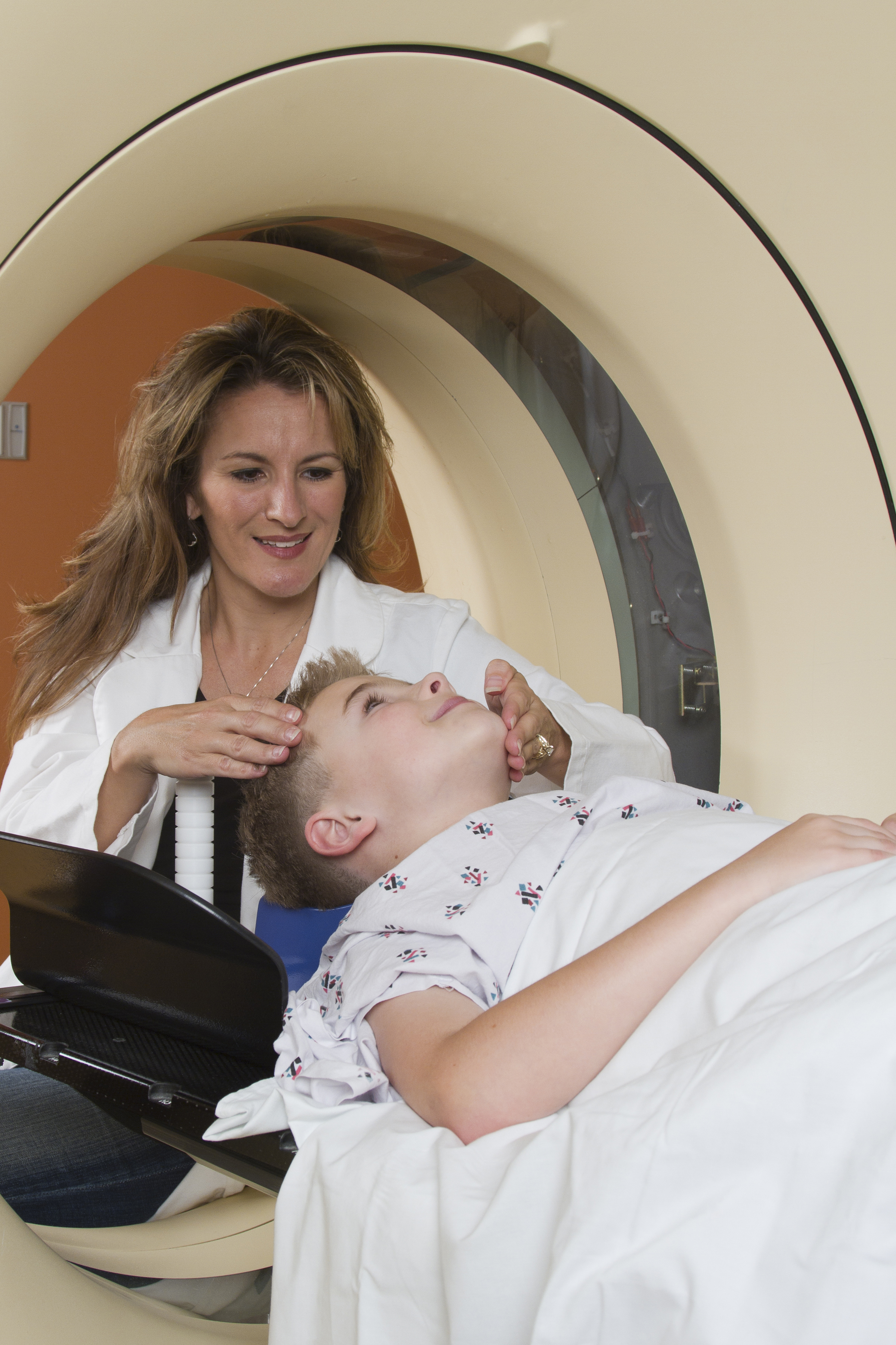
In a meta-analysis of eight prospective cohort studies including a total of 601 children published in Pediatrics, researchers looked at the association between S100B serum levels and CT findings in 373 patients. The median serum concentrations of S100B were 0.47 mcg/L for patients with intracerebral lesions and 0.21 mcg/L for those without lesions (P less than .001).
Additionally, researchers collected data from 358 individuals included in two studies for the origin of mTBI. The median concentrations of S100B were 0.39 mcg/L for road accidents, 0.29 mcg/L for domestic accidents, and 0.18 mcg/L for sport-related accidents. The difference was statistically significant between the road accidents group and the domestic accidents group (P less than .001) and the difference between the road accidents group and the sport-related accidents group (P less than .001). It is noted that S100B specificity could be higher after a sport-related trauma.
“S100B protein serum levels, in combination with the PECARN [Pediatric Emergency Care Applied Research Network] algorithm, could reduce the need for CT scans by one-third. In our additional analysis, based on 373 children, the importance of taking a blood sample 3 hours or less after trauma was underscored,” the researchers said.
“S100B represents a promising biomarker with 100% sensitivity. The limited specificity of S100B could be reevaluated for future research by using a combination of different brain biomarkers,” Dr. Oris and her colleagues concluded.
SOURCE: Oris C et al. Pediatrics. 2018. doi: 10.1542/peds.2018-0037.
according to Charlotte Oris, PharmD, of the University Hospital of Clermont-Ferrand, France, and her associates.
In a meta-analysis of eight prospective cohort studies including a total of 601 children published in Pediatrics, researchers looked at the association between S100B serum levels and CT findings in 373 patients. The median serum concentrations of S100B were 0.47 mcg/L for patients with intracerebral lesions and 0.21 mcg/L for those without lesions (P less than .001).
Additionally, researchers collected data from 358 individuals included in two studies for the origin of mTBI. The median concentrations of S100B were 0.39 mcg/L for road accidents, 0.29 mcg/L for domestic accidents, and 0.18 mcg/L for sport-related accidents. The difference was statistically significant between the road accidents group and the domestic accidents group (P less than .001) and the difference between the road accidents group and the sport-related accidents group (P less than .001). It is noted that S100B specificity could be higher after a sport-related trauma.
“S100B protein serum levels, in combination with the PECARN [Pediatric Emergency Care Applied Research Network] algorithm, could reduce the need for CT scans by one-third. In our additional analysis, based on 373 children, the importance of taking a blood sample 3 hours or less after trauma was underscored,” the researchers said.
“S100B represents a promising biomarker with 100% sensitivity. The limited specificity of S100B could be reevaluated for future research by using a combination of different brain biomarkers,” Dr. Oris and her colleagues concluded.
SOURCE: Oris C et al. Pediatrics. 2018. doi: 10.1542/peds.2018-0037.
Does Migraine Transiently Open the Blood–Brain Barrier?
STOWE, VT—Experimental evidence suggests that migraine, particularly migraine with aura, is associated with cortical spreading depression (CSD) that transiently disrupts the blood–brain barrier (BBB). For some, the question is not whether this disruption occurs, but whether it is clinically relevant. For others, even the basic premise of a CSD-mediated disruption of the BBB is suspect. At the Headache Cooperative of New England’s 28th Annual Stowe Headache Symposium, a debate between two researchers from opposing sides of this controversy indicated that there was little common ground. 
“Experiments in mice suggest that CSD might open the BBB and activate pain-sensitive fibers. However, you have heard nothing about data from humans, and in fact, many migraine patients report aura symptoms without head pain,” said Messoud Ashina, MD, PhD, Director of the Human Migraine Research Unit at the Danish Headache Center in Glostrup.
On the contrary, the scientific evidence that CSD opens the BBB is “incontrovertible,” according to Cenk Ayata, MD, PhD, Director of the Neurovascular Research Unit at Massachusetts General Hospital in Charlestown. “It is not a matter of whether the BBB opens, it is a matter of magnitude,” he said. “The critical question is whether it is clinically relevant.”
This controversy is of particular current interest. Although CSD has long been suspected to mediate migraine pain, a transient opening of the BBB may greatly affect the efficacy of monoclonal antibodies in development for migraine prophylaxis and treatment. Monoclonal antibodies that target the calcitonin gene-related peptide (CGRP) have shown promise in ongoing trials, but an opportunity to target the CNS directly could improve their efficacy. Monoclonal antibodies are too large to cross the BBB without increasing the latter’s permeability.
What Is CSD?
CSD is a wave of intense neurologic depolarization that is considered the electrophysiologic substrate of migraine aura, according to Dr. Ayata. The “massive transmembrane ion and water shifts hit the cerebral vasculature during CSD like a tsunami,” he said. Citing more than two decades’ worth of research, Dr. Ayata explained that CSD is accompanied by upregulation of multiple neurotransmitters and neuromodulators that trigger large blood flow responses and disrupt cerebrovascular reflexes, such as neurovascular coupling.
Initial studies of the role of CSD and its potential for disrupting the BBB were primarily related to stroke and head trauma. Numerous studies have supported the hypothesis that CSD is the underlying mechanism of migraine aura and a putative trigger for migraine headache. The specific sequence of events leading to CSD in migraine remains unclear, but Dr. Ayata and colleagues have conducted experiments that suggest that “CSD alone, without attendant tissue injury, can produce a transient opening of the BBB.”
In murine models of migraine, Evans blue leaked into the CNS after noninvasive induction of CSD. No leakage was observed in control animals that underwent a sham induction. Other controlled experiments that included objective measures of brain edema after CSD corroborated the association between CSD and BBB disruption.
“The opening of the BBB starts sometime between three and six hours [after induction of CSD], reaches a peak at six to 12 hours, and then gradually declines over the next 24 hours or so,” said Dr. Ayata. The BBB is completely restored at 48 hours, he added.
The Potential Role of Transcytosis
The BBB consists of astrocytes, pericytes, and endothelial cells with tight junctions that prevent most blood-borne substances from crossing into the CNS. Various processes can breach the BBB, and Dr. Ayata’s group is focusing on transcytosis. They hypothesize that ion pumps and other transporters cannot explain their observations that molecules as large as 70 kDa can pass through the BBB.
Transcytosis permits macromolecules from the luminal side of the endothelial cell to be brought into the cell by pinocytotic vesicles and to be released on the other side. It is a plausible mechanism for passage through the BBB, according to Dr. Ayata. Electron microscopy studies of BBB tissue from murine models strongly support this hypothesis.
“We found a significant increase in pinocytotic vesicles starting between three and six hours, but then a gradual decline to normal levels over the next 48 hours. This time course is exactly what we found in terms of leakage,” said Dr. Ayata. There was no evident change in endothelial tight junctions or in any other structure likely to provide an alternative mechanism for the observed BBB disruption, he added.
Although BBB disruption in an animal model of migraine is not proof of the same phenomenon in humans, Dr. Ayata suggested that the results are consistent with clinical observations. For example, a series of papers from 1985 to the present, including studies undertaken with gadolinium enhancement, found an association between severe migraine and documented edema. A twin study and studies of familial hemiplegic migraine also showed that CSD contributes to migraine pathogenesis and causes disruption of the BBB, he said.
Human Data Are Lacking
Although early studies provide a basis for the hypothesis that CSD is associated with migraine and might disrupt the BBB, clinical studies have consistently failed to link CSD with evidence of BBB disruption, said Dr. Ashina. In 1981, Olesen and colleagues reported cerebral hypoperfusion, a potential sign of CSD, followed by transient increases in cerebral perfusion (ie, hyperemia) during experimentally induced migraine aura.
“In some patients, headache disappeared when the hyperemia was observed, so there was no correlation,” said Dr. Ashina. Moreover, no changes in blood flow were observed when the same studies were conducted in patients with migraine without aura, and none of the studies reported changes in the permeability of the BBB in migraineurs with and without aura, according to Dr. Ashina. He and his colleagues at the Danish Headache Center remain active in this research.
He cited a 2017 study by Hougaard et al of 19 migraineurs with aura and 19 migraineurs without aura. Tissue perfusion in various parts of the brain was measured to assess change in BBB permeability. Patients underwent 3-T MRI during and in the absence of migraine attacks. “In aura patients, we found hyperperfusion in the brainstem during the headache phase of migraine with aura, while the BBB remained intact during attacks of migraine with aura,” said Dr. Ashina. Using sensitivity analyses, they looked for changes as small as 15%, but found nothing.
Other studies have looked more indirectly at the likelihood that the BBB is disrupted during migraine, but these, such as one that evaluated extracranial arterial dilatation during migraine attacks, have also been negative, according to Dr. Ashina. Studies of putative mechanisms for BBB disruption, such as one that evaluated the upregulation of matrix metalloproteinases, have also failed to support BBB disruption. “Nothing out there provides any evidence whatsoever that relates directly to BBB opening during migraine attacks with or without aura,” said Dr. Ashina.
If the BBB undergoes a transient disruption during migraine, it remains unclear whether this disruption is clinically meaningful or provides new opportunities to time treatment. The introduction of monoclonal antibodies for migraine may inspire the research needed to resolve this question.
Dr. Ashina has financial relationships with Alder BioPharmaceuticals, Allergan, Amgen, Eli Lilly, Novartis, and Teva. Dr. Ayata has no financial relationships relevant to this topic.
—Ted Bosworth
Amin FM, Asghar MS, Hougaard A, et al. Magnetic resonance angiography of intracranial and extracranial arteries in patients with spontaneous migraine without aura: a cross-sectional study. Lancet Neurol. 2013;12(5):454-461.
Amin FM, Hougaard A, Cramer SP, et al. Intact blood-brain barrier during spontaneous attacks of migraine without aura: a 3T DCE-MRI study. Eur J Neurol. 2017;24(9):1116-1124.
Ashina M, Tvedskov JF, Lipka K, et al. Matrix metalloproteinases during and outside of migraine attacks without aura. Cephalalgia. 2010;30(3):303-310.
Ayata C, Lauritzen M. Spreading depression, spreading depolarizations, and the cerebral vasculature. Physiol Rev. 2015;95(3):953-993.
Hougaard A, Amin FM, Christensen CE, et al. Increased brainstem perfusion, but no blood-brain barrier disruption, during attacks of migraine with aura. Brain. 2017;140(6):1633-1642.
Olesen J, Larsen B, Lauritzen M. Focal hyperemia followed by spreading oligemia and impaired activation of rCBF in classic migraine. Ann Neurol. 1981;9(4):344-352.
STOWE, VT—Experimental evidence suggests that migraine, particularly migraine with aura, is associated with cortical spreading depression (CSD) that transiently disrupts the blood–brain barrier (BBB). For some, the question is not whether this disruption occurs, but whether it is clinically relevant. For others, even the basic premise of a CSD-mediated disruption of the BBB is suspect. At the Headache Cooperative of New England’s 28th Annual Stowe Headache Symposium, a debate between two researchers from opposing sides of this controversy indicated that there was little common ground.
“Experiments in mice suggest that CSD might open the BBB and activate pain-sensitive fibers. However, you have heard nothing about data from humans, and in fact, many migraine patients report aura symptoms without head pain,” said Messoud Ashina, MD, PhD, Director of the Human Migraine Research Unit at the Danish Headache Center in Glostrup.
On the contrary, the scientific evidence that CSD opens the BBB is “incontrovertible,” according to Cenk Ayata, MD, PhD, Director of the Neurovascular Research Unit at Massachusetts General Hospital in Charlestown. “It is not a matter of whether the BBB opens, it is a matter of magnitude,” he said. “The critical question is whether it is clinically relevant.”
This controversy is of particular current interest. Although CSD has long been suspected to mediate migraine pain, a transient opening of the BBB may greatly affect the efficacy of monoclonal antibodies in development for migraine prophylaxis and treatment. Monoclonal antibodies that target the calcitonin gene-related peptide (CGRP) have shown promise in ongoing trials, but an opportunity to target the CNS directly could improve their efficacy. Monoclonal antibodies are too large to cross the BBB without increasing the latter’s permeability.
What Is CSD?
CSD is a wave of intense neurologic depolarization that is considered the electrophysiologic substrate of migraine aura, according to Dr. Ayata. The “massive transmembrane ion and water shifts hit the cerebral vasculature during CSD like a tsunami,” he said. Citing more than two decades’ worth of research, Dr. Ayata explained that CSD is accompanied by upregulation of multiple neurotransmitters and neuromodulators that trigger large blood flow responses and disrupt cerebrovascular reflexes, such as neurovascular coupling.
Initial studies of the role of CSD and its potential for disrupting the BBB were primarily related to stroke and head trauma. Numerous studies have supported the hypothesis that CSD is the underlying mechanism of migraine aura and a putative trigger for migraine headache. The specific sequence of events leading to CSD in migraine remains unclear, but Dr. Ayata and colleagues have conducted experiments that suggest that “CSD alone, without attendant tissue injury, can produce a transient opening of the BBB.”
In murine models of migraine, Evans blue leaked into the CNS after noninvasive induction of CSD. No leakage was observed in control animals that underwent a sham induction. Other controlled experiments that included objective measures of brain edema after CSD corroborated the association between CSD and BBB disruption.
“The opening of the BBB starts sometime between three and six hours [after induction of CSD], reaches a peak at six to 12 hours, and then gradually declines over the next 24 hours or so,” said Dr. Ayata. The BBB is completely restored at 48 hours, he added.
The Potential Role of Transcytosis
The BBB consists of astrocytes, pericytes, and endothelial cells with tight junctions that prevent most blood-borne substances from crossing into the CNS. Various processes can breach the BBB, and Dr. Ayata’s group is focusing on transcytosis. They hypothesize that ion pumps and other transporters cannot explain their observations that molecules as large as 70 kDa can pass through the BBB.
Transcytosis permits macromolecules from the luminal side of the endothelial cell to be brought into the cell by pinocytotic vesicles and to be released on the other side. It is a plausible mechanism for passage through the BBB, according to Dr. Ayata. Electron microscopy studies of BBB tissue from murine models strongly support this hypothesis.
“We found a significant increase in pinocytotic vesicles starting between three and six hours, but then a gradual decline to normal levels over the next 48 hours. This time course is exactly what we found in terms of leakage,” said Dr. Ayata. There was no evident change in endothelial tight junctions or in any other structure likely to provide an alternative mechanism for the observed BBB disruption, he added.
Although BBB disruption in an animal model of migraine is not proof of the same phenomenon in humans, Dr. Ayata suggested that the results are consistent with clinical observations. For example, a series of papers from 1985 to the present, including studies undertaken with gadolinium enhancement, found an association between severe migraine and documented edema. A twin study and studies of familial hemiplegic migraine also showed that CSD contributes to migraine pathogenesis and causes disruption of the BBB, he said.
Human Data Are Lacking
Although early studies provide a basis for the hypothesis that CSD is associated with migraine and might disrupt the BBB, clinical studies have consistently failed to link CSD with evidence of BBB disruption, said Dr. Ashina. In 1981, Olesen and colleagues reported cerebral hypoperfusion, a potential sign of CSD, followed by transient increases in cerebral perfusion (ie, hyperemia) during experimentally induced migraine aura.
“In some patients, headache disappeared when the hyperemia was observed, so there was no correlation,” said Dr. Ashina. Moreover, no changes in blood flow were observed when the same studies were conducted in patients with migraine without aura, and none of the studies reported changes in the permeability of the BBB in migraineurs with and without aura, according to Dr. Ashina. He and his colleagues at the Danish Headache Center remain active in this research.
He cited a 2017 study by Hougaard et al of 19 migraineurs with aura and 19 migraineurs without aura. Tissue perfusion in various parts of the brain was measured to assess change in BBB permeability. Patients underwent 3-T MRI during and in the absence of migraine attacks. “In aura patients, we found hyperperfusion in the brainstem during the headache phase of migraine with aura, while the BBB remained intact during attacks of migraine with aura,” said Dr. Ashina. Using sensitivity analyses, they looked for changes as small as 15%, but found nothing.
Other studies have looked more indirectly at the likelihood that the BBB is disrupted during migraine, but these, such as one that evaluated extracranial arterial dilatation during migraine attacks, have also been negative, according to Dr. Ashina. Studies of putative mechanisms for BBB disruption, such as one that evaluated the upregulation of matrix metalloproteinases, have also failed to support BBB disruption. “Nothing out there provides any evidence whatsoever that relates directly to BBB opening during migraine attacks with or without aura,” said Dr. Ashina.
If the BBB undergoes a transient disruption during migraine, it remains unclear whether this disruption is clinically meaningful or provides new opportunities to time treatment. The introduction of monoclonal antibodies for migraine may inspire the research needed to resolve this question.
Dr. Ashina has financial relationships with Alder BioPharmaceuticals, Allergan, Amgen, Eli Lilly, Novartis, and Teva. Dr. Ayata has no financial relationships relevant to this topic.
—Ted Bosworth
Amin FM, Asghar MS, Hougaard A, et al. Magnetic resonance angiography of intracranial and extracranial arteries in patients with spontaneous migraine without aura: a cross-sectional study. Lancet Neurol. 2013;12(5):454-461.
Amin FM, Hougaard A, Cramer SP, et al. Intact blood-brain barrier during spontaneous attacks of migraine without aura: a 3T DCE-MRI study. Eur J Neurol. 2017;24(9):1116-1124.
Ashina M, Tvedskov JF, Lipka K, et al. Matrix metalloproteinases during and outside of migraine attacks without aura. Cephalalgia. 2010;30(3):303-310.
Ayata C, Lauritzen M. Spreading depression, spreading depolarizations, and the cerebral vasculature. Physiol Rev. 2015;95(3):953-993.
Hougaard A, Amin FM, Christensen CE, et al. Increased brainstem perfusion, but no blood-brain barrier disruption, during attacks of migraine with aura. Brain. 2017;140(6):1633-1642.
Olesen J, Larsen B, Lauritzen M. Focal hyperemia followed by spreading oligemia and impaired activation of rCBF in classic migraine. Ann Neurol. 1981;9(4):344-352.